. 2010 Oct 8;40(1):75-86.
doi: 10.1016/j.molcel.2010.09.010.

ATM- and NEMO-dependent ELKS ubiquitination coordinates TAK1-mediated IKK activation in response to genotoxic stress

Affiliations

ATM- and NEMO-dependent ELKS ubiquitination coordinates TAK1-mediated IKK activation in response to genotoxic stress

Zhao-Hui Wu et al. Mol Cell. .

Abstract

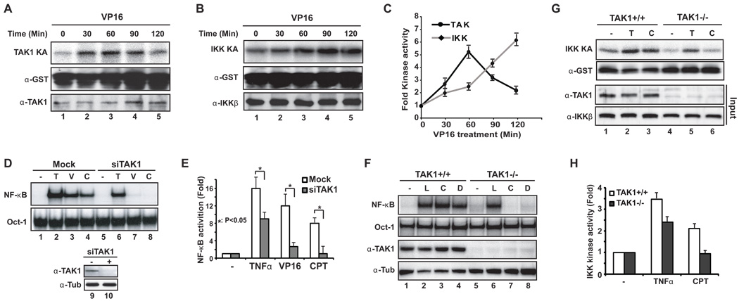
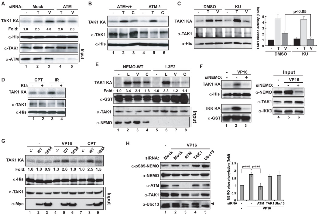
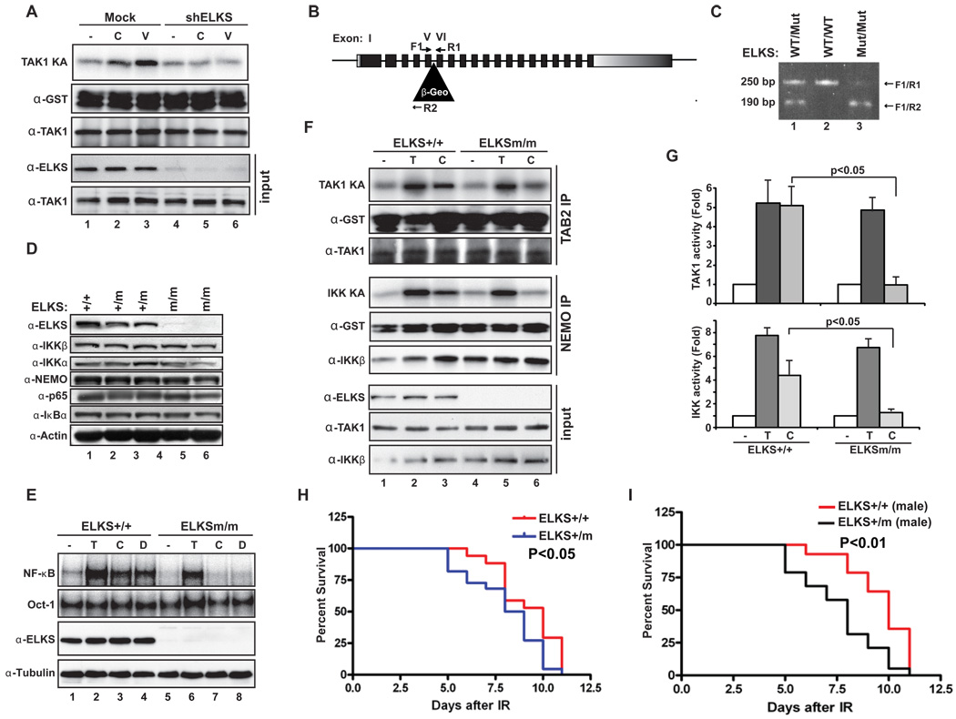
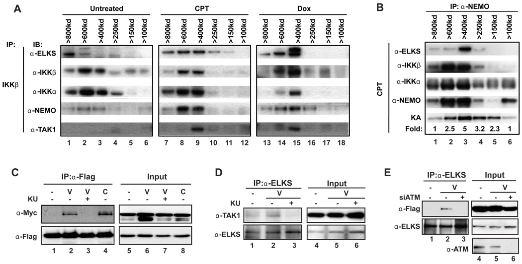
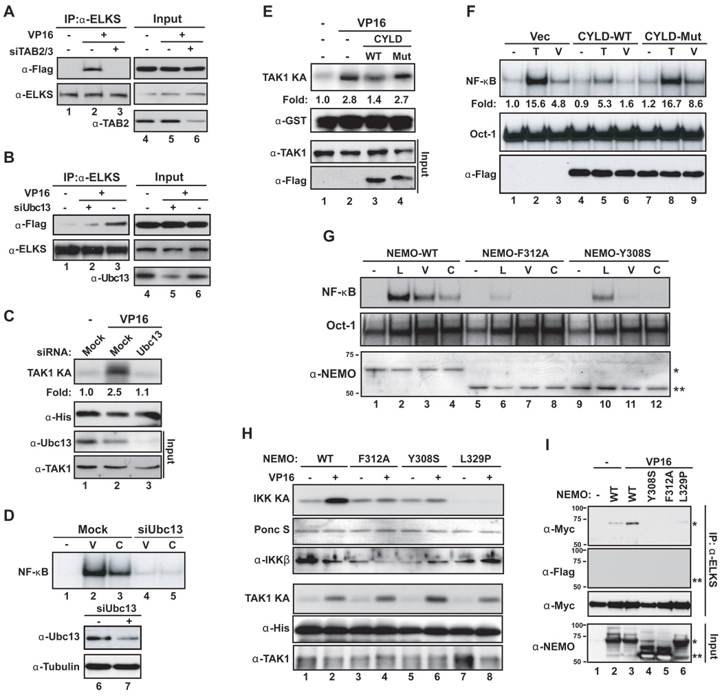
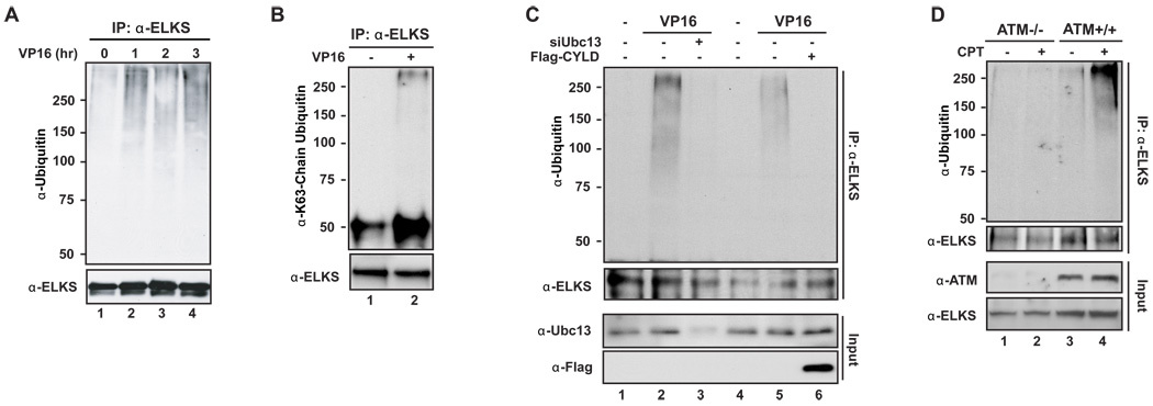
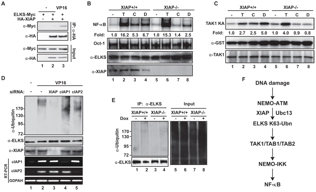
Activation of the transcription factor NF-κB by multiple genotoxic stimuli modulates cancer cell survival. This response is mediated by a conserved pathway involving the nuclear ATM kinase and cytoplasmic IκB kinase (IKK); however, the molecular link between them remains incompletely understood. Here we show that ATM activates the IKK kinase TAK1 in a manner dependent on IKKγ/NEMO and ELKS (a protein rich in glutamate, leucine, lysine, and serine). K63-linked polyubiquitination of ELKS, dependent on the ubiquitin ligase XIAP and the conjugating enzyme UBC13, allows ELKS association with TAK1 via its ubiquitin-binding subunits TAB2/3. Although NEMO mutants defective in ubiquitin binding permit ATM-dependent TAK1 activation, they block NEMO association with ELKS and IKK activation. Thus, ATM- and NEMO-dependent ubiquitination of ELKS leads to the ubiquitin-dependent assembly of TAK1/TAB2/3 and NEMO/IKK complexes, resulting in IKK and NF-κB activation following genotoxic stimuli.


Figures

Figure 1
Figure 1. TAK1 is required for NF-κB activation by DNA-damaging agents
(A) HEK293 cells were treated with VP16 (10 µM) for time as indicated. Whole cell lysates were used to precipitate TAB2 complex and subjected to a TAK1 kinase assay using His-MKK6 as the substrate. Western blotting with antibodies against His and TAK1 shows substrate and TAK1 present in each kinase reaction. (B) Similar cell treatment was carried our as in (A) by an IKK kinase assay using GST-IκBα as the substrate, and western blotting with antibodies as indicated. (C) A graph showing data obtained from three independent TAK1 or IKK kinase assays performed as in (A) and (B) are shown as Mean ± SD. (D) HEK293 cells were mock transfected or transfected with siRNA targeting TAK1, and treated with TNFα (10 ng/ml, 0.5 h), VP16 (10 µM, 2 h), CPT (10 µM, 2 h) or left untreated. Total cell extracts were analyzed by EMSA using 32P-labeled Igκ and Oct-1 probes, and by western blotting with antibodies as shown. (E) A graph showing data from three independent experiments as in (D) quantified by Phosphoimaging and analyzed by student-t test, shown as Mean ± SD. (F) TAK1+/+ and TAK−/− MEFs were treated with LPS (10 µg/ml, 0.5 h), CPT or Doxorubicin (5 µM, 2 h), and analyzed as in (D). (G) TAK1+/+ and TAK−/− MEFs were treated with TNFα, CPT or left untreated. IKK assay and blotting were done as in (B). (H) A graph showing data from two independent experiments as in (G) were plotted and shown as Mean ± SD. See also Figure S1.
Figure 2
Figure 2. ATM and NEMO are upstream of TAK1 activation
(A) HEK293 cells mock transfected or transfected with siRNA targeting ATM were treated with TNFα, VP16 or left untreated and whole cell lysates were analyzed by TAK1 kinase assay and western blotting. (B) TNFα or CPT induced TAK1 kinase activity in ATM−/− and ATM+/+ MEFs was measured as in (A). (C) HEK293 cell were treated as indicated, with or without KU55933 (10 µM), and analyzed by TAK1 kinase assay. The graph shows data from three independent experiments (analyzed by student-t test, mean+/−SD). (D) HEK293 cells were treated with CPT or IR (2 h post 10 Gy) with or without KU55933 and analyzed by TAK1 kinase assay. (E) NEMO-deficient 1.3E2 cells and those reconstituted with NEMO-WT were treated as indicated and analyzed by TAK1 kinase assay. Relative intensities were analyzed by Phosphorimager. Input (3%) was probed with indicated antibodies. (F) HEK293 cells were mock transfected or transfected with NEMO siRNA, treated with VP16 (1.5 h), and analyzed by TAK1 and IKK kinase assays. (G) 1.3E2 cells and those reconstituted with NEMO-WT or NEMO-S85A were treated and analyzed as in (E). (H) HEK293 cells were mock transfected or transfected with siRNA targeting ATM, TAK1 or Ubc13, and treated with VP16 (1 h) or DMSO alone. Phospho-S85-NEMO band was quantified by ImageJ analysis from three independent experiments and plotted with mean +/− SD. See also Figure S2.
Figure 3
Figure 3. ELKS mediates activation of TAK1 and IKK
(A) HEK293 cells were mocked transfected or transfected with shRNA targeting ELKS, treated with VP16 or CPT, then analyzed by TAK1 kinase assay using GST-IKKβ as the substrate. (B) Schematic drawing of the ELKS gene-trap where a fusion gene of β-galactosidase and neomycin phosphotransferase (β-Geo) was inserted between exon V and VI. The positions of primers (F1, R1, and R2) used in PCR genotyping are indicated. (C) PCR genotyping of ELKS+/+ (WT/WT), ELKS+/mut (WT/Mut), and ELKSmut/mut (Mut/Mut) mutant embryos. The size of PCR fragments is 250bp and 190 bp for WT and Mut alleles, respectively. (D) Whole cell lysates from indicated ELKS MEFs were analyzed by immunoblotting with antibodies as shown. (E) Indicated MEFs were treated with TNFα, CPT, Doxorubicin or left untreated and analyzed by EMSA and immunoblotting as indicated. (F) Indicated MEFs were treated as in (E), and analyzed by parallel TAK- and IKK-kinase assay using GST-IKKβ and GST-IκBα as the substrate, respectively. Input (2%) was probed as shown. (G) A graph showing data from three independent experiments as in (F) (mean+/−SD), analyzed with student-t test. (H) ELKS+/mut (male=13, female=9) and ELKS+/+ (male=11, female=6) mice were exposed to IR (11 Gy) and survival was plotted as shown. (I) Survival data of male ELKS+/+ (n=14) and ELKS+/mut (n=19) mice pooled from 3 independent experiments as in (H) and plotted. Significance was analyzed by Graphpad Prism 4. See also Figure S3.
Figure 4
Figure 4. ATM is required for ELKS association with TAK1
(A) Hela S3 cells were treated with PT (3 h), Doxorubicin (2 ug/ml, 3 h), or untreated, and whole cell extracts were run on Superdex 200 column and eluted fractions were analyzed by IP-western for IKKβ-associated protein complexes. The relative size of the complexes were inferred from the positions of molecular weight markers for the gel filtration: ribonuclease A (13.7 kDa), carbonic anhydrase (29 kDa), ovalbumin (43 kDa), conalbumin (75 kDa), aldolase (158 kDa), ferritin (440 kDa), thyroglobulin (669 kDa). (B) CPT-treated samples as in (A) were subjected to IKK kinase assay (KA) using full length IκBα as substrate and the relative kinase activity quantified by Phosphoimager is shown. (C) Flag-TAK1 and ELKS-Myc cotransfected HEK293 cells were left untreated or treated with CPT or VP16 with or without KU55933. Whole cell lysates were analyzed by IP with anti-Flag antibody followed by immunoblotting using antibodies as shown. (D) Endogenous ELKS from HEK293 cells treated as indicated was analyzed with IP using anti-ELKS antibody followed by immunoblotting with anti-TAK1 antibody. (E) HEK293 cells were transfected with Flag-TAK1 alone or cotransfected with ATM siRNA and analyzed as in (C). See also Figure S4.
Figure 5
Figure 5. K63-linked polyubiquitination mediates TAK1, ELKS and IKKs complex formation
(A) Endogenous ELKS from HEK293 cells co-transfected with Flag-TAK1 and siRNAs against TAB2 and 3 and treated as indicated was analyzed with IP-western analysis. (B) Experiments as in (A) were performed using UBC13 siRNAs. (C) Mock and Ubc13-depleted HEK293 treated with VP16 were analyzed by TAK1 kinase assay using His-MKK6 as the substrate. (D) Mock and Ubc13-depleted HEK293 treated with VP16 or CPT were analyzed by EMSA. (E) HEK293 cells were transfected with vector alone, CYLD-WT or catalytically inactive CYLD-Mut, and analyzed by TAK1 kinase assay as in (C). (F) HEK293 cells were transfected and treated as in (E) along with TNFα control and analyzed by EMSA as shown. (G) 1.3E2 cells stably reconstituted with NEMO-WT, -Y308S or -F312A mutants were treated as indicated and analyzed by EMSA. Expression of Myc6x-NEMO (*) and Flag-NEMO (**) is detected by western blotting. (H) Stable 1.3E2 cells reconstituted with indicated NEMO mutants were treated and analyzed by either IKK or TAK1 kinase assay. The loading of the substrates and kinases were visualized by either Ponceau S staining or western blotting. (I) HEK293 cells were transfected with NEMO-WT, -Y308S, -F312A or -L329P mutant along with ELKS-Myc construct, treated and analyzed by anti-ELKS IP followed by western blotting using indicated antibodies. * and ** are noted as in (G). Some additional NEMO products are also visible in the input panel. See also Figure S5.
Figure 6
Figure 6. ATM-dependent induction of ELKS K63-ubiquitination
(A) HEK293 cells were treated as indicated and endogenous ELKS was analyzed by anti-ELKS IP followed by immunoblotting using antiubiquitin antibody. (B) A sample (2 h) as in (A) was analyzed by an K63-linkage-specific ubiquitin antibody. (C) Cells were treated and analyzed as in (A) following mock transfected or transfected with either Ubc13 siRNA or Flag-CYLD wt. (D) ATM+/+ and ATM−/− MEFs cells were treated with CPT or DMSO and ELKS ubiquitination was analyzed as in (A). See also Figure S6.
Figure 7
Figure 7. XIAP facilitates ELKS ubiquitination
(A) HEK293 cells were transfected with HA-XIAP and ELKS-Myc, treated as indicated and subjected to anti-HA IP followed by anti-Myc blotting. (B, C) XIAP+/+ and XIAP−/− MEFs were treated with TNFα (T), CPT (C), Doxorubicin (D) and analyzed by EMSA (B) or TAK1 kinase assay (C). (D) HEK293 cells were mock transfected or transfected with XIAP siRNA and ELKS ubiquitination was analyzed by anti-ELKS IP followed by anti-Ub blotting. (E) XIAP+/+ and XIAP−/− MEFs were treated with Doxorubicin or left untreated and ELKS ubiquitination was analyzed as in (D). (F) A model depicting NF-κB activation upon genotoxic stress. See Discussion for details.

Similar articles

Cited by

References

    1. Biswas SK, Bist P, Dhillon MK, Kajiji T, Del Fresno C, Yamamoto M, Lopez-Collazo E, Akira S, Tergaonkar V. Role for MyD88-independent, TRIF pathway in lipid A/TLR4-induced endotoxin tolerance. J Immunol. 2007;179:4083–4092. - PubMed
    1. Chen ZJ, Bhoj V, Seth RB. Ubiquitin, TAK1 and IKK: is there a connection? Cell Death Differ. 2006;13:687–692. - PubMed
    1. Chen ZJ, Sun LJ. Nonproteolytic Functions of Ubiquitin in Cell Signaling. Mol Cell. 2009;33:275–286. - PubMed
    1. Chew J, Biswas S, Shreeram S, Humaidi M, Wong ET, Dhillion MK, Teo H, Hazra A, Fang CC, López-Collazo E, Bulavin DV, Tergaonkar V. WIP1 phosphatase is a negative regulator of NF-kappaB signalling. Nat Cell Biol. 2009;11:659–666. - PubMed
    1. Conze DB, Wu C-J, Thomas JA, Landstrom A, Ashwell JD. Lys63-Linked Polyubiquitination of IRAK-1 Is Required for Interleukin-1 Receptor- and Toll-Like Receptor-Mediated NF-κB Activation. Mol Cell Biol. 2008;28:3538–3547. - PMC - PubMed

Publication types

MeSH terms

Substances