. 2010 Dec 15;432(3):417-27.
doi: 10.1042/BJ20101440.

Mycobacterium tuberculosis WhiB1 is an essential DNA-binding protein with a nitric oxide-sensitive iron-sulfur cluster

Affiliations

Mycobacterium tuberculosis WhiB1 is an essential DNA-binding protein with a nitric oxide-sensitive iron-sulfur cluster

Laura J Smith et al. Biochem J. .

Abstract

Mycobacterium tuberculosis is a major pathogen that has the ability to establish, and emerge from, a persistent state. Wbl family proteins are associated with developmental processes in actinomycetes, and M. tuberculosis has seven such proteins. In the present study it is shown that the M. tuberculosis H37Rv whiB1 gene is essential. The WhiB1 protein possesses a [4Fe-4S]2+ cluster that is stable in air but reacts rapidly with eight equivalents of nitric oxide to yield two dinuclear dinitrosyl-iron thiol complexes. The [4Fe-4S] form of WhiB1 did not bind whiB1 promoter DNA, but the reduced and oxidized apo-WhiB1, and nitric oxide-treated holo-WhiB1 did bind to DNA. Mycobacterium smegmatis RNA polymerase induced transcription of whiB1 in vitro; however, in the presence of apo-WhiB1, transcription was severely inhibited, irrespective of the presence or absence of the CRP (cAMP receptor protein) Rv3676, which is known to activate whiB1 expression. Footprinting suggested that autorepression of whiB1 is achieved by apo-WhiB1 binding at a region that overlaps the core promoter elements. A model incorporating regulation of whiB1 expression in response to nitric oxide and cAMP is discussed with implications for sensing two important signals in establishing M. tuberculosis infections.


Figures
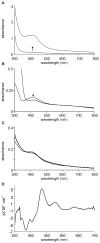
Figure 1
Figure 1. The M. tuberculosis WhiB1 protein iron-sulphur cluster can be reconstituted under anaerobic conditions and is O2 stable
(A) Analysis of the WhiB1 (250 μM) reconstitution reaction under anaerobic conditions by UV-visible spectroscopy. The spectra shown are before (lower line) and after (upper line) reconstitution. The arrow indicates the increase in absorbance at 420 nm with time. (B) Spectrum of reconstituted WhiB1 (25 μM cluster) in 20 mM sodium phosphate buffer (pH 7.4) containing 0.5 M NaCl and 1 mM DTT before and after treatment with sodium dithionite (0.2 mM for 15 min at 20°C). The arrow indicates the decrease in absorbance at 420 nm. (C) Spectra of WhiB1 (10 μM cluster) in 25 mM Tris-HCl buffer (pH 7.4) containing 0.5 M NaCl, 10% glycerol and 1 mM DTT before (thin line) and after (thicker line) incubation for 2 h in the presence of 110 μM O2. (D) CD spectrum of reconstituted WhiB1 (196 μM cluster) in 20 mM sodium phosphate buffer (pH 7.4) containing 0.5 M NaCl.
Figure 2
Figure 2. The WhiB1 iron-sulphur cluster reacts with NO
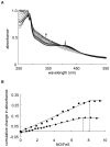
(A) Reconstituted WhiB1 (19 μM cluster) was exposed to increasing amounts of proline NONOate under anaerobic conditions and spectra were obtained 10 min after each addition. The arrows indicate the decrease in absorbance at 420 nm and the increase in absorbance at 355 nm as the concentration of proline NONOate was increased. (B) The cumulative changes in absorbance at 355 nm (corresponding to the formation of DNICs) from two representative NO titrations under anaerobic conditions in the absence of DTT were calculated taking into account the dilution associated with successive additions of proline NONOate. These values were plotted against the total NO added (μM) divided by the initial concentration of iron-sulphur cluster (μM) in the sample. The dashed vertical lines indicate the points at which further addition of NONOate failed to change the absorbance at 355 nm. All samples were in 25 mM Tris-HCl buffer (pH 7.4) containing 0.5 M NaCl and 10% glycerol.
Figure 3
Figure 3. EPR spectra of WhiB1 and the response to NO
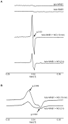
(A) From top to bottom: apo-WhiB1; anaerobically reconstituted holo-WhiB1; reconstituted WhiB1 treated with a 27-fold molar excess of proline NONOate for 10 min under anaerobic conditions; reconstituted WhiB1 treated with a 27-fold molar excess of proline NONOate for 2 h under anaerobic conditions. Spectra were recorded at 20 K. (B) EPR spectra (g = ~2 region) of the EPR active samples shown in panel A measured at 77 K for quantification. In all cases the WhiB1 protein concentration was 17 μM. The spectra were obtained using a continuous-wave EPR spectrometer with a microwave frequency of 9.68 GHz operating at a 100 kHz magnetic field modulation. All samples were in 25 mM Tris-HCl buffer (pH 7.4) containing 1 mM DTT.
Figure 4
Figure 4. Stability of the DNIC form of WhiB1
Trace 1 shows the spectrum of holo-WhiB1 (4.2 μM cluster). This protein was treated with 400 μM proline NONOate under anaerobic conditions resulting in spectrum 2 (~10 mM DNIC), which was unchanged after 2 h. The cuvette was then uncapped and the contents were exposed to air yielding spectrum 3 after 2 h and spectrum 4 after >24 h. The temperature was 25°C, and all samples were in 25 mM Tris-HCl buffer (pH 7.4) containing 0.5 M NaCl, 1 mM DTT and 10% glycerol.
Figure 5
Figure 5. WhiB1 is a DNA-binding protein
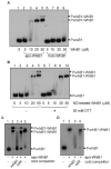
(A) Binding of WhiB1 at the whiB1 promoter (PwhiB1) region. Radiolabelled PwhiB1 was incubated with the indicated amounts of apo- or holo-WhiB1 before separation of protein-DNA complexes (PwhiB1-WhiB1) by electrophoresis. (B) Binding of NO-treated holo-WhiB1 to PwhiB1. A 20-fold molar excess of NO (as proline NONOate) was added to holo-WhiB1 and after incubation for 1 min at 20°C an aliquot was removed and DTT was added to a final concentration of 30 mM. After further incubation at 20°C for 3 min EMSAs were done using the DTT-treated and untreated samples as described in Materials and Methods. The final concentrations of WhiB1 protein are indicated. (C) Oxidation of WhiB1 results in the formation of a second retarded species. Binding of oxidized apo-WhiB1 (20 μM, pre-treated with 5 mM diamide) to radiolabelled PwhiB1 (lane 2) was challenged by the presence of 100-fold excess unlabelled PwhiB1 DNA (lane 3) or 100-fold excess unlabeled PrpfA DNA (lane 4). Lane 5 shows the EMSA with reduced (1 mM DTT) apo-WhiB1 and PwhiB1. (D) Apo-WhiB1 binding at PwhiB1 is specific. Binding of apo-WhiB1 (20 μM) in the presence of DTT (1 mM) to radiolabelled PwhiB1 (lane 2) was challenged by the presence of 100-fold excess unlabelled PwhiB1 DNA (lane 3) or 100-fold excess unlabeled PrpfA DNA (lane 4).
Figure 6
Figure 6. DNase I footprint of apo-WhiB1 at PwhiB1
(A) Increasing concentrations of apo-WhiB1, as indicated above each lane, were incubated with PwhiB1 before DNase I digestion as described in Materials and Methods. Hypersensitive sites associated with interaction with apo-WhiB1 are arrowed; numbering is relative to the transcriptional start site. The locations of apo-WhiB1 interaction (W1), the previously identified Rv3676 (CRPMt) binding sites (CRP1 and CRP2), and the −10 element are indicated. Lane 5 shows a Maxam and Gilbert G track. (B) Nucleotide sequence of PwhiB1 showing: the transcript start (+1, block arrow), the −10 and −35 elements (underlined), the tandem Rv3676 (CRPMt) binding sites (boxed, CRP1 and CRP2), the hypersensitive sites that characterize interaction with apo-WhiB1 (bold upper case), the location of the protected region (W1; overlined), and two related DNA sequences within the protected region (bold lower case) are indicated.
Figure 7
Figure 7. Apo-WhiB1 inhibits transcription of whiB1 in vitro
Reactions contained: 0.1 pmoles PwhiB1, 1 pmole M. smegmatis RNAP, 40 mM Tris-Cl pH 8.0, 10 mM MgCl2, 70 mM NaCl, 1 mM EDTA, 1 mM DTT, 250 μg ml−1 BSA, 5% glycerol. Proteins (Rv3676 and apo-WhiB1) were pre-incubated with PwhiB1 for 10 min at 37°C before addition of nucleotide triphosphates and further incubation for 10 min at 37°C. The location of the whiB1 transcript, the loading control, and the concentrations of the Rv3676 and apo-WhiB1 proteins, are indicated.
Figure 8
Figure 8. Alignment of the conserved cysteine-rich motif in Wbl proteins and the O2-sensor FNR
The protein sequences aligned are the Wbl-family proteins WhiB1 and WhiB3 from M. tuberculosis, WhiD from S. coelicolor, and the O2-sensing transcription factor FNR from E. coli. The sequences are shown in single letter amino acid code and the Cys residues are labelled 1-3 for ease of identification in the text.
Figure 9
Figure 9. Nitric oxide- and cAMP-mediated regulation of whiB1 expression
The forms of WhiB1 are represented by labelled circles, where no specific form is indicated this represents a mixture of oxidized and reduced apo-WhiB1 and DNIC-WhiB1. The whiB1 promoter DNA (PwhiB1), Rv3676 (labelled circles), NO and iron-sulphur cluster biosynthesis machinery are also indicated. From bottom left, when cAMP levels are low and NO is absent, transcription of whiB1 occurs at basal levels. At intermediate concentrations of cAMP, expression of whiB1 is activated by the M. tuberculosis CRP protein (Rv3676) binding at class I site to allow increased synthesis of apo-WhiB1 [5]. At high concentrations of cAMP, Rv3676 occupies tandem sites at PwhiB1 and represses whiB1 expression [5]. Apo-WhiB1 protein acquires a [4Fe-4S]2+ cluster by the action of the iron-sulphur cluster biosynthesis system (Figure 1), resulting in the non-DNA-binding form. Each WhiB1 [4Fe-4S]2+ cluster reacts extremely rapidly with 8NO (Figure 2) to produce a mixture of mononuclear and dinuclear dinitrosyl-iron complexes (DNIC; Figures 2 and 3). In the presence of O2, the DNIC form of WhiB1 slowly degrades to yield apo-WhiB1 (Figure 4). DNA-binding activity has been demonstrated for reduced and oxidized apo-WhiB1 and NO-treated WhiB1 (Figures 5 and 6), but it is not yet known whether the minority mononuclear and majority dinuclear DNIC forms possess DNA-binding activity, or whether they are intermediate species that yield the proven DNA-binding form, apo-WhiB1; however, the relative stability of the DNIC form (Figure 4) and the EPR spectra (Figure 3), suggest that the EMSAs with NO-treated WhiB1 contain mostly dinuclear DNIC WhiB1. The DNA-binding apo-WhiB1 severely represses whiB1 expression in vitro in the presence and absence of the M. tuberculosis CRP, Rv3676, by interactions at site(s) overlapping the −10 and −35 elements of the promoter (Figures 6 and 7). Apo-WhiB1 can be switched off by the action of the iron-sulphur cluster assembly machinery to regenerate the non-DNA-binding holo-WhiB1 resulting in derepression of whiB1 expression and synthesis of apoWhiB1 protein.

Similar articles

Cited by

References

    1. World Health Organization . Global Tuberculosis Control: a short update to the 2009 report. WHO Press; Geneva Switzerland: 2009. WHO/HTM/TB/2009.426.
    1. Russell DG. Mycobacterium tuberculosis: here today, and here tomorrow. Nat. Rev. Mol. Cell Biol. 2001;2:569–577. - PubMed
    1. Stewart GR, Robertson BD, Young DB. Tuberculosis: a problem with persistence. Nat. Rev. Microbiol. 2003;1:97–105. - PubMed
    1. Rickman L, Scott C, Hunt DM, Hutchinson T, Menendez MC, Whalan R, Hinds J, Colston MJ, Green J, Buxton RS. A member of the cAMP receptor protein family of transcription regulators in Mycobacterium tuberculosis is required for virulence in mice and controls transcription of the rpfA gene coding for a resuscitation promoting factor. Mol. Microbiol. 2005;56:1274–1286. - PMC - PubMed
    1. Stapleton MR, Haq I, Hunt DM, Arnvig KB, Artymiuk PJ, Buxton RS, Green J. Mycobacterium tuberculosis cAMP receptor protein (Rv3676) differs from the Escherichia coli paradigm in its cAMP binding and DNA binding properties and transcription activation properties. J. Biol. Chem. 2010;285:7016–7027. - PMC - PubMed

Publication types

MeSH terms