. 2010 Oct 4;1(1):10.
doi: 10.1186/2041-9139-1-10.

Genomic insights into Wnt signaling in an early diverging metazoan, the ctenophore Mnemiopsis leidyi

Affiliations

Genomic insights into Wnt signaling in an early diverging metazoan, the ctenophore Mnemiopsis leidyi

Kevin Pang et al. Evodevo. .

Abstract

Background: Intercellular signaling pathways are a fundamental component of the integrating cellular behavior required for the evolution of multicellularity. The genomes of three of the four early branching animal phyla (Cnidaria, Placozoa and Porifera) have been surveyed for key components, but not the fourth (Ctenophora). Genomic data from ctenophores could be particularly relevant, as ctenophores have been proposed to be one of the earliest branching metazoan phyla.

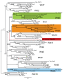
Results: A preliminary assembly of the lobate ctenophore Mnemiopsis leidyi genome generated using next-generation sequencing technologies were searched for components of a developmentally important signaling pathway, the Wnt/β-catenin pathway. Molecular phylogenetic analysis shows four distinct Wnt ligands (MlWnt6, MlWnt9, MlWntA and MlWntX), and most, but not all components of the receptor and intracellular signaling pathway were detected. In situ hybridization of the four Wnt ligands showed that they are expressed in discrete regions associated with the aboral pole, tentacle apparati and apical organ.

Conclusions: Ctenophores show a minimal (but not obviously simple) complement of Wnt signaling components. Furthermore, it is difficult to compare the Mnemiopsis Wnt expression patterns with those of other metazoans. mRNA expression of Wnt pathway components appears later in development than expected, and zygotic gene expression does not appear to play a role in early axis specification. Notably absent in the Mnemiopsis genome are most major secreted antagonists, which suggests that complex regulation of this secreted signaling pathway probably evolved later in animal evolution.


Figures

Figure 1
Figure 1
Non-bilaterian animal relationships. (A) Representative images of non-bilaterian animals, Ctenophora (Mnemiopsis leidyi), Cnidaria (Nematostella vectensis), Placozoa (Trichoplax adhaerens) and Porifera (Dysidea spp.). Photos courtesy of William E. Browne and Eric Roettinger. (B-G) Alternate hypotheses on early animal evolution and the placement of the ctenophores, based on (B) morphological data, (C) 18S ribosomal RNA results, (D-F) different phylogenomic analyses and (G) a combined morphological and phylogenomic approach.
Figure 2
Figure 2
Ctenophore development and body plan. (A) Early cleavage from egg to 60-cell stage, based on Martindale and Henry [20] and others. The top row shows the view from the aboral (or vegetal) pole and the bottom row shows the lateral view, with the oral pole at the bottom. The first two divisions are equal and meridional, and the third cleavage is unequal and oblique, giving rise to middle (M) and end (E) macromeres. Subsequent divisions are unequal, with micromeres given off at the aboral pole. (B) Basic ctenophore body plan, as shown during the cydippid stage. The oral pole is the location of the mouth, which opens to the pharynx. The pharynx leads internally to the gut and endodermal canals (yellow). Also shown are paired tentacle bulbs (from which the tentacles grow), the eight comb rows, and the apical sensory organ located at the aboral pole.
Figure 3
Figure 3
Overview of Wnt/β-catenin signaling Ctenophore pathway. (A) When Wnt signaling is inactive, cytoplasmic β-catenin protein is bound by the 'destruction complex' of axin, glycogen synthase kinase 3 (GSK-3) and adenomatous polyposis coli (APC). While sequestered, GSK-3 phosphorylates β-catenin, which targets β-catenin for ubiquitination and degradation. (B) In the presence of a Wnt ligand, the pathway is activated. Wnt binds to the seven-transmembrane receptor Frizzled and its co-receptor lipoprotein receptor-related protein 5/6 (LRP5/6), which causes Dishevelled (Dsh) to be activated. Dsh inhibits GSK-3, thereby allowing β-catenin to accumulate in the cytoplasm. Eventually, β-catenin gets translocated to the nucleus, where it interacts with the transcription factor T-cell-specific transcription factor/lymphoid enhancer binding factor (TCF/LEF) to activate target genes. The diffusible antagonists (Secreted Frizzled-related (Sfrp), Dickkopf (DKK), Wnt Inhibitory Factor (WIF) and Cerberus (CER)) can modulate Wnt activity by preventing the binding of Wnt to its receptors.
Figure 4
Figure 4
Wnt gene orthology. Bayesian analysis to determine orthology of Mnemiopsis Wnt genes. Shown is a consensus tree of four independent runs of 5 million generations each. Posterior probability support is shown at each node, as well as maximum likelihood (PhyML with WAG model) bootstrap support (posterior probability/ML bootstrap). Asterisks at each node indicate ≥ 99%. support. A dash (-) indicates that the node was not supported by maximum likelihood analyses. Mnemiopsis Wnt genes are shown shaded and marked with arrows. Ate = Archaearanea tepidariorum; Bfl = Branchiostoma floridae; Cte = Capitella teleta; Hsa = Homo sapiens; Mle = Mnemiopsis leidyi; Nve = Nematostella vectensis; Pdu = Platynereis dumerilii; Sko = Saccoglossus kowalevskii; Spu = Strongylocentrotus purpuratus; Tca = Tribolium castaneum.
Figure 5
Figure 5
Wnt gene structure and domains of Wnt pathway members. (A) Intron-exon structure of the four Mnemiopsis Wnt transcripts that were cloned. Turquoise shading indicates the coding region and the diagonal lines show the 5' and 3' untranslated regions. The start (ATG) is indicated, and the vertical lines represent intron positions. The conserved intron positions are marked with arrows. (B) Predicted protein domains present in the other Wnt components that were cloned out. Specific domains and other regions of interest are colored and named.
Figure 6
Figure 6
Wnt gene expression during development. Whole- mount in situ hybridization analyses of all Wnt genes during development. The timeline at the top depicts hours post-fertilization (HPF). All images are oriented laterally, unless otherwise specified (aboral). For lateral views, the oral pole is at the bottom and aboral pole at the top, with an asterisk (*) marking the blastopore/mouth. For the aboral views, the tentacular plane is horizontal and the sagittal plane vertical. Gene expression is detected colorimetrically and shown by the blue/purple staining. (A) MlWnt9 is first detected after gastrulation in four aboral regions of the future tentacle bulb (arrows). After approximately 9 HPF, these four groups of cells converge along the tentacular plane and form two groups of cells within the tentacle bulb. (B) MlWntA is also expressed in four groups of cells of the forming tentacle bulb, slightly more internal than MlWnt9. It remains expressed in these four groups of cells at the periphery of the tentacle bulb. (C) MlWnt6 is expressed in both the tentacle bulb and the floor of the apical organ (ao). The tentacular staining is fainter in the cydippid stage; however, the apical organ staining remains prominent. (D) MlWntX is expressed both in the apical organ floor and in the ciliated groove (cg), which is the structure that connects the apical organ to the individual comb rows.
Figure 7
Figure 7
Expression of Wnt pathway components. Whole-mount in situ hybridization of other members of the Wnt pathway, including (A) MlFzdA, (B) MlFzdB, (C) MlSfrp, (D) MlDsh, (E) MlBcat and (F) MlTcf. The timeline above the images denotes the different stages of embryos below, from 0-3 hours post-fertilization (if applicable) to 24 HPF or the cydippid stage. Unless noted, all images are lateral views, with the asterisk marking the blastopore or mouth. Blue/purple staining shows where the genes are expressed. (A) MlFzdA is detected uniformly from egg, through early cleavage stages and gastrulation. From 9 HPF onward, it is expressed mainly in the tentacle bulb (arrows) and pharynx (ph). (B) MlFzdB is not detected until 3-4 HPF in cells of the ectoderm (ec). At 5-6 HPF, it is expressed in the tentacle bulb and around the blastopore, in cells that will invaginate to form the pharynx. Later, it is additionally expressed in the trans-tentacular muscle (white arrow), which connects the two tentacles. (C) MlSfrp is expressed in the invaginating pharynx and in the presumptive mesoderm (mes). This mesodermal expression becomes confined to two regions of the tentacle bulb, which becomes barely detectable in the cydippids. The pharyngeal expression is also not detected in cydippid stages. (D) MlDsh is expressed uniformly from egg to cydippid stages. (E) MlBcat is first detected after gastrulation (about 4 HPF) in ectodermal cells around the blastopore. This blastoporal expression continues however, at 6 HPF there is MlBcat expression everywhere, except in the cells that form the comb plates (arrowheads). (F) MlTcf is expressed primarily in the ectoderm after gastrulation but excluded from cells that form the comb plates. At cydippid stages, it is expressed in discrete regions of the apical organ floor (ao) and in the tentacle bulbs.

Similar articles

Cited by

References

    1. Nielsen C, Scharff N, Eibye-Jacobsen D. Cladistic analyses of the animal kingdom. Biol J Linn Soc. 1996;57:385–410. doi: 10.1111/j.1095-8312.1996.tb01857.x. - DOI
    1. Zrzavy J, Mihulka S, Kepka P, Bezdek A, Tietz D. Phylogeny of the Metazoa based on morphological and 18S ribosomal DNA evidence. Cladistics. 1998;14:249–285. - PubMed
    1. Collins AG. Evaluating multiple alternative hypotheses for the origin of Bilateria: an analysis of 18S rRNA molecular evidence. Proc Natl Acad Sci USA. 1998;95:15458–15463. doi: 10.1073/pnas.95.26.15458. - DOI - PMC - PubMed
    1. Kim J, Kim W, Cunningham CW. A new perspective on lower metazoan relationships from 18S rDNA sequences. Mol Biol Evol. 1999;16:423–427. - PubMed
    1. Podar M, Haddock SHD, Sogin ML, Harbison GR. A molecular phylogenetic framework for the phylum Ctenophora using 18S rRNA genes. Mol Phylogenet Evol. 2001;21:218–230. doi: 10.1006/mpev.2001.1036. - DOI - PubMed

LinkOut - more resources