. 2010 Nov;177(5):2622-34.
doi: 10.2353/ajpath.2010.091075. Epub 2010 Oct 1.

Oncogenic role of the E3 ubiquitin ligase NEDD4-1, a PTEN negative regulator, in non-small-cell lung carcinomas

Affiliations

Oncogenic role of the E3 ubiquitin ligase NEDD4-1, a PTEN negative regulator, in non-small-cell lung carcinomas

Nicola Amodio et al. Am J Pathol. 2010 Nov.

Abstract

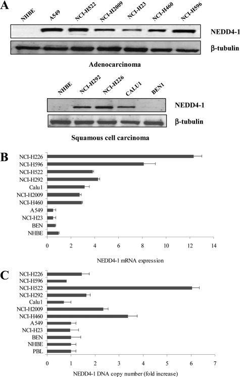
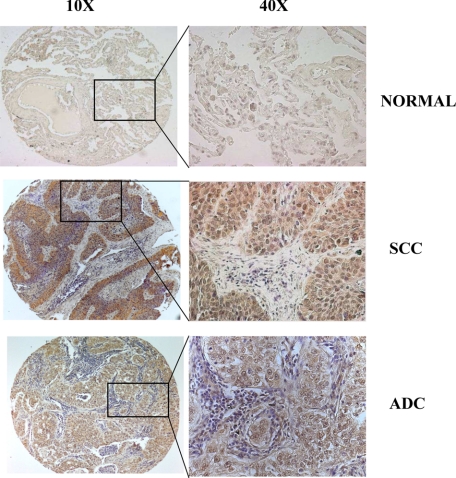
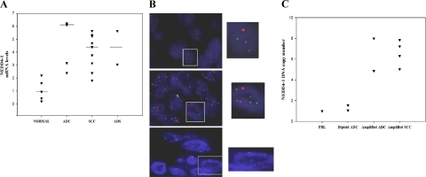
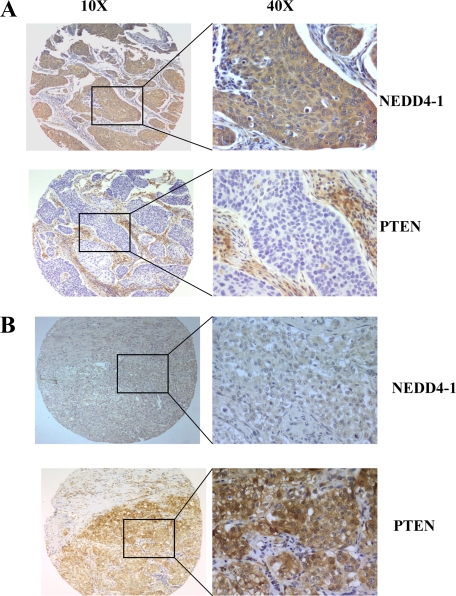
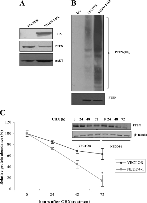
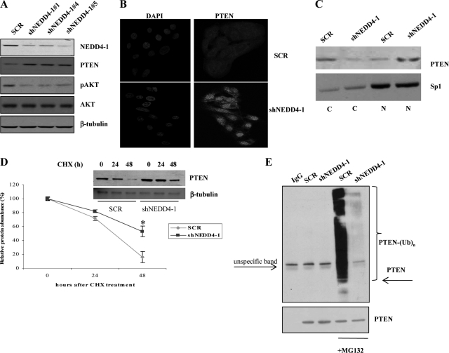
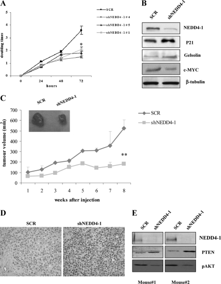
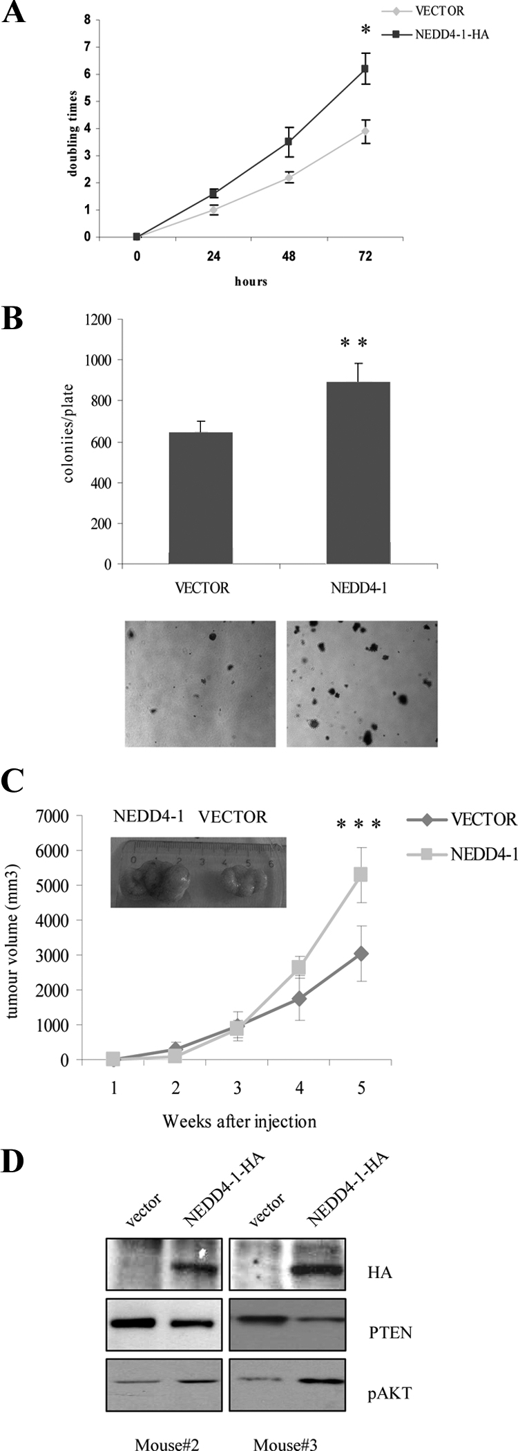
Loss of the PTEN tumor suppressor gene occurs frequently in non-small-cell lung carcinoma (NSCLC), although neither genetic alterations nor epigenetic silencing are significant predictors of PTEN protein levels. Since recent reports implicated neural precursor cell expressed, developmentally down-regulated 4-1 (NEDD4-1) as the E3 ubiquitin ligase that regulates PTEN stability, we investigated the role of NEDD4-1 in the regulation of PTEN expression in cases of NSCLC. Our findings indicate that NEDD4-1 plays a critical role in the development of NSCLC and provides novel insight on the mechanisms that contribute to inactivate PTEN in lung cancer. Immunohistochemical analysis on tissue microarrays containing 103 NSCLC resections revealed NEDD4-1 overexpression in 80% of tumors, which correlated with the loss of PTEN protein (n=98; P<0.001). Accordingly, adoptive NEDD4-1 expression in NSCLC cells decreased PTEN protein stability, whereas knock-down of NEDD4-1 expression decreased PTEN ubiquitylation and increased PTEN protein levels. In 25% of cases, NEDD4-1 overexpression was due to gene amplification at 15q21. In addition, manipulation of NEDD4-1 expression in different lung cell systems demonstrated that suppression of NEDD4-1 expression significantly reduced proliferation of NSCLC cells in vitro and tumor growth in vivo, whereas NEDD4-1 overexpression facilitated anchorage-dependent and independent growth in vitro of nontransformed lung epithelial cells that lack pRB and TP53 (BEAS-2B). NEDD4-1 overexpression also augmented the tumorigenicity of lung cancer cells that have an intact PTEN gene (NCI-H460 cells).


Figures

Figure 1
Figure 1
NEDD4-1 expression in human NSCLC cell lines. A: Immunoblot analysis of NEDD4-1 in human NSCLC-derived cell lines. NHBE, normal human lung epithelial cells; ADC-derived cell lines: A549, NCI-H522, NCI-H2009, NCI-H23, NCI-H460, and NCI-H596; SCC-derived cell lines: NCI-H292, NCI-H226, CALU1, and BEN1. B: NEDD4-1 relative mRNA expression by quantitative RT-PCR. C: NEDD4-1 relative gene number by Q-PCR. DNA copy number from normal PBLs was set arbitrarily to 1.
Figure 2
Figure 2
NEDD4-1 expression in human primary NSCLC by TMA immunostaining. Expression of NEDD4-1 in NSCLC: representative immunostaining of NEDD4-1 in normal and pathological lung tissues. Serial formalin-fixed paraffin-embedded sections from normal lung (number 11520), SCC (number 2252), or ADC (number 3044) were analyzed for NEDD4-1 expression. Epithelial cells from normal lung tissue are negative for NEDD4-1 expression (upper panels); cancer cells show intense NEDD4-1 expression in SCC and ADC. Original magnification: left column, ×10; right column, ×40.
Figure 3
Figure 3
NEDD4-1 expression in human primary NSCLC by quantitative RT-PCR. A: NEDD4-1 relative mRNA expression by quantitative RT-PCR in primary NSCLC. The mean NEDD4-1 expression of normal lung tissues (n = 5) was set to 1. B: FISH analysis of primary NSCLC: normal diploid cells (upper row, left picture), tumors with five and seven signals of NEDD4-1 gene (middle and lower row left picture, respectively). Red, NEDD4-1 gene; green, chromosome 15 centromere. Nuclei were visualized under an Olympus BX61 microscope using a 60× objective; an enlargement of the inset is shown in the row right picture. C: NEDD4-1 relative gene copy number by Q-PCR. DNA copy number from normal PBLs was set arbitrarily to 1.
Figure 4
Figure 4
Inverse correlation between NEDD4-1 and PTEN expression in NSCLC. A: Immunostaining of NEDD4-1 and PTEN in a SCC (number 335) with high NEDD4-1 and low PTEN expression. B: Immunostaining of NEDD4-1 and PTEN in a SCC (number 4510) that shows low NEDD4-1 and high PTEN expression. Original magnification: left column, ×10; right column, ×40.
Figure 5
Figure 5
NEDD4-1 induces PTEN degradation and increased ubiquitination in NSCLC (NCI-H460) cells. A: Immunoblot analysis of HA-tagged NEDD4-1, PTEN, and phosphorylated S473 (pAKT) in vector-transfected and NEDD4-1-transfected NCI-H460 cells. B: Effects of NEDD4-1 on PTEN ubiquitylation in NSCLC cells (NCI-H460 cells). Lysates from vector- or NEDD4-1-HA-transduced NCI-H460 were immunoprecipitated with PTEN antibodies (except for lane 1, in which control normal goat serum was used) and then probed by immunoblot with anti-ubiquitin antibody (upper panel). C: Vector or NEDD4-1-HA-transduced NCI-H460 cells were incubated with 25 μg/ml cycloheximide for the indicated time, and cells were harvested, lysed, and analyzed by immunoblotting. Half-life of PTEN protein is shown in the graph. *P < 0.005, compared with control at 72 hours.
Figure 6
Figure 6
Block of NEDD4-1 expression reduces PTEN ubiquitylation, increases PTEN nuclear expression, and suppresses AKT activation in human NSCLC (NCI-H292) cells. A: Immunoblot analysis of NEDD4-1, PTEN, phosphorylated S473 (pAKT), total AKT, and β-tubulin in NCI-H292 cells transduced with scrambled oligos (SCR) or with three different NEDD4-1-specific shRNA lentiviruses (shNEDD4-1). B: Indirect immunofluorescence for PTEN in NCI-control H292 cells (SCR) or transduced with shRNA to NEDD4-1 (shNEDD4-1). PTEN-positive cells were evidentiated by using fluorescein isothiocyanate-conjugated secondary antibody. Cell nuclei were identified by 4′,6-diamidino-2-phenylindole staining. Fluorescence was visualized under a Leica TCS-SP2 confocal microscopy with a 63× magnification. C: Immunoblot analysis of PTEN localization in NCI-H292 cells (SCR or shNEDD4-1) subjected to nuclear-cytoplasmic fractionation. Sp1 was used as loading control of nuclear proteins. D: SCR or shNEDD4-1-transduced NCI-H292 cells were incubated with 25 μg/ml cycloheximide for the indicated time, to determine the half-life of PTEN protein. Bars indicate SDs of a representative experiment carried out with two different shRNA to NEDD4-1. *P = 0.005, compared with control at 48 hours. E: Effects of NEDD4-1 on PTEN ubiquitylation in NCI-H292 cells. Lysates from SCR or shNEDD4-1-transduced NCI-H292 cells were immunoprecipitated with PTEN antibodies (except for lane 1, in which control normal goat serum was used) and then probed by immunoblot with anti-ubiquitin antibody (upper panel).
Figure 7
Figure 7
Block of NEDD4-1 expression decreases growth and tumorigenesis of human NSCLC (NCI-H292) cells. A: Trypan blue exclusion assay of SCR and three different shNEDD4-1 transduced NCI-H292 cells. Cells (2.5 × 10⁵) were seeded in 6-well plates, and the number of viable cells excluding trypan blue was measured every day for three days. *P < 0.005 for shNEDD4-1 cells, compared with control at 72 hours. B: Immunoblot analysis of NEDD4-1, p21, c-MYC, gelsolin, and β-tubulin in SCR- and shNEDD4-1-transduced NCI-H292 cells. A representative blot relative to SCR-control cells and cells transduced with one of the three shNEDD4-1 is shown. C: SCR and shNEDD4-1-transduced NCI-H292 cells were subcutaneously injected into the flank of athymic nude mice, and the growth of xenotransplated tumor was measured as described in Material and Methods. A representative image of a SCR- and a shNEDD4-1-NCI-H292 xenotransplanted tumor is shown in the inset. **P < 0.005, compared with control. D: IHC-analysis of PTEN localization in NCI-H292 tumor xenografts 8 weeks after the injection (original magnification, ×40). E: Immunoblot analysis of NEDD4-1, PTEN, and phosphorylated S473 (pAKT) in lysates obtained from two different NCI-H292 tumor xenografts eight weeks after injection.
Figure 8
Figure 8
NEDD4-1 overexpression increases in vitro growth rate of human nontumorigenic lung cells (BEAS-2B). A: Immunoblot analysis of HA-tagged NEDD4-1 in vector-transduced and NEDD4-1-transduced BEAS-2B cells. B: Trypan blue exclusion assay of NEDD4-1-HA-transduced BEAS-2B cells. Cells (2.5 × 10⁵) were seeded in 6-well plates, and the number of viable cells excluding trypan blue was measured every day for four days. *P < 0.05, compared with control at 72 hours. C: Colony formation assay of vector- or NEDD4-1-HA-transduced BEAS-2B cells. Experiments were carried out in triplicate, and colonies were visualized with 0.1% crystal violet staining. **P < 0.005, compared with control.
Figure 9
Figure 9
NEDD4-1 overexpression increases growth rate and tumorigenesis of human non-NSCLC cells (NCI-H460). A: Trypan blue exclusion assay of vector transduced or NEDD4-1-HA-transduced NCI-H460 cells. Cells (2.5 × 10⁵) were seeded in 6-well plates, and the number of viable cells excluding trypan blue was measured every day for three days. *P < 0.05, compared with control at 72 hours. B: Colony formation assay of vector-transduced or NEDD4-1-HA-transduced NCI-H460 cells. Experiments were carried out in triplicate, and colonies were visualized with 0.1% crystal violet staining. **P < 0.05, compared with control. C: Control vector and NEDD4-1-HA-transduced NCI-H460 cells were subcutaneously injected into the flank of athymic nude mice, and the growth of xenotransplated tumor was measured as described in Material and Methods. A representative image of a vector- and a NEDD4-1-NCI-H460 xenotransplanted tumor is shown in the inset. ***P < 0.005, compared with control. D: Immunoblot analysis of NEDD4-1, PTEN, and phosphorylated S473 (pAKT) in lysates obtained from two different NCI-H460 tumor xenografts five weeks after injection.

Similar articles

Cited by

References

    1. Mountain CF. Revisions in the International System for Staging Lung Cancer. Chest. 2007;111:1710–1717. - PubMed
    1. Gabrielson E. Worldwide trends in lung cancer pathology. Respirology. 2006;11:533–538. - PubMed
    1. Jemal A, Siegel R, Ward E, Murray T, Xu J, Thun MJ. Cancer statistics. CA Cancer J Clin. 2007;57:43–66. - PubMed
    1. Minna JD, Roth JA, Gazdar AF. Focus on lung cancer. Cancer Cell. 2002;1:49–52. - PubMed
    1. Altomare DA, Testa JR. Perturbations of the AKT signaling pathway in human cancer. Oncogene. 2005;24:7455–7464. - PubMed

Publication types

MeSH terms

Substances