. 2010 Sep 23;6(9):e1001137.
doi: 10.1371/journal.pgen.1001137.

Loss of maternal ATRX results in centromere instability and aneuploidy in the mammalian oocyte and pre-implantation embryo

Affiliations

Loss of maternal ATRX results in centromere instability and aneuploidy in the mammalian oocyte and pre-implantation embryo

Claudia Baumann et al. PLoS Genet. .

Abstract

The α-thalassemia/mental retardation X-linked protein (ATRX) is a chromatin-remodeling factor known to regulate DNA methylation at repetitive sequences of the human genome. We have previously demonstrated that ATRX binds to pericentric heterochromatin domains in mouse oocytes at the metaphase II stage where it is involved in mediating chromosome alignment at the meiotic spindle. However, the role of ATRX in the functional differentiation of chromatin structure during meiosis is not known. To test ATRX function in the germ line, we developed an oocyte-specific transgenic RNAi knockdown mouse model. Our results demonstrate that ATRX is required for heterochromatin formation and maintenance of chromosome stability during meiosis. During prophase I arrest, ATRX is necessary to recruit the transcriptional regulator DAXX (death domain associated protein) to pericentric heterochromatin. At the metaphase II stage, transgenic ATRX-RNAi oocytes exhibit abnormal chromosome morphology associated with reduced phosphorylation of histone 3 at serine 10 as well as chromosome segregation defects leading to aneuploidy and severely reduced fertility. Notably, a large proportion of ATRX-depleted oocytes and 1-cell stage embryos exhibit chromosome fragments and centromeric DNA-containing micronuclei. Our results provide novel evidence indicating that ATRX is required for centromere stability and the epigenetic control of heterochromatin function during meiosis and the transition to the first mitosis.


Conflict of interest statement

The authors have declared that no competing interests exist.

Figures

Figure 1
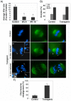
Figure 1. Oocyte-specific ablation of the ATRX protein in a transgenic RNAi mouse model.
(A) Nucleus of control oocyte showing bright ATRX staining (red, left image) at heterochromatin domains (arrow) as well as γ-tubulin signals (green) towards the nuclear periphery (arrowheads and inset). No ATRX staining is detectable in the nucleus of transgenic oocytes (arrow, right image). However, γ -tubulin staining remained unaffected (arrowheads and inset). Note that ATRX protein is expressed in surrounding somatic granulosa cells in both control and transgenic animals (bold arrows). The position of the nucleolus is indicated by (*). (B) Partial co-localization of ATRX and CREST signals at pericentric heterochromatin domains in control oocytes (arrow and inset, left image). By contrast, while localization of CREST signals is unaffected in transgenic oocytes (arrow and inset, right image), ATRX is not detectable at DAPI-bright heterochromatin domains. (C) Control MII oocyte immunostained with ATRX (red, arrow) and β-tubulin (green) showing proper chromosome alignment at the metaphase II spindle (left image, arrow, inset shows ATRX/β-tubulin merge only). Lack of ATRX in transgenic oocytes (right image, inset) results in abnormal chromosome alignment. (D) Proportion of in vivo matured MII stage oocytes that exhibited ATRX at centromeric heterochromatin. (E–F) Quantitative analysis of Atrx transcripts by real-time PCR revealed a significant reduction (p<0.001) in the levels of Atrx mRNA in transgenic (Tg) oocytes compared with controls (non-Tg). Analysis of β-actin mRNA was used as a housekeeping control. All data are presented as the mean ± s.d. of three independent experiments. Scale bars  = 10 µm.
Figure 2
Figure 2. Lack of ATRX function disrupts chromosome alignment at the metaphase II spindle and results in severe sub fertility.
(A) Control and transgenic female founders were continuously housed with fertile males for a period of 7 months. Transgenic females presented a significant reduction in average litter size (P<0.001). (B) Proportion of oocytes at the germinal vesicle (GV), metaphase I (MI), or metaphase II (MII) stage following 14 h of in vitro maturation. Selective ablation of ATRX has no effect on the progression of meiosis. (C) Control in vitro matured oocytes show proper chromosome alignment at the metaphase II spindle. In contrast, transgenic oocytes exhibit a spectrum of chromosome segregation defects following maturation in vitro and in vivo including (D) presence of single chromatids (insets) and (E) formation of chromosome bridges during premature anaphase II onset (arrowhead, inset shows overexposed DAPI image) as well as lagging chromosomes (arrow and inset). Asterisks indicate the position of the polar body. (F) Proportion of in vitro matured oocytes with misaligned chromosomes. Data are presented as the mean ± s.d. of three independent experiments. Scale bar  = 10 µm.
Figure 3
Figure 3. Abnormal chromosome condensation and high incidence of aneuploidy in ATRX deficient oocytes.
(A) Chromosome spread from a control oocyte at the metaphase II stage showing prominent ATRX staining at pericentric heterochromatin (arrows). (B) Lack of ATRX staining at pericentric heterochromatin (thin arrow) is associated with abnormal chromosome condensation (bold arrows) and chromatid breaks (C, arrowhead). (D) In vivo matured transgenic ATRX-RNAi oocytes exhibit a high incidence of aneuploidy. Chromosome spread exhibiting a hyperploid karyotype of 20 chromosomes and an additional single chromatid (circled) is shown. Data are presented as the mean ± s.d. of three independent experiments. Scale bar  = 10 µm.
Figure 4
Figure 4. ATRX is required to recruit the transcriptional regulator (DAXX) to pericentric heterochromatin in mammalian oocytes.
(A) Top Panel: Nucleus of a control oocyte showing a precise co-localization of DAXX (red) with bright, DAPI-stained pericentric heterochromatin domains (arrows). The position of the centromere is indicated by CREST (green). Lower Panel: Analysis of transgenic oocytes demonstrated that in the absence of ATRX, the transcriptional regulator DAXX fails to associate with pericentric heterochromatin domains while nucleoplasmic expression of DAXX persists. The position of the nucleolus is indicated by (*). (B) Proportion of germinal vesicle (GV) stage oocytes showing pericentric DAXX localization. More than 80% of transgenic oocytes fail to recruit DAXX to pericentric heterochromatin. Data are presented as the mean ± s.d. of three independent experiments. CREST immunolocalization (green) was conducted as an experimental control for centromeric-kinetochore integrity. Scale bars  = 10 µm.
Figure 5
Figure 5. Reduced phosphorylation of histone H3 at serine 10 (H3S10ph) in the chromosomes of ATRX-deficient oocytes.
(A) Phosphorylation of H3S10 (red) was visualized in metaphase II chromosome spreads from control (upper panel) and transgenic (lower panel) oocytes. Control oocytes exhibit prominent chromosome-wide H3S10ph staining. In contrast, a high proportion of transgenic oocytes exhibit negligible H3S10ph staining at pericentric heterochomatin and throughout the chromatids. CREST immunochemistry (green) served as an experimental control. (B) Proportion of oocytes with reduced phosphorylation of H3S10 in transgenic oocytes and controls. Data are presented as the mean ± s.d. of three independent experiments (p<0.05). Scale bar  = 10 µm.
Figure 6
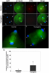
Figure 6. Chromosome fragmentation in ATRX deficient oocytes during the transition to the first mitosis.
(A) Strontium chloride-activated control and (B) transgenic oocytes at late anaphase II stained with ATRX (red) and β-tubulin (green). Numerous chromosomal fragments (arrows) are evident in the transgenic oocyte. Asterisks indicate the position of the polar body. Scale bar  = 50 µm. (D) Transgenic oocyte with a multi-polar spindle (arrows, right image) and a control (C, left image). Scale bar  = 10 µm. (E) Proportion of artificially activated oocytes presenting chromosome fragmentation. Data are presented as the mean ± s.d. of three independent experiments.
Figure 7
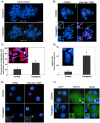
Figure 7. Centromere instability and micronuclei formation following fertilization of ATRX-deficient oocytes.
(A) Transmission of aneuploidy from the oocyte to the pre-implantation embryo in chromosome spreads obtained from transgenic zygotes analyzed by CREST (green) immunochemistry. Upper panel: transgenic zygote spread with hypoploid karyotype (2N = 39); lower panel: hyperploid karyotype showing 41 chromosomes (micrographs of chromosome complements of this zygote were taken individually (white line) to display at a comparable magnification with the panel above). (B) Partial chromosome spreads from zygotes undergoing the first mitotic division. Chromosomes were immunostained with CREST (green) and subsequently subjected to fluorescence in situ hybridization using a pan-centromeric DNA probe (red). Transgenic zygotes frequently presented chromosomal breaks at pericentric heterochromatin as indicated by detachment of a chromosomal fragment exhibiting a CREST signal and presence of major satellite DNA (red) at the proximal as well as at the distal fragment (arrow, right inset). Note that satellite DNA sequences in some chromosomes exhibit excessive stretching (arrowhead). (C) High incidence (P<0.005) of illegitimate centromere mitotic recombination in transgenic parthenogenetic zygotes as indicated by changes in lateral asymmetry of major satellite sequences (arrow). A centromeric break within the same chromosome complement is marked by an arrowhead. (D) Proportion of zygotes that exhibit centromeric breaks at the first mitotic division. (E) Pan-centromeric FISH revealed that chromosomal instability in transgenic zygotes results in the formation of centromeric DNA-containing micronuclei (arrow, insets) in ATRX deficient 2-cell embryos. (F) Distribution of kinetochore domains in 2-cell stage embryos. CREST signals (green) are detectable at perinuleolar regions in the nuclei of control and transgenic embryos (arrowheads, insets). In contrast, micronuclei in transgenic 2-cell stage embryos are CREST-signal negative (arrow, inset), therefore originating from DNA fragmentation and subsequent missegregation of acentric chromosomal material. Scale bars  = 10 µm.
Figure 8
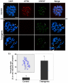
Figure 8. Lack of ATRX function during oogenesis results in severe genomic instability in the pre-implantation embryo.
(A) Developmental potential of ATRX deficient oocytes. Control and transgenic oocytes exhibit similar rates of blastocyst formation at 96 hours following in vitro fertilization (hpf). (B,C) Presence of micronuclei throughout pre-implantation development in ATRX deficient embryos. Although ATRX protein can be detected at the 8–16 cell stage following fertilization of ATRX deficient oocytes, a high proportion of ATRX deficient embryos exhibit micronuclei formation (arrow) as evidence of chromosome instability at all stages evaluated. Data are presented as the mean ± s.d. of three independent experiments. Scale bar  = 10 µm.

Similar articles

Cited by

References

    1. Festenstein R, Sharghi-Namini S, Fox M, Roderick K, Tolaini M, et al. Heterochromatin protein 1 modifies mammalian PEV in a dose- and chromosomal-context-dependent manner. Nat Genet. 1999;23:457–461. - PubMed
    1. Bernard P, Maure J, Partridge J, Genier S, Javerzat J, et al. Requirement of heterochromatin for cohesion at centromeres. Science. 2001;294:2539–2542. - PubMed
    1. Bernard P, Allshire R. Centromeres become unstuck without heterochromatin. Trends Cell Biol. 2002;12:419–424. - PubMed
    1. Dillon N, Festenstein R. Unravelling heterochromatin: competition between positive and negative factors regulates accessibility. Trends Genet. 2002;18:252–258. - PubMed
    1. Cleveland D, Mao Y, Sullivan K. Centromeres and kinetochores: from epigenetics to mitotic checkpoint signaling. Cell. 2003;112:407–421. - PubMed

Publication types

MeSH terms

LinkOut - more resources