. 2010 Dec;21(23):4141-50.
doi: 10.1091/mbc.E10-05-0424. Epub 2010 Sep 29.

Oxysterol binding protein-dependent activation of sphingomyelin synthesis in the golgi apparatus requires phosphatidylinositol 4-kinase IIα

Affiliations

Oxysterol binding protein-dependent activation of sphingomyelin synthesis in the golgi apparatus requires phosphatidylinositol 4-kinase IIα

Sangeeta Banerji et al. Mol Biol Cell. 2010 Dec.

Abstract

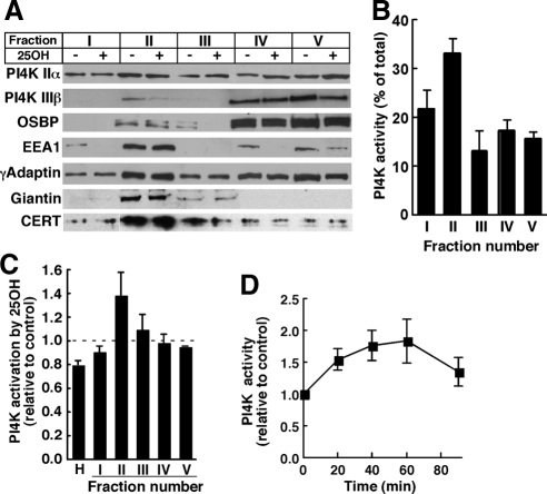
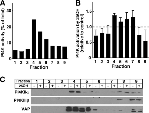
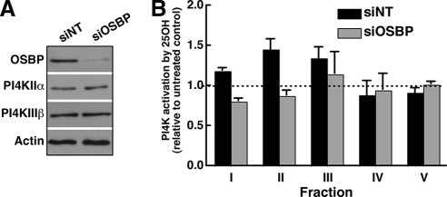
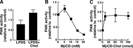
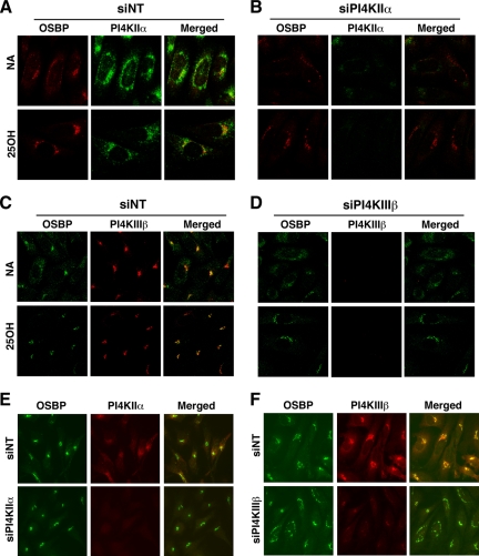
Cholesterol and sphingomyelin (SM) associate in raft domains and are metabolically coregulated. One aspect of coordinate regulation occurs in the Golgi apparatus where oxysterol binding protein (OSBP) mediates sterol-dependent activation of ceramide transport protein (CERT) activity and SM synthesis. Because CERT transfer activity is dependent on its phosphatidylinositol 4 phosphate [PtdIns(4)P]-specific pleckstrin homology domain, we investigated whether OSBP activation of CERT involved a Golgi-associated PtdIns 4-kinase (PI4K). Cell fractionation experiments revealed that Golgi/endosome-enriched membranes from 25-hydroxycholesterol-treated Chinese hamster ovary cells had increased activity of a sterol-sensitive PI4K that was blocked by small interfering RNA silencing of OSBP. Consistent with this sterol-requirement, OSBP silencing also reduced the cholesterol content of endosome/trans-Golgi network (TGN) fractions containing PI4KIIα. PI4KIIα, but not PI4KIIIβ, was required for oxysterol-activation of SM synthesis and recruitment of CERT to the Golgi apparatus. However, neither PI4KIIα nor PI4KIIIβ expression was required for 25-hydroxycholesterol-dependent translocation of OSBP to the Golgi apparatus. The presence of OSBP, CERT, and PI4KIIα in the TGN of oxysterol-stimulated cells suggests that OSBP couples sterol binding or transfer activity with regulation of PI4KIIα activity, leading to CERT recruitment to the TGN and increased SM synthesis.


Figures

Figure 1.
Figure 1.
Analysis of an oxysterol-activated PI4K in the Golgi and endosomes. (A) Homogenates of CHO cells, cultured in the absence (−) or presence (+) of 25-hydroxycholesterol (25OH; 2.5 μg/ml) for 1 h, were fractionated on discontinuous sucrose gradients. Equivalent volumes of fractions I–V were resolved by SDS-PAGE and immunoblotted for the indicated proteins as described in Materials and Methods. (B) Sucrose gradient fractions of untreated CHO cell homogenates were assayed for PI4K activity and expressed as a percentage of total. Results are the mean and SEM of three experiments. (C) Homogenates (H) of CHO cells treated with 25-hydroxycholesterol (2.5 μg/ml) or control solvent for 1 h were separated on discontinuous sucrose gradients, and individual fractions were assayed for PI4K activity and expressed relative to controls. Results are the mean and SEM of three experiments. (D) Fraction II was isolated from cells treated with 25-hydroxycholesterol for the indicated times and assayed for PI4K activity. Results are expressed relative to time-matched untreated controls (mean and SEM of 6 experiments).
Figure 2.
Figure 2.
PI4K activity in detergent-resistant membranes is stimulated by 25-hydroxycholesterol. (A) Untreated CHO cells were solubilized in β-OG/DOC buffer, pH 11.0, separated on discontinuous sucrose gradients, and individual fractions were assayed for PI4K activity. (B) β-OG/DOC extracts of CHO cells treated with 25-hydroxycholesteorl (25OH) or control solvent were separated on sucrose gradients and assayed for PI4K activity. Results are expressed relative to solvent-treated controls and are the mean and SEM of four experiments. (C) Equivalent volumes of fractions prepared as described in B were immunoblotted for PI4KIIα, PI4KIIIβ, and VAP.
Figure 3.
Figure 3.
PI4K activation by 25-hydroxycholesterol is dependent on OSBP. (A) CHO cells were transfected with siNT or siOSBP for 48 h and subsequently cultured in the absence or presence of 25-hydroxycholesterol (2.5 μg/ml) for 1 h. Expression of OSBP, PI4KIIα, or PI4KIIIβ in cell homogenates from siNT- and siOSBP-transfected CHO cells was determined by immunoblotting. (B) Homogenates of control and OSBP-depleted cells, treated with or without 25-hydroxycholesterol, were separated on sucrose gradients and fractions were assayed for PI4K activity. Activation of PI4K activity by oxysterol is expressed relative to solvent-treated controls (mean and SEM of 4 experiments).
Figure 4.
Figure 4.
In vitro regulation of Golgi/endosomal PI4K activity by cholesterol. (A) CHO cells were treated with or without 5 mM MβCD in DMEM for 30 min, washed twice in DMEM, and incubated in DMEM with 5% LPDS with or without MβCD-cholesterol (100 μM) complex (LPDS+Chol). After 2 h, cells were harvested, and fraction II was isolated and assayed for PI4K activity. PI4K activity is expressed relative to cells that were not treated with MβCD. Results are the mean and SEM of three experiments. (B and C) Golgi-enriched fraction II from CHO cells was treated with the indicated concentrations of MβCD (B) or MβCD-cholesterol complex (C) for 30 min at 37°C before assaying for PI4K activity. Results are expressed relative to buffer-treated controls and are the mean and SEM of three experiments.
Figure 5.
Figure 5.
OSBP depletion reduces the cholesterol content of Golgi/endosomal membranes containing PI4KIIα. (A) The localization of cholesterol was determined by filipin staining of CHO cells transiently expressing PI4KIIα-GFP and treated with or without 25-hydroxycholesterol (2.5 μg/ml) for 60 min. (B) Equivalent amounts (2–2.5 mg protein) of the postnuclear supernatants from CHO cells expressing shOSBP or a nontargeting control (shNT) were fractionated on an Opti-Prep gradient and assayed for unesterified cholesterol content as described in Materials and Methods. The cholesterol content of individual fraction is expressed as a percentage of the total cholesterol recovered from the gradient and is the mean and SEM of three experiments (*p < 0.05). (C) Distribution of the organelle markers NPC1 (late endosomes), TGN38 (Golgi), VAP (ER), and caveolin (plasma membrane/endosomes), as well as OSBP and PI4KIIα, were determined by immunoblotting of equivalent volumes of each fraction.
Figure 6.
Figure 6.
siRNA silencing of PI4KIIα prevents 25-hydroxycholesterol activation of SM synthesis. (A) CHO cells were transfected with siNT, siPI4KIIα, or siPI4KIIIβ for 48 or 72 h. Cells were then treated with solvent (gray bars) or 25-hydroxycholesterol (2.5 μg/ml; black bars) for 6 h. During the last 2 h of oxysterol treatment, cells were pulse labeled with [3H]serine. and isotope incorporation into SM, ceramide and GlcCer was quantified as described in Materials and Methods. Results are the mean and SEM of three to six separate experiments. The extent of PI4KIIα and PI4KIIIβ knockdown was determined by immunoblotting of whole cell lysates by using actin as a load control. *p < 0.05 compared with oxysterol-treated siNT. (B) HeLa cells were transfected with two different siRNAs against PI4KIIα or PI4KIIIβ, or siOSBP, for 48 h. 25-Hydroxycholesterol–stimulated SM synthesis was then measured as described in A. Results are expressed relative to solvent treated controls and are the mean and SEM of three separate experiments.
Figure 7.
Figure 7.
Knockdown of PI4KIIα prevents oxysterol-mediated translocation of GFP-CERT to the Golgi apparatus. (A) Knockdown of PI4KIIα or PI4KIIIβ in CHO cells stably expressing GFP-CERT was confirmed by immunoblotting as described in Materials and Methods. (B) GFP-CERT was visualized by confocal microscopy in CHO cells in which PI4KIIα or PI4KIIIβ was knocked down by >90%. Images are single optical sections (0.4–0.2 mM) captured using an LSM510/AxioVert 100M inverted microscope equipped with a 100× oil immersion objective (NA 1.4). In some experiments, the absence of PI4KIIα and PI4KIIIβ expression also was confirmed by coimmunofluorescence as described in the legend to Figure 9. (C) Localization of GFP-CERT to perinuclear Golgi structures was quantified in 63× wide-field images of control and oxysterol-treated CHO cells transfected with siNT, siPI4KIIα, or siPI4KIIIβ as described in Materials and Methods. Results are the mean and SEM of three experiments.
Figure 8.
Figure 8.
Colocalization of OSBP, CERT, PI4KIIα, and PI4KIIIβ at the TGN. (A) CHO cells were treated with 25-hydroxycholesterol (2.5 μg/ml) for 30 min followed by addition of nocodazole (2 μg/ml) for an additional 30 min. Endogenous OSBP was detected with a polyclonal antibody followed by goat anti-rabbit Alexa Fluor488- or Alexa Fluor594-conjugated secondary antibodies. Cells were costained for giantin or PI4KIIIβ by using corresponding Alexa Fluor488- or Alexa Fluor594-conjugated secondary antibodies. CHO cells transiently expressing GFP-CERT were immunostained for OSBP as described above using a goat anti-rabbit Alexa Fluor594-conjugated secondary antibody. (B) HeLa cells were immunostained with a sheep anti-goat TGN46 antibody and a goat anti-sheep Alexa488-conjugated secondary antibody. This was followed by polyclonal or monoclonal antibodies against OSBP, PI4KIIα, or PI4KIIβ and appropriate Alexa Fluor594-conjugated secondary antibodies. Images are single confocal sections (0.2–0.4 μm) obtained as described in Materials and Methods and the legend to Figure 7.
Figure 9.
Figure 9.
Knockdown of PI4KIIα or PI4KIIIβ does not prevent oxysterol-dependent Golgi localization of OSBP. CHO cells transfected with siNT (A and C), siPI4KIIα (B), or siPI4KIIIβ (D) were treated with ethanol solvent or 25-hydroxycholesterol (25OH; 2.5 μg/ml) for 1 h. Cells were then fixed and incubated with an OSBP polyclonal antibody and goat anti-rabbit Alexa Fluor488 (C and D) or Alexa Fluor594 (A and B) secondary antibodies, followed by PI4KIIα or PI4KIIIβ monoclonal antibodies and a goat anti-mouse Alexa Fluor594 C and D) or Alexa Fluor488 (A and B) secondary antibodies. Images are single confocal sections (0.4–0.2 μM) taken on an LSM510/AxioVert 100M inverted microscope equipped with a 100× oil immersion objective (NA 1.4). HeLa cells were transiently transfected with siPI4KIIα (E) or siPI4KIIIβ (F), treated with 25-hydroxycholesterol for 1 h, and immunostained for the corresponding PI4K and OSBP. Images were captured on an Axiovert 200M fluorescence microscope equipped with a 63× objective (NA 1.4).

Similar articles

Cited by

References

    1. Balch W. E., Dunphy W. G., Braell W. A., Rothman J. E. Reconstitution of the transport of protein between successive compartments of the Golgi measured by the coupled incorporation of N-acetylglucosamine. Cell. 1984;39:405–416. - PubMed
    1. Balla A., Tuymetova G., Barshishat M., Geiszt M., Balla T. Characterization of type II phosphatidylinositol 4-kinase isoforms reveals association of the enzymes with endosomal vesicular compartments. J. Biol. Chem. 2002;277:20041–20050. - PubMed
    1. Balla A., Tuymetova G., Tsiomenko A., Varnai P., Balla T. A plasma membrane pool of phosphatidylinositol 4-phosphate is generated by phosphatidylinositol 4-kinase type-III alpha: studies with the PH domains of the oxysterol binding protein and FAPP1. Mol. Biol. Cell. 2005;16:1282–1295. - PMC - PubMed
    1. Barylko B., Gerber S. H., Binns D. D., Grichine N., Khvotchev M., Sudhof T. C., Albanesi J. P. A novel family of phosphatidylinositol 4-kinases conserved from yeast to humans. J. Biol. Chem. 2001;276:7705–7708. - PubMed
    1. Bonifacino J. S., Traub L. M. Signals for sorting of transmembrane proteins to endosomes and lysosomes. Annu. Rev. Biochem. 2003;72:395–447. - PubMed

Publication types

Substances

LinkOut - more resources