. 2011 Jan;10(1):M110.001172.
doi: 10.1074/mcp.M110.001172. Epub 2010 Sep 27.

A QUICK screen for Lrrk2 interaction partners--leucine-rich repeat kinase 2 is involved in actin cytoskeleton dynamics

Affiliations

A QUICK screen for Lrrk2 interaction partners--leucine-rich repeat kinase 2 is involved in actin cytoskeleton dynamics

Andrea Meixner et al. Mol Cell Proteomics. 2011 Jan.

Abstract

Mutations in human leucine-rich repeat kinase 2 (Lrrk2), a protein of yet unknown function, are linked to Parkinson's disease caused by degeneration of midbrain dopaminergic neurons. The protein comprises several domains including a GTPase and a kinase domain both affected by several pathogenic mutations. To elucidate the molecular interaction network of endogenous Lrrk2 under stoichiometric constraints, we applied QUICK (quantitative immunoprecipitation combined with knockdown) in NIH3T3 cells. The identified interactome reveals actin isoforms as well as actin-associated proteins involved in actin filament assembly, organization, rearrangement, and maintenance, suggesting that the biological function of Lrrk2 is linked to cytoskeletal dynamics. In fact, we demonstrate Lrrk2 de novo binding to F-actin and its ability to modulate its assembly in vitro. When tested in intact cells, knockdown of Lrrk2 causes morphological alterations in NIH3T3 cells. In developing dopaminergic midbrain primary neurons, Lrrk2 knockdown results in shortened neurite processes, indicating a physiological role of Lrrk2 in cytoskeletal organization and dynamics of dopaminergic neurons. Hence, our results demonstrate that molecular interactions as well as the physiological function of Lrrk2 are closely related to the organization of the actin-based cytoskeleton, a crucial feature of neuronal development and neuron function.


Figures
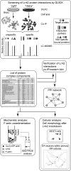
Fig. 1.
Fig. 1.
Schematic overview of the experimental procedure. The interactome of Lrrk2 was analyzed by QUICK using NIH3T3 cells. A list of putative Lrrk2 complex components was obtained and a selected subset was verified by Western blot analysis following co-IP. The identified Lrrk2 interacting proteins were mapped into a PPI network, integrating interaction data stored in the HPRD and BioGRID database as well as interactions identified through experimental results. The resulting protein network suggests a biological role of Lrrk2 within actin cytoskeletal dynamics. The physiological relevance of the hypothesis was analyzed through biochemical and cellular assays.
Fig. 2.
Fig. 2.
Verification of Lrrk2 interaction partners identified by QUICK using co-immunoprecipitation. Equal volumes of cell lysates with identical protein amounts from wt and LVmiB3-transduced NIH3T3 were immunoprecipitated using the anti-Lrrk2 antibody cross-linked to Protein G Sepharose. 50% of the immunoprecipitates and a lysate volume corresponding to 2% of the IP-input were separated by SDS-PAGE. Selected Lrrk2 protein complex components found through QUICK were verified by Western blotting using the indicated antibodies. Anti-GAPDH Western blotting assured equal protein loading of the input samples. IP, immunoprecipitation antibody; WB, Western blotting antibody.
Fig. 3.
Fig. 3.
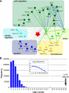
Network of Lrrk2 interacting proteins. Known interactions among the proteins identified by QUICK were analyzed and visualized by Pathway Palette. The obtained network displays significant physical cohesiveness. A, Out of the 36 proteins identified as specific Lrrk2 interactors, 20 had previously been described as connected through 36 interactions. Interactions of proteins with Lrrk2 were added manually, on the basis of their identification in the QUICK assay and, in part, verification by co-IP experiments. Proteins within the network are depicted by star-shaped nodes and colored according to their molecular function as classified in Table I. Solid lines denote known protein interactions from the HPRD and BioGRID database prioritized and qualified in an evidence-based fashion, referring to the PPI detection method, as reflected by a corresponding color-scheme of the edges: “BioGRID: Low-Throughput” or “HPRD: in vivo” type (green) and “BioGRID: HTP/Complex” or “HPRD: in vitro” type (blue). Manually added interactions are figured as dashed gray lines. Proteins are indicated by their gene names, with their full names shown in Table I. B, Distribution of interactions within a million randomly selected sets of proteins (set size 37) with the same node degree as those of the QUICK set. The graph insert represents the logarithmic depiction of interaction frequencies. The Lrrk2 data set gave significantly more interactions compared with the randomly selected sets.
Fig. 4.
Fig. 4.
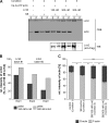
Lrrk2 binds directly to F-actin and affects its polymerization/depolymerization in vitro. A, F-actin cosedimentation experiments were performed by incubating 3 μm Ca-G-ATP actin under polymerizing conditions with 150 nm (condition 3) or 300 nm (condition 4) SF-TAP tagged Lrrk2 purified from HEK293 cells in storage buffer (SB). Samples without Lrrk2 and SB (condition 1), with only SB (condition 2) or containing 300 nm Lrrk2 in the absence of actin (condition 5) served as controls. Samples with SB or Lrrk2/SB always contained a constant amount of the storage buffer components ensuring identical conditions. G-actin was allowed to polymerize until equilibrium was reached and sedimented by high speed centrifugation. The pellets (P) and supernatants (S) were analyzed by SDS-PAGE followed by Coomassie brilliant blue staining (upper panel) or Western blot using an anti-Flag antibody (lower panel). The results of a representative experiment are shown. Lrrk2 cosedimented with actin (condition 3 and 4), whereas in the absence of actin, Lrrk2 was only detected in the supernatant (condition 5). CBB: Coomassie brilliant blue; WB: Western blot. B, Relative amounts of Lrrk2 in the pellet upon incubation of 150 or 300 nm Lrrk2 with 3 μm actin for three experiments (Exp1–Exp3) using two different protein purifications (batch#1 and batch #2), based on quantification of Western blots as in A (lower panel). At the higher concentration, an increased amount of cosedimented Lrrk2 was consistently detected. C, Quantification of actin in the supernatant (G-actin) and pellet (F-actin) in the absence of SB (control, condition1), presence of SB (SB control, condition 2) or Lrrk2 (150 or 300 nm) in SB (condition 3 and 4). The addition of Lrrk2 led to a significant shift of actin from the pellet fraction to the supernatant (** p < 0.01, *** p < 0.001, n = 4, one-way ANOVA, Holm-Sidak post-hoc test). Whereas in Lrrk2 deficient samples the majority of actin was detected in the pellet fraction, a decrease in sedimented actin accompanied by a corresponding increase in unpolymerized actin in the supernatant was found in the presence of Lrrk2.
Fig. 5.
Fig. 5.
Knockdown of Lrrk2 alters the morphology of NIH3T3 cells. NIH3T3 cells were plated onto 6-well plates and transduced the following day with lentiviral vectors encoding Lrrk2 targeting shRNA (miB3) or the transfer vector LVTH alone as indicated. Five days after transduction, cells were replated at a density of 5%–10% on glass coverslips, serum-starved for 24 h and analyzed. A, To visualize cell shape, cells were immunostained for F-actin. LVmiB3-transduced NIH3T3 cells displayed cell body elongation and narrowing. Scale bar: 50 μm. B, To quantify the observed alterations in cell shape the perimeter to area ratio (P:A-ratio) was determined. Silencing of Lrrk2 resulted in a significant increase of this ratio (** p < 0.01 versus wt, n = 4, one-way ANOVA, Tukey‘s post-hoc test). C, Relative frequency distributions of P:A-ratios for wt, LVTH- or LVmiB3-transduced NIH3T3 cells. Ratios were grouped into consecutive bins increasing by 0.05 μm−1, ranging from 0.05 to > 0.4 μm−1, and were calculated as percentage of cells with a given ratio. The knockdown of Lrrk2 led to a shift of the P:A-ratio toward higher values, denotative for an increase in the amount of cells exhibiting an enhanced P:A-ratio.
Fig. 6.
Fig. 6.
Knockdown of Lrrk2 in primary VM cultures. VM tissue was isolated from E12.5 mouse embryos and cells were plated onto 8-well chamber slides. Cultures were transduced at DIV1 with LVTH, LVmiB3 and LVmiB4 and analyzed at DIV9 or DIV14. A, Semiquantitative real-time PCR displayed reduced Lrrk2 mRNA levels in primary VM cultures transduced with the two independent shRNA-constructs LVmiB3 and LVmiB4. B, Western blot analysis revealed that the transduction with LVmiB3 and LVmiB4 resulted in decreased Lrrk2 protein levels at both time points. GFP and GAPDH Western blots served as controls for transduction efficiency and protein load, respectively.
Fig. 7.
Fig. 7.
Lentiviral-mediated knockdown of Lrrk2 in primary VM cultures leads to an impaired neurite outgrowth of developing DA neurons. Lrrk2 expression in primary VM cultures was depleted by lentiviral delivery of two independent silencing constructs (LVmiB3 and LVmiB4). Non-transduced (wt) or LVTH-transduced cells served as negative controls and cultures were analyzed at DIV9 or DIV14. A, Cell counts of TH-ir cells per well. No differences in the amount of DA neurons was evident at DIV9, whereas the knockdown of Lrrk2 resulted in a decrease in TH-ir cell counts at DIV14 (** p < 0.01 versus wt DIV14, n = 3–5, one-way ANOVA, Tukey‘s post-hoc test). B, Quantification of neurite length of TH-ir neurons. Knockdown of Lrrk2 led to a significant decrease in the average neurite length at DIV9 and DIV14 (*** p < 0.001 versus wt DIV9, n = 4–5, +++ p < 0.001 versus wt DIV14, n = 3–5, one-way ANOVA, Tukey‘s post-hoc test). C, Corresponding frequency distributions of neurite length for the different treatment groups (wt, LVTH, LVmiB3, and LVmiB4) at DIV9 and DIV14. Absolute neurite lengths were grouped into 40 μm bins of increasing size and the percentage of DA neurites of given length was calculated. The expression of miB3 and miB4 resulted at both time points in a shifted distribution toward shorter processes compared with wt and LVTH-control cultures, reflecting an increase in the number of shorter neurites and a decrease of longer neurites.
Fig. 8.
Fig. 8.
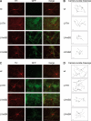
TH-ir neurons expressing Lrrk2 mRNA targeting shRNAs. Primary VM progenitor cultures were transduced with lentiviruses encoding Lrrk2 silencing shRNA-constructs (LVmiB3 and LVmiB4) or containing the transfer vector (LVTH) alone as indicated. (A and C) DA neurons were visualized using immunofluorescent labeling for tyrosine hydroxylase (TH) (left columns) at DIV9 (A) or DIV14 (C). GFP fluorescence (center columns) indicated successfully transduced neurons whereas the combination of TH-immunostaining and GFP fluorescence (right panel; merge) demonstrated the efficient transduction of DA neurons. (B and D) Camera lucida drawing of representative, randomly selected GFP-positive TH-ir cells at DIV9 (B) and DIV14 (D). Drawings were obtained by manually tracing of the neuron in NeuronJ. TH-ir neurons in miB3- and miB4-expressing cultures exhibited shorter neurites compared with non-transduced (wt) or LVTH-transduced neurons at DIV9 and DIV14. Scale bar: 50 μm.

Similar articles

Cited by

References

    1. Elbaz A. (2008) LRRK2: bridging the gap between sporadic and hereditary Parkinson's disease. Lancet Neurol. 7, 562–564 - PubMed
    1. Paisán-Ruiz C. (2009) LRRK2 gene variation and its contribution to Parkinson disease. Hum. Mutat. 30, 1153–1160 - PubMed
    1. Wszolek Z. K., Pfeiffer R. F., Tsuboi Y., Uitti R. J., McComb R. D., Stoessl A. J., Strongosky A. J., Zimprich A., Müller-Myhsok B., Farrer M. J., Gasser T., Calne D. B., Dickson D. W. (2004) Autosomal dominant parkinsonism associated with variable synuclein and tau pathology. Neurology 62, 1619–1622 - PubMed
    1. Zimprich A., Biskup S., Leitner P., Lichtner P., Farrer M., Lincoln S., Kachergus J., Hulihan M., Uitti R. J., Calne D. B., Stoessl A. J., Pfeiffer R. F., Patenge N., Carbajal I. C., Vieregge P., Asmus F., Müller-Myhsok B., Dickson D. W., Meitinger T., Strom T. M., Wszolek Z. K., Gasser T. (2004) Mutations in LRRK2 cause autosomal-dominant parkinsonism with pleomorphic pathology. Neuron 44, 601–607 - PubMed
    1. Taylor J. P., Mata I. F., Farrer M. J. (2006) LRRK2: a common pathway for parkinsonism, pathogenesis and prevention? Trends Mol. Med. 12, 76–82 - PubMed

Publication types

MeSH terms

LinkOut - more resources