. 2010 Nov 19;285(47):36401-9.
doi: 10.1074/jbc.M110.172866. Epub 2010 Sep 17.

Transcriptional repressor E4-binding protein 4 (E4BP4) regulates metabolic hormone fibroblast growth factor 21 (FGF21) during circadian cycles and feeding

Affiliations

Transcriptional repressor E4-binding protein 4 (E4BP4) regulates metabolic hormone fibroblast growth factor 21 (FGF21) during circadian cycles and feeding

Xin Tong et al. J Biol Chem. .

Abstract

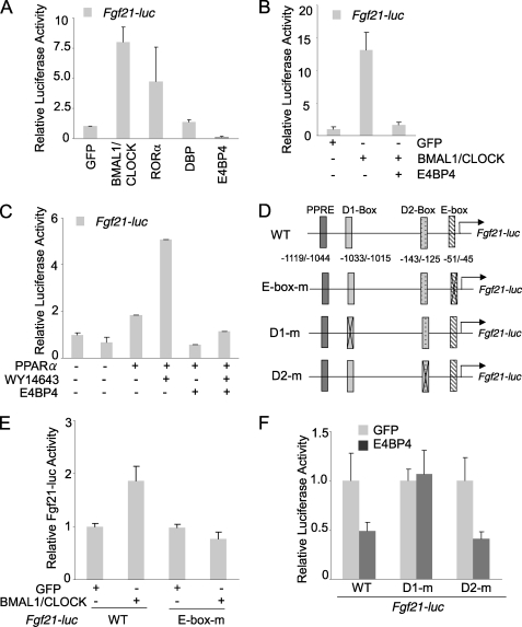
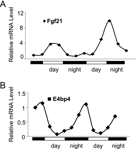
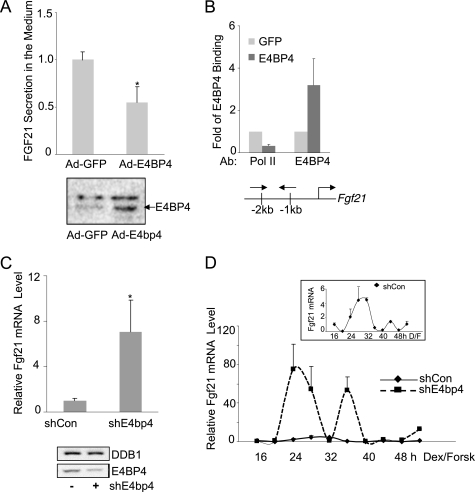
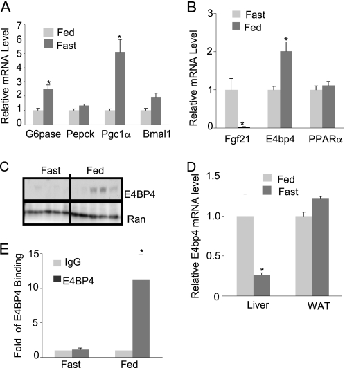
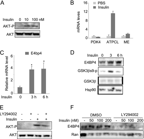
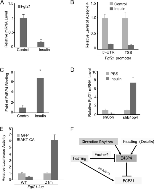
Fibroblast growth factor 21 (FGF21) is a potent antidiabetic and triglyceride-lowering hormone whose hepatic expression is highly responsive to food intake. FGF21 induction in the adaptive response to fasting has been well studied, but the molecular mechanism responsible for feeding-induced repression remains unknown. In this study, we demonstrate a novel link between FGF21 and a key circadian output protein, E4BP4. Expression of Fgf21 displays a circadian rhythm, which peaks during the fasting phase and is anti-phase to E4bp4, which is elevated during feeding periods. E4BP4 strongly suppresses Fgf21 transcription by binding to a D-box element in the distal promoter region. Depletion of E4BP4 in synchronized Hepa1c1c-7 liver cells augments the amplitude of Fgf21 expression, and overexpression of E4BP4 represses FGF21 secretion from primary mouse hepatocytes. Mimicking feeding effects, insulin significantly increases E4BP4 expression and binding to the Fgf21 promoter through AKT activation. Thus, E4BP4 is a novel insulin-responsive repressor of FGF21 expression during circadian cycles and feeding.


Figures

FIGURE 1.
FIGURE 1.
Mouse Fgf21 is a circadian output gene regulated by the circadian protein E4BP4. A, mouse Fgf21 promoter is differentially regulated by clock proteins. A luciferase vector driven by the mouse Fgf21 promoter was cotransfected with the expression vectors encoding various clock proteins in Hepa1c1c-7 cells. The relative luciferase activities were calculated based on the value of GFP group set as 1. Results are expressed as mean ± S.D. (error bars) (n = 3). B, E4BP4 overexpression blocks transcription activation of the Fgf21-luc by BMAL1-CLOCK. The mouse Fgf21 promoter-driven luciferase reporter was co-transfected with expression vectors of various clock proteins in Hepa1c1c-7 cells. The relative luciferase activities were calculated based on the value of GFP group set as 1. Results are expressed at mean ± S.D. (n = 3). C, E4BP4 abolishes the activation of the Fgf21-luc by PPARα ligand WY14643. 24 h after co-transfection with the mouse Fgf21-luc and expression vectors of E4BP4 and PPARα, Hepa1c1c-7 cells were treated with either DMSO or PPARα ligand WY14643 (10 μm) for 16 h. The relative luciferase activities are expressed as mean ± S.D. (n = 3). D, schematic diagram shows the putative binding sites (PPAR-response element (PPRE), E box, and D box) within the 1.5 kb Fgf21 promoter upstream of the transcription starting site. The E-box and D-box deletion mutants are also indicated. E, mouse Fgf21 promoter contains a functional E-box element. Deletion of the E-box binding site (−51 to −45) abolished the BMAL1-CLOCK-mediated activation of the Fgf21 promoter. F, a D-box-binding element is required for E4BP4-mediated repression of the Fgf21 promoter. The Fgf21-luc mutant D1m (−1033 to −1015 deleted) was not repressed by E4BP4 in the co-transfection assay. Both D2m mutant (−143 to −125 deleted) and wild-type Fgf21-luc respond to E4BP4 in similar manners.
FIGURE 2.
FIGURE 2.
The circadian expression of Fgf21 and E4bp4 in the mouse liver. The liver mRNA levels of Fgf21 (A) and E4bp4 (B) are shown during two circadian cycles. The cDNA of each time point was derived from the pooled liver samples of three mice.
FIGURE 3.
FIGURE 3.
E4BP4 directly represses Fgf21 and is required for its circadian oscillation in mouse liver cells. A, overexpression of E4BP4 suppresses the secretion of FGF21 in culture medium of primary mouse hepatocytes. Cells were infected with either Ad-GFP or Ad-E4BP4 for 48 h and the culture medium was then collected for ELISA analysis of FGF21. Results are expressed as mean ± S.D. (error bars) of triplicates. A representative result of three individual experiments is shown. The levels of total E4BP4 protein in primary hepatocytes transduced with either Ad-GFP or Ad-E4BP4 were detected by Western blot. B, chromatin immunoprecipitation was performed to detect E4BP4 binding to the mouse Fgf21 promoter in Hepa1c1c-7 cells transfected with either GFP or E4BP4 expression plasmid. The recruitment of RNA polymerase II was examined as well. Results are expressed as mean ± S.D. of three experiments. The locations of ChIP primers are shown as well. C, Hepa1c1c-7 cells were transfected with the control or E4bp4 shRNA for 72 h. Cells were synchronized by 50% horse serum for 2 h and incubated in serum-free medium for another 16 h. Cells then were harvested for mRNA analysis of the Fgf21 gene by Q-PCR. Results are expressed as mean ± S.D. of at least three independent experiments. *, p < 0.05. Knockdown efficiency of E4BP4 protein knockdown is shown below. D, knockdown of E4BP4 alters the circadian oscillation of mouse Fgf21 in synchronized Hepa1c1c-7 cells. After synchronization treatment with both dexamethasone (100 nm) and forskolin (10 μm), the Hepa1c1c-7 cells stably expressing either control shRNA or E4bp4 shRNA were collected for mRNA analysis at the indicated time points. The value given for the amount of mRNA present at the 16 h time point was set as 1. Error bars, ±range (n = 2). Inset, circadian oscillation of Fgf21 in the shRNA control group.
FIGURE 4.
FIGURE 4.
E4BP4 mediates feeding-induced FGF21 gene suppression. A, the mRNA levels of gluconeogenic genes, including G6pase, Pepck, and Pgc1α, in the fasted or fed liver tissues isolated from wild-type C57BL6 mice (n = 5). Data are presented as mean ± S.E. (error bars). *, p < 0.05. B, Q-PCR analysis for the mRNA level of E4bp4 in the same set of liver tissues (n = 5). The expression levels of Fgf21 and PPARα were measured as well. Data are presented as mean ± S.E. *, p < 0.05. C, immunoblotting analysis for the liver E4BP4 protein level from the same tissue samples. D, Q-PCR analysis of the E4bp4 mRNA levels in both the liver and adipose tissues isolated from fasted or fed wild-type C57BL6 mice (n = 5). Data are presented as mean ± S.E. *, p < 0.05. E, ChIP assay for the occupancy of E4BP4 on the Fgf21 promoter in both fasting and feeding conditions. Liver tissues (n = 3) harvested after fasting or feeding for 24 h were used as input materials. Data are presented as mean ± S.E. *, p < 0.05.
FIGURE 5.
FIGURE 5.
Insulin up-regulates the expression of E4BP4 through AKT activation in liver cells. A, insulin-induced AKT phosphorylation in Hepa1c1c-7 cells. Both total and phosphorylated AKT proteins were detected by immunoblotting. B, Q-PCR analysis of the mRNA levels of Pdk4, ATP citrate lyase, and malic enzyme (ME) in the insulin-treated Hepa1C1C-7 cells. C, insulin up-regulates the gene expression of E4bp4 in a time-dependent manner in Hepa1c1c-7 cells. Confluent Hepa1c1c-7 cells were treated with insulin at 200 nm for 0, 3, and 6 h before harvest. The mRNA level of E4bp4 was determined by Q-PCR. Results are expressed as mean ± S.D. (error bars) (n = 3). *, p < 0.05. D, insulin up-regulates the E4BP4 protein levels in freshly isolated primary mouse hepatocytes. Cells were treated with insulin at 200 nm for 0, 3, and 6 h. The protein levels of E4BP4, GSK3β, GSK3βs9p, and loading control Ran were detected by specific antibodies. E, pretreatment with PI3K inhibitor LY294002 (50 μm) blocks AKT phosphorylation by insulin in Hepa1c1c-7 cells. The control was treated with DMSO. F, AKT inhibition blocks the insulin-induced E4BP4 protein expression in Hepa1C1C-7 cells. Cells were treated with either DMSO or LY294002 for 2 h before insulin treatment at various concentrations for 12 h. An immunoblot was used for detecting E4BP4 protein levels.
FIGURE 6.
FIGURE 6.
Insulin suppresses Fgf21 via the induction of E4BP4 in liver cells. A, insulin suppresses the Fgf21 expression in Hepa1c1c-7 cells. Confluent Hepa1c1c-7 cells were first synchronized by 50% horse serum for 2 h and incubated at serum-free medium for another 6 h. Cells were then treated with 200 nm insulin for 12 h before Fgf21 Q-PCR. Results are expressed as mean ± S.D. (error bars) (n = 3). *, p < 0.05. B, insulin treatment increases the levels of acetylated H4 around the Fgf21 promoter regions. Confluent Hepa1c1c-7 cells were treated as in A before ChIP analysis with specific antibodies. Results are expressed as mean ± S.D. (n = 3). C, insulin treatment stimulates the recruitment of E4BP4 onto the Fgf21 promoter. Confluent Hepa1c1c-7 cells were treated as in A before a ChIP assay with anti-E4BP4 antibody. D, E4BP4 is required for the insulin-induced suppression of Fgf21 in liver cells. After synchronization by 50% horse serum for 2 h and incubation in serum-free medium for another 6 h, Hepa1c1c-7 cells stably expressing either shRNA or E4bp4 shRNA were then treated with insulin at 200 nm for 12 h before Fgf21 mRNA analysis. Results are expressed at mean ± S.D. (n = 3). *, p < 0.05. E, AKT activation represses the luciferase activity driven by the wild type Fgf21 promoter but not by the E4BP4 binding site-deleted mutant. A constitutively active AKT was co-transfected into Hepa1c1c-7 cells along with either WT Fgf21-luc or D1m Fgf21-luc. The relative luciferase activities 48 h post-transfection are presented as mean ± S.D. (n = 3). F, model for the regulation of hepatic FGF21 expression by E4BP4. Both insulin and feeding induce E4bp4 to regulate the Fgf21 circadian oscillation upon food intake. E4BP4 directly binds to a distal D-box element of the Fgf21 promoter to suppress its expression. As a first order circadian output gene, E4BP4 links the circadian rhythm to liver metabolism.

Similar articles

Cited by

References

    1. Dibner C., Schibler U., Albrecht U. (2010) Annu. Rev. Physiol. 72, 517–549 - PubMed
    1. Green C. B., Takahashi J. S., Bass J. (2008) Cell 134, 728–742 - PMC - PubMed
    1. Schibler U. (2009) J. Biol. Rhythms 24, 3–15 - PubMed
    1. Takahashi J. S., Hong H. K., Ko C. H., McDearmon E. L. (2008) Nat. Rev. Genet. 9, 764–775 - PMC - PubMed
    1. Ceriani M. F., Hogenesch J. B., Yanovsky M., Panda S., Straume M., Kay S. A. (2002) J. Neurosci. 22, 9305–9319 - PMC - PubMed

Publication types

MeSH terms

Substances