. 2010 Oct 15;185(8):4835-45.
doi: 10.4049/jimmunol.1001032. Epub 2010 Sep 15.

IL-21 deficiency influences CD8 T cell quality and recall responses following an acute viral infection

Affiliations

IL-21 deficiency influences CD8 T cell quality and recall responses following an acute viral infection

John S Yi et al. J Immunol. .

Abstract

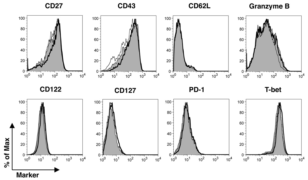
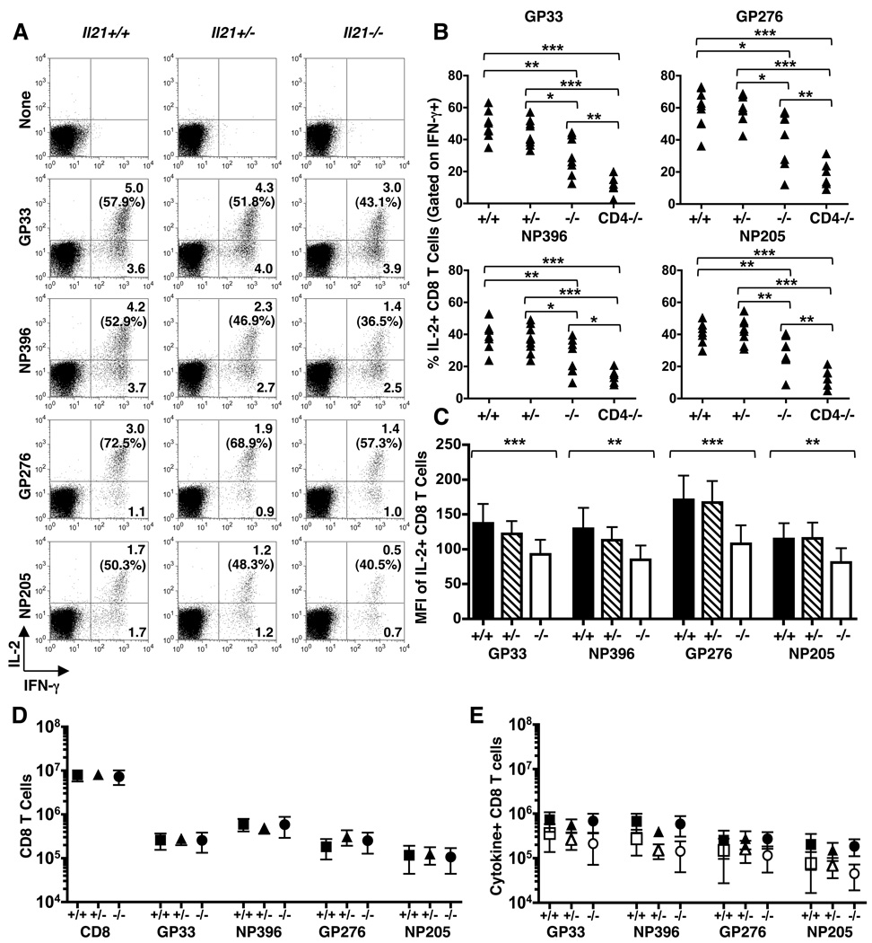
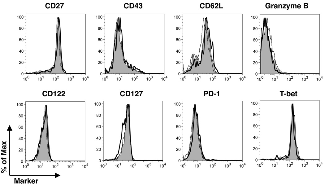
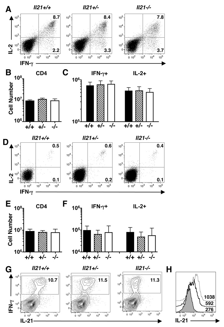
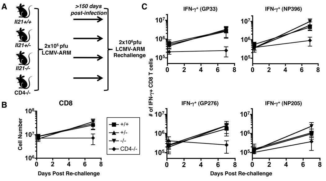
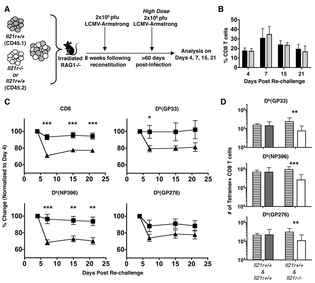
CD4 T cells are principal producers of IL-21 and are often required for optimal CD8 T cell responses. Therefore, we investigated the importance of IL-21 in determining the phenotypic attributes, functional quality, and maintenance of antiviral CD8 T cells following acute infection with the prototypic mouse pathogen lymphocytic choriomeningitis virus. Previous reports have documented an obligatory role for IL-21 in sustaining CD8 T cell responses during chronic infections. Here we show that the requirements for IL-21 are less stringent following acute infections; however, in the absence of IL-21, the capacity of CD8 T cells to attain the polyfunctional trait of IL-2 production is consistently reduced during both the effector and memory phases. This is further supported by in vitro studies showing that the addition of IL-21 promotes the differentiation of IL-2-producing CD8 T cells. Although the generation of memory CD8 T cells, which are capable of mounting protective recall responses, proceeds independently of IL-21, we demonstrate that IL-21 does function to support secondary responses, especially under competitive conditions. Collectively, these studies highlight the potential roles of IL-21 in determining the quality of CD8 T cell responses postinfection.


Figures

FIGURE 1
FIGURE 1
Reduced IL-2-producing effector CD8 T cell responses in the absence of IL-21. IFN-γ and IL-2 production by CD8 T cells specific for four distinct viral epitopes were evaluated at 8 days following acute LCMV-Armstrong infection of IL-21+/+, IL-21+/−, IL-21−/− mice. A, Intracellular cytokine analysis for IFN-γ and IL-2 following stimulation with various antigenic peptides. Gated CD8 T cells are shown and the percentages of CD8+, IFN-γ+ cells that co-produce IL-2 are reported in parentheses. B, Composite data from individual mice showing the fraction of IFN-γ, IL-2-co-producing CD8 T cells specific for individual viral epitopes. C, The MFI of IL-2 staining by epitope-specific CD8 T cells from the various cohorts. D, The total numbers of CD8 T cells and viral-epitope-specific CD8 T cells were determined by flow cytometric analysis using a panel of MHC class I tetramers. E, Epitope-specific IFN-γ producing (black symbols) and IL-2 producing (white symbols) CD8 T cells were enumerated following intracellular cytokine analyses. Error bars are s.d. For B–E, statistical significance is represented by * p<0.05, ** p<0.01, *** p< 0.001. Results are shown from 4 independent experiments analyzing a total of 9–13 mice per group.
FIGURE 2
FIGURE 2
Expression of phenotypic attributes associated with effector CD8 T cell generation is unaffected by IL-21. At 8 days following acute LCMV-Armstrong infection the expression of CD27, CD43, CD62L, CD122, CD127, PD-1, granzyme B and T-bet by LCMV- GP33-specific CD8 T cells from IL-21+/+ (shaded), IL-21+/− (thin line), and IL-21−/− (dark line) mice was determined by flow cytometry. Plots show gated GP33 tetramer+ CD8 T cells. Representative results from 4 independent experiments analyzing a total of 9–13 mice per group.
FIGURE 3
FIGURE 3
Altered IL-2 production by CD8 T cells during the memory phase in IL-21−/− mice. IFN-γ and IL-2 production by CD8 T cells specific for four distinct viral epitopes were evaluated at days 120–273 following acute LCMV-Armstrong infection of IL-21+/+, IL-21+/−, IL-21−/− mice. A, Intracellular cytokine analysis for IFN-γ and IL-2 following stimulation with various antigenic peptides. Gated CD8 T cells are shown and the percentages of CD8+, IFN-γ+ cells that co-produce IL-2 are reported in parentheses. B, Composite data from individual mice showing the fraction of IFN-γ, IL-2-co-producing CD8 T cells specific for individual viral epitopes. C, The MFI of IL-2 staining by epitope-specific CD8 T cells from the various cohorts. D, The total numbers of CD8 T cells and viral-epitope-specific CD8 T cells were determined by flow cytometric analysis using a panel of MHC class I tetramers. E, Epitope-specific IFN-γ producing (black symbols) and IL-2 producing (white symbols) CD8 T cells were enumerated following intracellular cytokine analyses. Error bars are s.d. For B and C, statistical significance is represented by * p<0.05, **p<0.01 and ***p<0.001. Results are shown from 3 independent experiments analyzing a total of 6–9 mice per group.
FIGURE 4
FIGURE 4
Expression of phenotypic attributes associated with memory CD8 T cell generation is unaffected by IL-21. At 120 days following acute LCMV-Armstrong infection the expression of CD27, CD43, CD62L, CD122, CD127, PD-1, and T-bet by LCMV GP33-specific CD8 T cells from IL-21+/+ (shaded), IL-21+/− (thin line), and IL-21−/− (dark line) mice was determined by flow cytometric analysis. Granzyme B levels were measured at day 44 following acute LCMV-Armstrong infection. Plots show gated GP33 tetramer+ CD8 T cells. Representative results from 2–3 independent experiments are shown for a total of 6–9 mice analyzed per group.
FIGURE 5
FIGURE 5
Reduced frequencies of IL-2-producing CD8 T cells in the liver, lungs and circulation of IL-21−/− mice. IFN-γ and IL-2 production by LCMV-specific CD8 T cells in the liver, lungs, and blood were evaluated at day 9 (A–B) and at day 32 (C–D) following acute LCMV-Armstrong infection of IL-21+/+ and IL-21−/− mice. A and C, Intracellular cytokine analysis for IFN-γ and IL-2 following stimulation with the GP33 peptide epitope. The values represent the percentage of gated CD8 T cells present in each quadrant. B and D, Bar graphs show the fraction of virus-specific, IFN-γ producing, CD8 T cells which co-produce IL-2 in IL-21+/+ (black bars) and IL-21−/− (white bars) mice. Error bars are s.d. For B and D, statistical significance is represented by *p<0.05, **p<0.01, ***p<0.001. Representative or composite results are from two independent experiments at each timepoint analyzing a total of 8 mice per group.
FIGURE 6
FIGURE 6
Th1-associated virus-specific CD4 T cell responses are IL-21 independent. CD4 T cell responses were evaluated at 8 days (A–C and G–H) and at days 120–273 (D–F) following acute (LCMV-Armstrong) infection. A and D, The production of IFN-γ and IL-2 was determined by intracellular cytokine staining following a brief period of stimulation with the GP61 peptide epitope. The values represent the percentage of gated CD4 T cells present in each quadrant. B and E, Enumeration of total splenic CD4 T cells, and (C and F) IFN-γ+ and IL-2+ GP61-specific CD4 T cells, following infection of IL-21+/+ (black bars), IL-21+/− (striped bars), IL-21−/− (white bars) mice. G, IL-21 and IFN-γ production by GP61-specific CD4 T cells and (H) histograms depicting the MFI of IL-21 staining on gated IFN-γ+ CD4 T cells following GP61 peptide stimulation from a representative IL-21+/+ (thin line), IL-21+/− (dark line), IL-21−/− (shaded) mouse. Mean values ± s.d are shown from 3–4 independent experiments analyzing a total of 6–13 mice per group.
FIGURE 7
FIGURE 7
IL-21 promotes IL-2+ CD8 T cell generation in vitro. A and B, CFSE labeled splenocytes from naïve P14 (LCMV-specific) TCR transgenic mice were activated in vitro with GP33 peptide and co-cultured with: IL-15, IL-21, or IL-21 together with IL-15. C–F, Sorted naïve (CD44lo) CD8 T cells from IL-21R+/− [(C) and (E)] or IL-21R−/− [(D) and (F)] P14 TCR transgenic mice were CFSE labeled and co-cultured together with GP33 peptide and irradiated feeder cells in the absence or presence of exogenous IL-21. After ~5 days of culture, responder cells were briefly restimulated and the production of cytokines by CD8 T cells analyzed by intracellular cytokine staining. The percentage of gated CD8 T cells which produce the indicated cytokines are reported. For each series of studies representative results from three independent experiments are shown.
FIGURE 8
FIGURE 8
Intact secondary CD8 T cell responses in the absence of IL-21. IL-21+/+ (squares), IL-21+/− (triangles), IL-21−/− (inverted triangles), and CD4−/− (diamonds) mice, which had undergone primary acute LCMV infection at least 150 days previously, were rechallenged and secondary responses analyzed 7 days later. A, Schematic of the experimental set-up. B, The number of total splenic CD8 T cells and (C) the number IFN-γ producing virus-specific CD8 T cells were determined by intracellular cytokine staining following peptide stimulation. The number of cells at day 0 represents the mean value in control cohorts that were not given a secondary challenge. Mean values ± s.d are shown for four independent experiments analyzing a total of 9–12 per group.
FIGURE 9
FIGURE 9
Diminished IL-2 production by memory IL-21−/− CD8 T cells following adoptive transfer. Populations of purified CD8 T cells normalized to contain 5×10⁴ Db(GP33)-specific memory IL-21+/+ or IL-21−/− CD8 T cells were transferred into CD45.1 mice and splenic secondary responses in the recipients were analyzed 6 days following rLM33 challenge. A, Schematic of the experimental set-up. B, Detection of IFN-γ production by donor (CD45.2+) and endogenous (CD45.2−) CD8 T cells following GP33 peptide stimulation. Gated CD8 T cells are shown and the values represent the percentages of CD8 T cells present within the indicated regions. C, Enumeration of IFN-γ and IL-2 producing donor memory CD8 T cells. D, The recoveries of donor GP33 and GP34 tetramer+ CD8 T cells from the challenged mice (IL-21+/+ donor cells: black bars; IL-21−/− donor cells: white bars). E, The production of IL-21 by the recipient LLO-specific CD4 T cells (bold line and thin line: the endogenous CD4 T cell response in the recipients receiving donor CD8 T cells from IL-21+/+ and IL-21−/− mice, respectively; the shaded line shows the control staining profile using α-Fc PE only. For C and D, statistical significance is represented by * p<0.05 and **p<0.01. Results are shown from 3 independent experiments analyzing a total of 5–16 mice per group.
FIGURE 10
FIGURE 10
A requirement for IL-21 during secondary CD8 T cell responses under competitive conditions. Mixed bone marrow chimeras were generated using combinations of IL-21R+/+ and IL-21R−/− cells or IL-21R+/+ and IL-21R+/+ cells as controls. Chimeras were rechallenged with LCMV at least 60 days following primary infection. Recall responses were subsequently analyzed. A, Schematic of the experimental set-up. B, The percentages of total CD8 T cells in the circulation at various times following secondary challenge of the control (IL-21R+/+ | IL-21R+/+) (black bars) and experimental (IL-21R+/+ | IL-21R−/−) chimeras (vertically striped bars). C, The fraction of CD45.2 IL-21R+/+ CD8 T cells in the control chimeras (squares) and of CD45.2 IL-21R−/− in the experimental chimeras (triangles) were evaluated in the circulation over time following secondary challenge. The proportion of total and epitope-specific CD8 T cells were normalized (100%) to the recovery at day 4 following rechallenge. D, Enumeration of splenic virus-specific CD8 T cells in the control and experimental chimeras at day 21 after secondary challenge. CD45.1 (IL-21R+/+): horizontal striped bars. CD45.2 (IL-21R+/+) in the control chimeras: gray bars. CD45.2 (IL-21R−/−) in the experimental chimeras: white bars. Statistical significance is represented by * p<0.05, **p<0.01 and ***p<0.001; error bars are s.e.m. Results are shown from 4 experiments analyzing a total of 12–18 mice per group.

Similar articles

Cited by

References

    1. Lau LL, Jamieson BD, Somasundaram T, Ahmed R. Cytotoxic T-cell memory without antigen. Nature. 1994;369:648–652. - PubMed
    1. Murali-Krishna K, Altman JD, Suresh M, Sourdive DJ, Zajac AJ, Miller JD, Slansky J, Ahmed R. Counting antigen-specific CD8 T cells: a reevaluation of bystander activation during viral infection. Immunity. 1998;8:177–187. - PubMed
    1. Homann D, Teyton L, Oldstone MB. Differential regulation of antiviral T-cell immunity results in stable CD8+ but declining CD4+ T-cell memory. Nat Med. 2001;7:913–919. - PubMed
    1. Kristensen NN, Christensen JP, Thomsen AR. High numbers of IL-2-producing CD8+ T cells during viral infection: correlation with stable memory development. J Gen Virol. 2002;83:2123–2133. - PubMed
    1. Sarkar S, Kalia V, Haining WN, Konieczny BT, Subramaniam S, Ahmed R. Functional and genomic profiling of effector CD8 T cell subsets with distinct memory fates. J Exp Med. 2008;205:625–640. - PMC - PubMed

Publication types