Review
. 2011 May;29(3):283-94.
doi: 10.1016/j.ijdevneu.2010.08.005. Epub 2010 Sep 15.

Three phases of DiGeorge/22q11 deletion syndrome pathogenesis during brain development: patterning, proliferation, and mitochondrial functions of 22q11 genes

Affiliations
Review

Three phases of DiGeorge/22q11 deletion syndrome pathogenesis during brain development: patterning, proliferation, and mitochondrial functions of 22q11 genes

D W Meechan et al. Int J Dev Neurosci. 2011 May.

Abstract

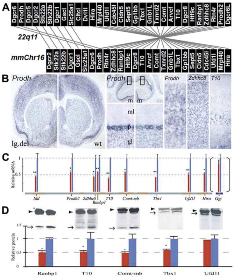
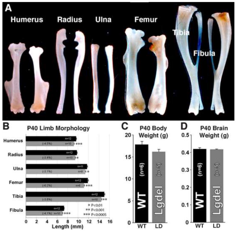
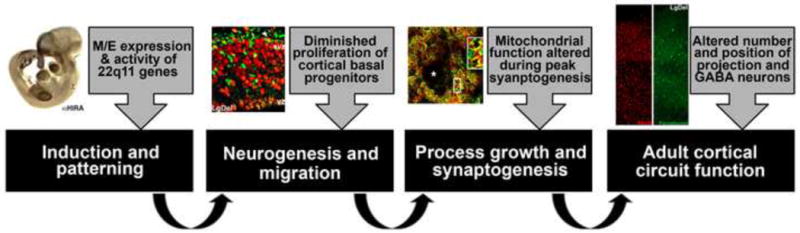
DiGeorge, or 22q11 deletion syndrome (22q11DS), the most common survivable human genetic deletion disorder, is caused by deletion of a minimum of 32 contiguous genes on human chromosome 22, and presumably results from diminished dosage of one, some, or all of these genes--particularly during development. Nevertheless, the normal functions of 22q11 genes in the embryo or neonate, and their contribution to developmental pathogenesis that must underlie 22q11DS are not well understood. Our data suggests that a substantial number of 22q11 genes act specifically and in concert to mediate early morphogenetic interactions and subsequent cellular differentiation at phenotypically compromised sites--the limbs, heart, face and forebrain. When dosage of a broad set of these genes is diminished, early morphogenesis is altered, and initial 22q11DS phenotypes are established. Thereafter, functionally similar subsets of 22q11 genes--especially those that influence the cell cycle or mitochondrial function--remain expressed, particularly in the developing cerebral cortex, to regulate neurogenesis and synaptic development. When dosage of these genes is diminished, numbers, placement and connectivity of neurons and circuits essential for normal behavior may be disrupted. Such disruptions likely contribute to vulnerability for schizophrenia, autism, or attention deficit/hyperactivity disorder seen in most 22q11DS patients.


Figures

Figure 1
Figure 1. 22q11 genes, 22q11 deletion, and the brain
A. A schematic of the 32 genes in the minimal critical deleted region associated with 22q11 Deletion Syndrome (22q11DS) on human chromosome 21, and the 26 murine orthologues of these genes found on mouse chromosome (mmChr) 16. The lines indicate either colinearity between orthologues, or reversal of 5′ and 3′ position on the chromosome. B. Brain expression of representative 22q11 genes in the large deletion mouse model (Lgdel) of 22q11DS or wild type controls, shown by in situ hybridization. There is widespread regional and cellular expression of 22q11 genes in the adult mouse brain. C. mRNA levels for multiple 22q11 orthologues are diminished by 50% in the adult (post natal day 60) Lgdel mouse brain. D. Protein levels of a subset of 22q11 genes are also diminished by 50% in the embryonic (shown here) as well as adult brain, in parallel with mRNA, except for Ufd1L, for which there is translational dosage compensation—protein levels return to 100% of wild type, despite a 50% decrement in message levels.
Figure 2
Figure 2. 22q11 genes and mesenchymal/epithelial (M/E) interactions
A. A PCR array evaluating multiple 22q11 genes from the 1.5 MB minimal critical deleted region in microdissected samples of four regions from E10.5 mouse embryos where M/E interactions drive morphogenesis: the frontonasal mass and forebrain (fnm/ fb), the branchial arches (ba), the aortic arches and heart (aah) and the forelimb bud. At the far right, four genes that are differentially associated with each region are included as controls to validate the identity of the microdissected material. B. Immuno- (brown label) or in situ hybridization localization of 5 22q11 genes showing selective or specific expression in the forelimb bud, the heart or aortic arches, the branchial arches, and the frontonasal mass/ forebrain. C. At left: littermate wild type and Lgdel mutant E10.5 embryos hybridized for Prodh (these two embryos were labeled simultaneously in the same reaction vials, and genotyped subsequently) show distinct diminished expression levels of 22q11 genes following heterozygous deletion, without appreciable change in patterned expression in the fnm/ fb (1), the ba (2), the aah (3) or the forelimb bud (4). At right: qPCR analysis of expression of 10 representative 22q11 genes in microdissected fnm/ fb from wild type and Lgdel E10.5 embryos shows consistent 50% decrease in mRNA levels. Ggt, a 22q11 gene in human that is not located on mmChr.16 with other 22q11 orthologues, is not diminished in dosage. D. Coincidence of 22q11 gene expression with neural crest-related genes in the mesenchyme at sites of M/ E interactions. At left: a wild type E10.5 embryo hybridized for mRNA for Snail2, which is selectively expressed in mesenchymal neural crest. Center: E10.5 section in situ preparations from the fnm/ fb and flb of wild type (+/ +) and Lgdel (+/ -) embryos showing the coincidence of Snail2 and ProdH2, a 22q11 gene, in the mesenchyme. At right: Snail2 mRNA levels in Lgdel embryos, measured using qPCR, approximate those seen in wild type embryos.
Figure 3
Figure 3. Morphometric assessment of brain and body phenotypes in the Lgdel mouse model of 22q11DS
A. There are visible differences in the lengths of major limb bones from the Lgdel young adult mouse compared to wild type littermates. B. The lengths of all of the major bones of the forelimb are significantly reduced compared to wild type littermates. C. Body weight does not differ significantly in Lgdel mouse. D. There is a trend for diminished brain weight; however this difference does not reach statistical significance.
Figure 4
Figure 4. 22q11 genes are robustly expressed in the proliferating zones of the embryonic cortex and proliferation is specifically disrupted in the svz of the embryonic Lgdel cortex
A. In-situ hybridization of wild-type embryonic mouse forebrain for the 22q11 genes, Ranbp1, Cdc45l, Htf9c, Ufd1l, Hira and Sept5. Signal intensity is most intense in the cortical proliferative zone for Ranbp1 and Cdc45l. Expression is more broadly distributed for Htf9c, Ufdl and Hira. Finally, Sept5 expression is absent from the proliferative zone and prominent in the cortical plate. B: Cartoon and immunohistochemical images indicate that the mitotic marker PH3 (phosphohistone 3) is reduced in the subventricular zone of embryonic day 13.5 Lgdel cortex but not in the ventricular zone. C. Similarly, the DNA synthesis mitotic marker, BrdU, is less frequently observed in subventricular zone cells (Tbr2+) of the Lgdel cortex. However, non-subventricular BrdU + mitotic cells are not disrupted (right panels).
Figure 5
Figure 5. Embryonic interneurons are altered in their location during migration and mature interneurons and projection neurons have altered laminar location/ frequency in the postnatal cortex
A. Staining for the interneuron marker, calbindin, in E13 Lgdel and wild-type cortex shows that these cells are less frequently observed in the Lgdel at dorsal cortical locations. B. By marking 5 equidistant Bins in the cortex (see A), we show that calbindin cells are less frequently observed in more dorsal cortical locations. C. Upper layer projection neurons (Cux1 +) are less frequent in postnatal Lgdel cortex compared to wild-type. D. In postnatal day 21 cortex, the distribution of the interneuron marker, parvalbumin, is altered in Lgdel animals.
Figure 6
Figure 6. A subset of 22q11 genes is associated with synaptic mitochondria
A. Six 22q11 gene products are localized to mitochondria. The localization of five proteins (Mrpl40, Prodh, Slc25a1, T10, Txnrd2) are illustrated by co-transfection of a GFP-labeled expression construct with a mitochondrially-targeted mCherry construct. Co-localized products appear yellow in merged images. Localization of Zdhhc8 is illustrated by immunostaining with anti-Zdhhc8 antiserum (green) and anti-Uqcrc1 antiserum (red). B. Mitochondrial localization of Zdhhc8 and Txnrd2 was also demonstrated by fractionating cortical tissue extracts to enrich for free mitochondria and synaptosomal mitochondria; western blot analysis shows an abundance of both proteins in synaptic mitochondrial fractions. C. Zdhhc8 is present in synapses, particularly those in glutamatergic synaptic terminals (which are recognized based on immunoreactivity for the vesicular glutamate transporter, Vglut1). The dense neuropil of the glomeruli of the olfactory bulb, which include both excitatory glutamatergic and inhibitory GABAergic terminals provide a clear illustration of the restricted localization of Zdhhc8 in the Vglut1 labeled glutamatergic terminals (presumably from olfactory receptor neuron axons) within the glomeruli. In contrast, Zdhhc8 is largely absent from the Gad67 and Parvalbumin labeled GABAergic subset of glomerular synaptic terminals.
Figure 7
Figure 7. Mitochondrial function of 22q11 genes
A. Controlled expression of HA-tagged Zdhhc8 protein in a tetracycline-inducible 3T3 cell line shows that high level overexpression of Zdhhc8 causes substantial apoptosis by 18 hours. B. Mitochondrial localization and induction of cell death by Zdhhc8 require the C-terminus of the protein, which includes the DHHC domain. C-E. Zdhhc8 over-expression induced cell death (shown in C) appears to be apoptotic, as it is robustly inhibited both by co-expression of the anti-apoptotic factor Bcl2 (D) and by the addition of an oxygen radical-scavenging agent PBN (E). F. A yeast two-hybrid analysis indicates that the Zdhhc8 tail domain (AA 215-762) can interact with the Uqcrc1 protein. Full length Uqcrc1 (AA 23-480) was co-transfected into yeast with deletion fragments of Zdhhc8. Right columns show growth of reporter yeast transformed with both plasmids on selective (-His) or non-selective (+His) growth media (-Leu/ -Trp). G. Reduced expression of 22q11 genes in the Lgdel mouse may influence ongoing function of cortical mitochondria. In the cortex of the Lgdel mouse, the expression of 22q11 genes, including Zdhhc8, is reduced by approximately 50%; however, several mitochondrial genes that are not on 22q11 show increased expression levels in the neonatal cortex. This increased expression of other mitochondrial genes (particularly Uqcrc1 and Uqcrc2, components of mitochondrial Complex III) may represent compensation for reduced expression of one or more 22q11 mitochondrial genes.
Figure 8
Figure 8. A summary of critical stages of brain development compromised by diminished dosage of 22q11 genes, based upon data summarized in this review
Our evidence suggests that 22q11 genes act normally to regulate each major step of neural development. Heterozygous deletion leads to altered dosage and function of critical subsets of 22q11 genes, some with maximal function during particular neurodevelopmental events. The consequence of these changes due to 22q11 deletion include altered frequency of cortical projection neurons in layers 2/ 3, particularly in anterior/ frontal association cortices, and parallel disruption of position of GABAergic interneurons.

Similar articles

Cited by

References

    1. Arnsten AF. Fundamentals of attention-deficit/hyperactivity disorder: circuits and pathways. J Clin Psychiatry. 2006;67(Suppl 8):7–12. - PubMed
    1. Bachiller D, Klingensmith J, Shneyder N, Tran U, Anderson R, Rossant J, De Robertis EM. The role of chordin/Bmp signals in mammalian pharyngeal development and DiGeorge syndrome. Development. 2003;130:3567–3578. - PubMed
    1. Bearden CE, van Erp TG, Dutton RA, Lee AD, Simon TJ, Cannon TD, Emanuel BS, McDonald-McGinn D, Zackai EH, Thompson PM. Alterations in midline cortical thickness and gyrification patterns mapped in children with 22q11.2 deletions. Cereb Cortex. 2009;19:115–126. - PMC - PubMed
    1. Bearden CE, van Erp TG, Dutton RA, Tran H, Zimmermann L, Sun D, Geaga JA, Simon TJ, Glahn DC, Cannon TD, Emanuel BS, Toga AW, Thompson PM. Mapping cortical thickness in children with 22q11.2 deletions. Cereb Cortex. 2007;17:1889–1898. - PMC - PubMed
    1. Beasley CL, Zhang ZJ, Patten I, Reynolds GP. Selective deficits in prefrontal cortical GABAergic neurons in schizophrenia defined by the presence of calcium-binding proteins. Biol Psychiatry. 2002;52:708–715. - PubMed

Publication types

MeSH terms