. 2011 Jan 28;286(4):2956-65.
doi: 10.1074/jbc.M110.159517. Epub 2010 Sep 8.

Structural studies and the assembly of the heptameric post-translational translocon complex

Affiliations

Structural studies and the assembly of the heptameric post-translational translocon complex

Yoichiro Harada et al. J Biol Chem. .

Abstract

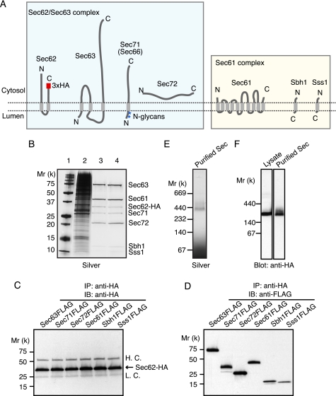
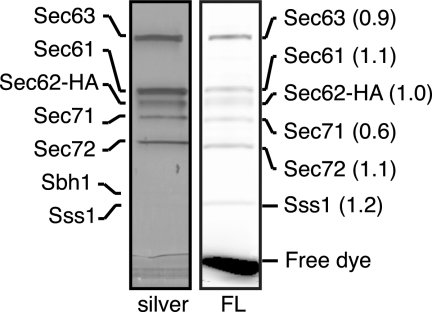
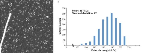
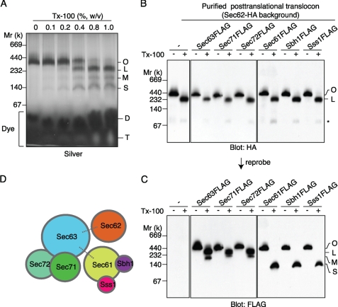
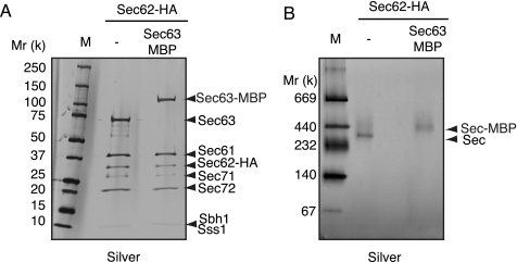
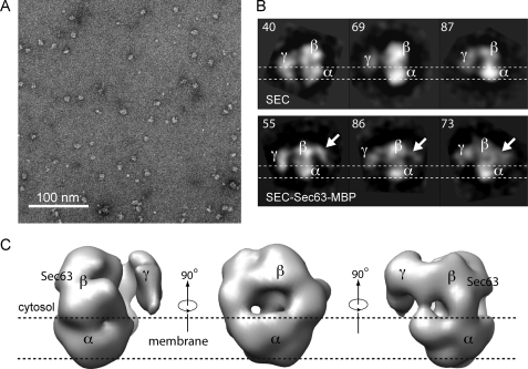
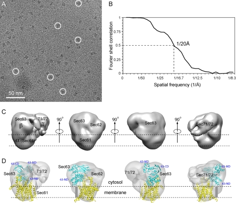
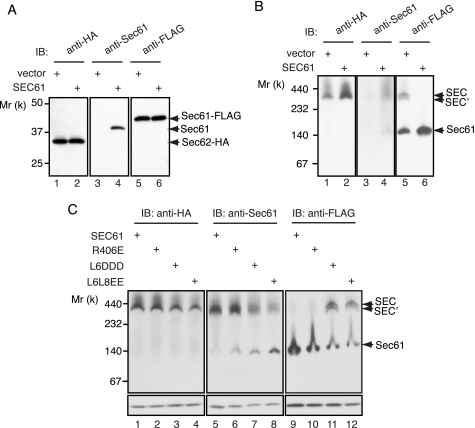
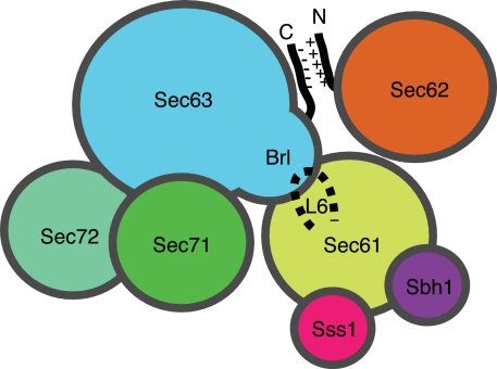
In Saccharomyces cerevisiae, some of the nascent chains can be post-translationally translocated into the endoplasmic reticulum through the heptameric post-translational translocon complex (post-translocon). This membrane-protein complex is composed of the protein-conducting channel and the tetrameric Sec62/63 complex. The Sec62/63 complex plays crucial roles in targeting of the signal recognition particle-independent protein substrate to the protein-conducting channel and in assembly of the post-translocon. Although the molecular mechanism of the post-translational translocation process has been well established, the structure of the post-translocon and how the channel and the Sec62/63 complex form the heptameric complex are largely uncharacterized. Here, we report a 20-Å resolution cryo-electron microscopy structure of the post-translocon. The purified post-translocon was found to have a mass of 287 kDa, which is consistent with the unit stoichiometry of the seven subunits as determined by a cysteine labeling experiment. We demonstrated that Triton X-100 dissociated the heptameric complex into three subcomplexes identified as the trimeric translocon Sec61-Sbh1-Sss1, the Sec63-Sec71-Sec72 trimer, and the heterotetramer Sec62-Sec63-Sec71-Sec72, respectively. Additionally, a role of the sixth cytosolic loop of Sec61 in assembly of the post-translocon was demonstrated. Mutations of conserved, positively charged amino acid residues in the loop caused decreased formation of the post-translocon. These studies provide the first architectural description of the yeast post-translocon.


Figures

FIGURE 1.
FIGURE 1.
Purification of the post-translocon from yeast. A, subunit composition of the post-translocon. The HA tag, transmembrane regions, and luminal and cytosolic locations of the N and C termini of each subunit and N-glycan (shown as asterisks) are diagrammed on the figure. Light green and light yellow boxes represent the Sec62/Sec63 complex and the Sec61 complex, respectively. B, SDS-PAGE analysis for purification steps of the post-translocon. The Sec62-HA was immunoprecipitated from the digitonin-soluble fraction of the microsomes using anti-HA beads (1 eq/lane, lane 2). The beads were washed and then eluted with HA peptide (800 eq/lane, lane 3). The eluate was subjected to Q-Sepharose anion exchange chromatography. After washing the beads, the Sec62/63 complex was eluted with 1.0 m NaCl (3,000 eq/lane, lane 4). The molecular weight markers are represented in lane 1. The gel was stained with silver. C and D, immunoprecipitation of the post-translocon containing Sec62-HA and either the FLAG-tagged Sec63, Sec71, Sec72, Sec61, Sbh1, or Sss1. The Sec62-HA was immunoprecipitated with anti-HA antibody from the digitonin-solubilized microsomes. The immunoprecipitate was analyzed by SDS-PAGE, followed by Western blot with anti-HA (C) and anti-FLAG (D) antibodies. H. C. and L. C., heavy and light chains of IgG, respectively. E and F, blue native-PAGE analysis for purification steps of the Sec62/63 complex. E, the HA peptide-eluate fraction from anti-HA beads was analyzed (300 eq/lane) and stained with silver. F, the fractions as shown in B (lanes 2 and 3) were subjected to blue native-PAGE and analyzed by Western blot using anti-HA antibody. IP, immunoprecipitate; IB, immunoblot.
FIGURE 2.
FIGURE 2.
Subunit stoichiometry of the post-translocon. The purified post-translocon was denatured, reduced, and subsequently labeled with the maleimide containing a fluorescent label. The labeled samples (1 pmol) were analyzed by SDS-PAGE followed either by silver staining (left panel) or by direct detection of the fluorescence intensity (right panel). The protein amount was estimated by SDS-PAGE/silver staining using BSA as a standard. Individual subunits were identified based on their molecular weight. The molar ratios of Cys, as indicated in parentheses, were calculated from fluorescent intensity of each subunit divided by the number of Cys in that subunit. Sec62-HA was set to 1.0.
FIGURE 3.
FIGURE 3.
STEM mass measurement of yeast post-translocon supports a unit stoichiometry of the heptameric complex. A, a selected STEM image with seven post-translocon particles encircled randomly as examples. TMV indicates the helical rod of the tobacco mosaic virus with a width of 180 Å. B, a histogram of the measured particle masses.
FIGURE 4.
FIGURE 4.
Subcomplex formation of the post-translocon. A, the purified post-translocon (130 eq) was incubated with 0–1.0% (w/v) of Triton X-100 (Tx-100) in the presence of 0.7% digitonin and analyzed by BN-PAGE/silver staining. Original (O), large (L), medium (M), and small (S) subcomplexes are indicated at the right of the panel. Digitonin (D) and Triton X-100 (T) in the purified post-translocon migrated to the positions indicated at the right of the panel. B and C, treatment of the purified post-translocon containing the FLAG-tagged subunit (30 eq) without (−) or with (+) 1.0% Triton X-100, followed by BN-PAGE/Western blot. The membrane was first probed with anti-HA antibody (B) and then reprobed with anti-FLAG antibody (C). An asterisk indicates the free Sec62. D, schematic model of subcomplexes of the post-translational translocon. The dashed lines represent a Triton X-100-sensitive interaction.
FIGURE 5.
FIGURE 5.
Production of MBP fusion complex for mapping Sec63 by EM. A, SDS-PAGE analysis of the non-MBP-fused and the MBP-fused post-translocon (300 eq/lane). The gel was stained with silver. The Sec subunits were identified based on their molecular weight and indicated at the right of the panel. B, BN-PAGE/silver stain of the non-MBP-fused and the MBP-fused post-translocon (150 eq/lane). They are indicated as Sec and Sec-MBP, respectively.
FIGURE 6.
FIGURE 6.
Negative stain EM of the post-translocon and the MBP-fused post-translocon. A, a typical area of the raw micrograph of the post-translocon in negative stain. B, two-dimensional class averages of the side views. Top panel, the post-translocon; bottom panel, the post-translocon with MBP fused to the C terminus of Sec63 subunit. The white arrows point to extra clouds of density that are attributed to the fused MBP. Three major density features (α, β, and γ) are labeled. The likely orientation of the particles with respect to the membrane bilayer is indicated. The numbers at the top left corner of each panel are the numbers of raw particle images used to generate the averages. The density α is chosen over density γ because transmembrane region because γ is too small to account for the predicted transmembrane mass of the complex. See detail in Fig. 7. C, three-dimensional reconstruction of the post-translocon from negatively stained EM images of the purified membrane complex. The three major domains are labeled. The approximate position of Sec63 is also labeled.
FIGURE 7.
FIGURE 7.
Cryo-EM of the post-translocon at ∼20-Å resolution. A, Raw image with six individual particles highlighted in white circles. B, Fourier shell correlation. C, surface rendered side views of the three-dimensional cryo-EM map. D, tentative docking of Sec61 heterotrimer in yellow and Sec63 cytosolic domain homolog structure in cyan. A gray bar represents a hypothetical transmembrane helices of Sec63 that precede the N-terminal subdomain (63-ND) of the cytosolic Brl domain of Sec63. The middle helical (63-MD) and the C-terminal immunoglobulin-like subdomains (63-CD) are labeled.
FIGURE 8.
FIGURE 8.
Effects of the cytosolic loops of Sec61 on formation of the post-translocon. A, SDS-PAGE/Western blot analyses of the digitonin-solubilized microsomes. The microsomes were prepared from cells expressing Sec62-HA and Sec61-FLAG that were transformed with either empty vector (vector; lanes 1, 3, and 5) or plasmid encoding wild type Sec61 (SEC61; lanes 2, 4, and 6). The PVDF membranes were blotted with anti-HA antibody for the Sec62-HA marker (left panel), anti-Sec61 antibody for the nontagged Sec61 marker (middle panel), and anti-FLAG antibody for the Sec61-FLAG marker (right panel). B, the same sample as in A was analyzed by BN-PAGE, followed by Western blot with anti-HA (left panel), anti-Sec61 (middle panel), and anti-FLAG (right panel) antibodies. The post-translocon, the Sec62-dissociated complex, and the Sec61 complex are indicated as SEC, SEC′, and Sec61, respectively. C, BN- and SDS-PAGE/Western blot analyses of the digitonin-solubilized microsomes from cells containing the wild type (SEC61; lanes 1, 5, and 9), R406E (lanes 2, 6, and 10), L6DDD (lanes 3, 7, and 11), and L6L8EE (lane 4, 8, and 12) mutants Sec61. Upper panels, BN-PAGE; lower panels, SDS-PAGE. IB, immunoblot.
FIGURE 9.
FIGURE 9.
Hypothetical model for formation of the post-translocon. A cytosolic view of the post-translocon is depicted. See details in main text.

Similar articles

Cited by

References

    1. Rapoport T. A. (2007) Nature 450, 663–669 - PubMed
    1. Stroud R. M., Walter P. (1999) Curr. Opin. Struct. Biol. 9, 754–759 - PubMed
    1. McClellan A. J., Brodsky J. L. (2000) Genetics 156, 501–512 - PMC - PubMed
    1. Wilkinson B. M., Critchley A. J., Stirling C. J. (1996) J. Biol. Chem. 271, 25590–25597 - PubMed
    1. Van den Berg B., Clemons W. M., Jr., Collinson I., Modis Y., Hartmann E., Harrison S. C., Rapoport T. A. (2004) Nature 427, 36–44 - PubMed

Publication types

MeSH terms

Substances