Review
. 2010:92:165-200.
doi: 10.1016/S0070-2153(10)92005-X.

Endocytosis and intracellular trafficking of Notch and its ligands

Affiliations
Review

Endocytosis and intracellular trafficking of Notch and its ligands

Shinya Yamamoto et al. Curr Top Dev Biol. 2010.

Abstract

Notch signaling occurs through direct interaction between Notch, the receptor, and its ligands, presented on the surface of neighboring cells. Endocytosis has been shown to be essential for Notch signal activation in both signal-sending and signal-receiving cells, and numerous genes involved in vesicle trafficking have recently been shown to act as key regulators of the pathway. Defects in vesicle trafficking can lead to gain- or loss-of-function defects in a context-dependent manner. Here, we discuss how endocytosis and vesicle trafficking regulate Notch signaling in both signal-sending and signal-receiving cells. We will introduce the key players in different trafficking steps, and further illustrate how they impact the signal outcome. Some of these players act as general factors and modulate Notch signaling in all contexts, whereas others modulate signaling in a context-specific fashion. We also discuss Notch signaling during mechanosensory organ development in the fly to exemplify how endocytosis and vesicle trafficking are effectively used to determine correct cell fates. In summary, endocytosis plays an essential role in Notch signaling, whereas intracellular vesicle trafficking often plays a context-dependent or regulatory role, leading to divergent outcomes in different developmental contexts.


Figures

Fig. 1
Fig. 1. Overview of Endocytosis and Vesicle Trafficking
Transmembrane proteins are made in the ER and traffic through the Golgi apparatus to reach the plasma membrane. From the cell surface, these proteins can re-enter the cell via various endocytosis pathways. Clathrin-dependent endocytosis is usually referred to as “canonical endocytosis”. Clathrin adaptor proteins, such as the AP-2 complex, recruit clathrin and cargo transmembrane proteins to the site of endocytosis. The clathrin-coated endocytic vesicle is pinched off by the action of Dynamin GTPase and the clathrin coat is then removed by molecular chaperone Hsc70 via the assistance of Auxilin. On the other hand, endocytosis can also occur without clathrin and is referred to as “non-canonical endocytosis” or “Clathrin-independent endocytosis”. After endocytosis, small GTPase Rab5 and SNARE protein Avalanche (Avl) mediate the fusion of endocytic vesicles with the early/sorting endosome. From the early endosome, endocytosed proteins can recycle back to the plasma membrane directly in a Rab4-dependent manner or indirectly through the recycling endosome in a Rab11-dependent manner. Alternatively, they can return back to the Golgi, or travel to the late endosome and lysosome for degradation. Proteins destined for degradation are sorted into Rab7-positive late endosome or multi vesicular bodies (MVB). Packaging of transmembrane proteins into intraluminal vesicles is mediated by the ESCRT complexes. In certain cell contexts, MVB can secrete their contents to extracellular regions. These secreted MVBs are referred to as exosomes. Finally, through HOPS and AP3 complexes, MVB/late endosomes fuse with the lysosome and transmembrane proteins are degraded by proteases and acid hydrolases.
Fig. 2
Fig. 2. Endocytosis and Trafficking of DSL Ligands in the Signal Sending Cell
DLS ligands are synthesized in the ER and traffics to the cell surface through the secretory pathway. Endocytosis is required in the signal sending cell for activation of the canonical Notch signaling pathway, but there are two non-mutually exclusive hypotheses to explain how endocytosis promotes DSL ligand activity. In the “Ligand Activation” theory, DSL proteins that have just been synthesized and reached the cell surface are still inactive and do not have the capacity to activate the Notch receptor on the signal receiving cell. DSL are endocytosed and sorted into a unique endocytic compartment where they become “activated”. The activated ligands return to the cell surface via the recycling pathway where they interact with and activate the Notch receptor. In contrast, the “pulling force” model insists that when DSL ligands and Notch receptor interact, endocytosis in the signal sending cell generates a mechanical force that leads to a conformational change in the Notch receptor. This force mediates the separation of the Notch heterodimer, and allows the S2 cleavage mediated by ADAM proteases. The extracellular portion of Notch is trans-endocytosed into the signal sending cell and assumed to be degraded through the lysosomal pathway along with the DSL ligands.
Fig. 3
Fig. 3. Endocytosis and Trafficking of Notch during Canonical Signal Activation
Canonical Notch signaling occurs in a cell-cell contact dependent manner, in which membrane-bounded DSL (Delta/Serrate/LAG-2) ligands activate Notch receptor on the neighboring cell. This interaction with ligands leads to a conformational change in Notch receptors and exposes the S2 cleavage site, which is cleaved by ADAM metalloprotease to produce Notch extracellular truncation (NEXT). NEXT then undergoes S3/S4 cleavage via γ-secretase to generate Notch intracellular domain (NICD). During this process, whether endocytosis is required for NEXT cleavage by γ-secretase is controversial. Two possible models are shown as dashed lines: (1) S3 cleavage takes place on the cell surface and does not require endocytosis. Instead, endocytosis might play a negative tuning role since γ-secretase prefers the generation of unstable form of NICD in endosomal compartments. (2) Endocytosis is required for S3 cleavage. Dynamin and two early endosomal proteins, Rab5 and Avl, are important in internalization and endocytic trafficking of NEXT fragment. Rbcn-3A, Rbcn-3B, and V-ATPase function in acidification of endosomal compartments where γ-secretase is more active and S3 cleavage is thus more efficient.
Fig. 4
Fig. 4. Endocytosis and Trafficking of Notch Receptor during Lysosomal Degradation and Non Canonical Activation
Inactive Notch receptors undergo constitutive endocytosis, endocytic trafficking, and are finally degraded in the lysosome. Full-length Notch receptors are firstly mono-ubiquitinated by E3 ligase Deltex, Su(dx), or DNedd4 for internalization. With the help of Rab5 and Avl, Notch receptors enter into the early endosome, from which they can recycle back to the cell surface or further progress into MVB/late endosomes. Sorting of Notch receptors into intraluminal vesicles is mediated by Hrs and ESCRT complexes (I, II, and III). Lgd, a C2-containing phospholipid binding protein, is placed between Hrs and ESCRT complexes based on epistatic analysis results. HOPS and AP-3 complexes are involved in endocytic trafficking/fusion between late endosome and lysosome. They are required for Dx-dependent Notch signaling activation. When endocytic trafficking of full-length Notch receptor for lysosomal degradation is disrupted, Notch receptor can undergo proteolytic cleavages in a ligand-independent manner (dashed line), which leads to ectopic activation of Notch signaling.
Fig. 5
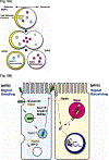
Fig. 5. Regulation of Notch Signaling via Endocytosis and Vesicle Trafficking during Mechanosensory Organ Development in Drosophila
A) During mitosis of the sensory organ precursor (SOP) cell, cell fate determinants Neuralized (Neur) and Numb are asymmetrically segregated into the anterior crescent which is determined through interactions between cell polarity factors. Upon cytokinesis, Neur and Numb are both inherited by the anterior pIIb cell, whereas the posterior cell fails lacks these factors. In parallel to the asymmetric segregation of the two cell fate determinants, Rab11-positive recycling endosomes become enriched in the pIIb cell, whereas Sara-positive endosomes are sorted into the pIIa cell. This asymmetric segregation of cell fate determinants and specific endocytic compartment biases the following Notch signaling between the pIIa and the pIIb cell B) In the pIIb cell, Neur promotes the endocytosis and sorting of Delta for activation. Activated Delta traffics through Rab11-positive endosomes and recycle back to the apical cell surface where there is an enrichment of actin filaments and microvilli, referred to as the ARS (apical Actin Rich Structure). Sec15, a Rab11 effector and component of the exocyst complex is required for this apical recycling of Delta. In addition, Arp2/3 and WASp, positive regulators of actin polymerization are also required for recycling of Delta, possibly through mobilization of Delta positive vesicles and/or facilitation of ARS formation. Activated Delta that returned to the cell surface interacts with and activates Notch in the pIIa cell. Sanpodo (Spdo), a four transmembrane domain protein is present at the cell surface of the pIIa cell to promote the reception of this signal. pIIa cell cannot signal to the pIIb since they are not able to activate Delta due to lack of Neur. In addition, pIIb cannot receive Notch signaling since Spdo is endocytosed by Numb and kept in an inactive form. As a third mechanism, Sara-positive endosome has been recently been proposed to bias Notch signaling by actively recruiting Notch and Delta into the pIIa cell. γ-secretase cleavage of Notch has been proposed to be happening at or before Notch entering the Sara-positive endosome, but the exact mechanism and role of Sara is not fully understood.

Similar articles

Cited by

References

    1. Acconcia F, Sigismund S, and Polo S (2009). Ubiquitin in trafficking: the network at work. Experimental cell research 315, 1610–1618. - PubMed
    1. Ahimou F, Mok LP, Bardot B, and Wesley C (2004). The adhesion force of Notch with Delta and the rate of Notch signaling. The Journal of cell biology 167, 1217–1229. - PMC - PubMed
    1. Babaoglan AB, O’Connor-Giles KM, Mistry H, Schickedanz A, Wilson BA, and Skeath JB (2009). Sanpodo: a context-dependent activator and inhibitor of Notch signaling during asymmetric divisions. Development (Cambridge, England) 136, 4089–4098. - PMC - PubMed
    1. Bardin AJ, Le Borgne R, and Schweisguth F (2004). Asymmetric localization and function of cell-fate determinants: a fly’s view. Current opinion in neurobiology 14, 6–14. - PubMed
    1. Bardin AJ, and Schweisguth F (2006). Bearded family members inhibit Neuralized-mediated endocytosis and signaling activity of Delta in Drosophila. Developmental cell 10, 245–255. - PubMed

Publication types

LinkOut - more resources