Comparative Study
. 2010 Aug 25;30(34):11259-69.
doi: 10.1523/JNEUROSCI.1949-10.2010.

Development and regeneration of sensory transduction in auditory hair cells requires functional interaction between cadherin-23 and protocadherin-15

Affiliations
Comparative Study

Development and regeneration of sensory transduction in auditory hair cells requires functional interaction between cadherin-23 and protocadherin-15

Andrea Lelli et al. J Neurosci. .

Abstract

Tip links are extracellular filaments that connect pairs of hair cell stereocilia and convey tension to mechanosensitive channels. Recent evidence suggests that tip links are formed by calcium-dependent interactions between the N-terminal domains of cadherin-23 (CDH23) and protocadherin-15 (PCDH15). Mutations in either CDH23 or PCDH15 cause deafness in mice and humans, indicating the molecules are required for normal inner ear function. However, there is little physiological evidence to support a direct role for CDH23 and PCDH15 in hair cell mechanotransduction. To investigate the contributions of CDH23 and PCDH15 to mechanotransduction and tip-link formation, we examined outer hair cells of mouse cochleas during development and after chemical disruption of tip links. We found that tip links and mechanotransduction with all the qualitative properties of mature transduction recovered within 24 h after disruption. To probe tip-link formation, we measured transduction currents after extracellular application of recombinant CDH23 and PCDH15 fragments, which included putative interaction domains (EC1). Both fragments inhibited development and regeneration of transduction but did not disrupt transduction in mature cells. PCDH15 fragments that carried a mutation in EC1 that causes deafness in humans did not inhibit transduction development or regeneration. Immunolocalization revealed wild-type fragments bound near the tips of hair cell stereocilia. Scanning electron micrographs revealed that hair bundles exposed to fragments had a reduced number of linkages aligned along the morphological axis of sensitivity of the bundle. Together, the data provide direct evidence implicating CDH23 and PCDH15 proteins in the formation of tip links during development and regeneration of mechanotransduction.


Figures
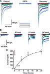
Figure 1.
Figure 1.
Recovery of transduction current after EGTA treatment. A, Representative families of transduction currents recorded from outer hair cells in control conditions (left), immediately after EGTA treatment (center), and 12 h after EGTA treatment and return to standard calcium (right). The cells were voltage clamped at −64 mV. Hair bundles were deflected, as indicated by the stimulation protocol shown below, with deflections that ranged between −0.2 and 1.2 μm in 0.2 μm increments. The scale bar applies to all three current families. B, Time course of current recovery. Representative families of transduction currents recorded 3, 6, 12, and 24 h after EGTA treatment. Deflections ranged between −0.2 and 1.2 μm in 0.2 μm increments. The scale bar applies to all four current families. C, Mean maximal transduction current amplitudes (squares) plotted as a function of recovery time. The number of cells at each time point is shown next to the data point. The data were fitted with a single exponential equation of the form: I = IMax(1 − e t) with IMax = −549 pA, τ = 10.6 h. For comparison, a single data point (circle) from age (P2) and region (base) matched control outer hair cells is reprinted from Lelli et al. (2009).
Figure 2.
Figure 2.
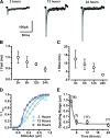
Properties of transduction currents during recovery. A, Representative transduction currents at 3, 12, and 24 h after EGTA treatment (black line). Current traces that represented ∼50% activation were fitted with a double exponential equation (green line). The scale bars apply to all current traces. B, C, Fast (B) and slow (C) time constants from double exponential fits were averaged for the time points shown. Error bars represent SE. The number of samples at each time point are shown in E. D, Representative stimulus-response curves at 3, 12, and 24 h after EGTA treatment. The data were fit with second-order Boltzmann equations. E, Operating range was taken as the stimulus amplitude required to activate transduction between 10 and 90%. Mean values ± SE are shown for each time point with the number of samples indicated above.
Figure 3.
Figure 3.
CDH23 and PCDH15 fragments interfere with transduction current recovery. Schematic drawing of pairs of stereocilia illustrating the experimental paradigm. CDH23 fragments (CDH23-His), PCDH15 fragments (PCDH15-His), or integrin fragments (INTG-His) were added to the culture media immediately after EGTA treatment (left). We hypothesize that CDH23-His and PCDH15-His bind the N terminus of its endogenous binding partner to prevent reformation of full-length tip links. A, A representative family of transduction currents recorded 12 h after EGTA treatment and application of CDH23-His (middle). Twenty-four hours after a second treatment with EGTA and a vigorous washout of CDH23-His, the currents recovered (right). The stimulus protocol is shown at the top. Deflections ranged between −0.2 and 1.2 μm in 0.2 μm increments. The protocol and scale bars apply to all current families. B, A representative family of transduction currents recorded 12 h after application of PCDH15-His (middle). Twenty-four hours after a second treatment with EGTA and a vigorous wash, the currents recovered (right). C, A representative family of transduction currents recorded after application of INTG-His.
Figure 4.
Figure 4.
Effects of mutations in CDH23 and PCDH15 fragments. A, A form of CDH23-His that carried the salsa mutation (E737V) was applied to hair bundles after the EGTA treatment. A representative family of transduction currents showed reduction in current amplitude. Presumably, the mutation in the seventh EC domain did not affect binding to endogenous PCDH15. Bundles were deflected in 0.2 μm steps that ranged between −0.2 and 1.2 μm. The scale bar and stimulus protocol apply to both families of transduction currents. B, In this case a PCDH15-Fc that carried a mutation (R139G) in the first cadherin domain was added after EGTA treatment. The equivalent mutation causes DFNB23 in humans. A representative family of transduction currents with normal amplitudes is shown on the right. It is hypothesized that the mutation prevented binding to the N terminus of CDH23, which allowed the endogenous molecules to reform functional tip links.
Figure 5.
Figure 5.
Summary bar graph and dose-response relation for CDH23 fragments. A, Bar graph that summarizes mean maximal transduction current amplitudes in the various conditions presented in Figures 1 –4. CTRL, Control (no fragments added); INTG-His, integrin fragments; CDH23-His, cadherin 23 fragments; CDH23 Wash, washout of CDH23-His; PCDH15-His, protocadherin 15 fragments; PCDH15 Wash, washout of PCDH15-His; CDH23-E737V, CDH23 fragments that carry the salsa mutation; and PCDH15-R139G, PCDH15 fragments that carry the DFNB23 mutation in EC1. The number of cells examined in each condition is shown above each bar. Error bars indicate SE. B, Mean maximal amplitude of currents recorded 12 h after EGTA treatment plotted as a function of the concentration of CDH23-His solution added to the media. Higher concentrations decreased the current amplitude in a dose-dependent manner. The zero concentration corresponds to control experiments. Data from CDH23-His and CDH23-E737V were pooled since their effect was indistinguishable. The number of cells examined in each condition is shown next to each data point. Error bars indicate SE. The data were fitted with a logistic curve: y = A 2 + (A 1A 2)/(1 + (x/x 0)p); A 1 = −401 pA; A2 = −58.4 pA; x 0 = 13.6 ng/μl; p = 0.53.
Figure 6.
Figure 6.
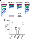
Transduction current development is impaired by CDH23 and PCDH15 fragments. A, Representative families of transduction currents recorded from apical outer hair cells. Cochleas were harvested before the onset of mechanotransduction (P0) and cultured with media supplemented with control solution, CDH23 fragments (CDH23-His), PCDH15 fragments (PCDH15-His), or PCDH15 that carried a DFNB23 mutation (PCDH15–R139G). Transduction currents were recorded after 3 d in culture. The stimulus protocol is shown at the bottom of each dataset: 0.2 μm steps from −0.2 μm to 1.2 μm. B, Bar graph illustrating the mean maximal transduction current amplitude in the different experimental conditions shown in A. The number of cells for each condition is shown above the bars. Error bars show SE.
Figure 7.
Figure 7.
Binding of CDH23 and PCDH15 fragments to hair bundles. Confocal images of cultures fixed 12 h after EGTA treatment and exposure to CDH23F11-Fc, PCDH15-Fc, or PCDH15-R139G. To increase the specificity for immunolocalization of the exogenous proteins, human IgG Fc fragments were used as a tag, which were detected by a specific FITC-conjugated secondary antibody. The left column shows F-actin visualized with Alexa Fluor 546 phalloidin. Scale bar, 5 μm. The second column shows the pattern of FITC fluorescence in samples treated with fragments. The third column shows the merge of the Alexa Fluor 546 channel and the FITC channel. The right-hand column shows a higher magnification view of representative hair bundles. Scale bars, 2 μm. A, The first 11 cadherin domains of CDH23 fused to an Fc domain. B, The entire ectodomain of PCDH15 fused to the Fc domain. C, PCDH15-R139G mutant form fused to the Fc domain.
Figure 8.
Figure 8.
Fragments prevent recovery of tip links after EGTA treatment. A, Scanning electron micrographs of outer hair cell bundles. Scale bars, 2 μm. B, Higher magnification views of the samples shown in A. Scale bars, 300 nm. Linkages (white arrows) connecting the tips of hair cell stereocilia were apparent in untreated samples (control), but were absent in samples fixed immediately after EGTA treatment (EGTA0h). Arrows indicate unoccupied tip-link sites. Several cultures were allowed 12 h of recovery after EGTA treatment (EGTA12h) and linkages reappeared (arrows). When CDH23-His and PCDH15-Fc were added during the recovery period, only a few links reformed, whereas links oriented along the axis of symmetry of the bundle were absent between most pairs of stereocilia (arrows). C, The percentage of occupied tip-link sites was quantified for each experimental condition: control (CTRL), immediately after EGTA treatment (EGTA0), 12 h after EGTA treatment (EGTA12), 12 h after EGTA, incubated with CDH23-His for 12 h after EGTA treatment, and incubated with PCDH15-Fc for 12 h after EGTA treatment. The number of bundles examined in each condition is indicated. Error bars indicate SE.

Similar articles

Cited by

References

    1. Ahmed ZM, Riazuddin S, Bernstein SL, Ahmed Z, Khan S, Griffith AJ, Morell RJ, Friedman TB, Riazuddin S, Wilcox ER. Mutations of the protocadherin gene PCDH15 cause Usher syndrome type 1F. Am J Hum Genet. 2001;69:25–34. - PMC - PubMed
    1. Ahmed ZM, Riazuddin S, Ahmad J, Bernstein SL, Guo Y, Sabar MF, Sieving P, Riazuddin S, Griffith AJ, Friedman TB, Belyantseva IA, Wilcox ER. PCDH15 is expressed in the neurosensory epithelium of the eye and ear and mutant alleles are responsible for both USH1F and DFNB23. Hum Mol Genet. 2003;12:3215–3223. - PubMed
    1. Ahmed ZM, Goodyear R, Riazuddin S, Lagziel A, Legan PK, Behra M, Burgess SM, Lilley KS, Wilcox ER, Riazuddin S, Griffith AJ, Frolenkov GI, Belyantseva IA, Richardson GP, Friedman TB. The tip link antigen, a protein associated with the transduction complex of sensory hair cells, is protocadherin-15. J Neurosci. 2006;26:7022–7034. - PMC - PubMed
    1. Alagramam KN, Yuan H, Kuehn MH, Murcia CL, Wayne S, Srisailpathy CR, Lowry RB, Knaus R, Van Laer L, Bernier FP, Schwartz S, Lee C, Morton CC, Mullins RF, Ramesh A, Van Camp G, Hageman GS, Woychik RP, Smith RJ, Hagemen GS. Mutations in the novel protocadherin PCDH15 cause Usher syndrome type 1F. Hum Mol Genet. 2001;10:1709–1718. - PubMed
    1. Assad JA, Shepherd GM, Corey DP. Tip link integrity and mechanical transduction in vertebrate hair cells. Neuron. 1991;7:985–994. - PubMed

Publication types

LinkOut - more resources