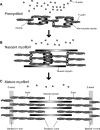
Dynamic regulation of sarcomeric actin filaments in striated muscle
- PMID: 20737540
- PMCID: PMC2963174
- DOI: 10.1002/cm.20476
Dynamic regulation of sarcomeric actin filaments in striated muscle
Abstract
In striated muscle, the actin cytoskeleton is differentiated into myofibrils. Actin and myosin filaments are organized in sarcomeres and specialized for producing contractile forces. Regular arrangement of actin filaments with uniform length and polarity is critical for the contractile function. However, the mechanisms of assembly and maintenance of sarcomeric actin filaments in striated muscle are not completely understood. Live imaging of actin in striated muscle has revealed that actin subunits within sarcomeric actin filaments are dynamically exchanged without altering overall sarcomeric structures. A number of regulators for actin dynamics have been identified, and malfunction of these regulators often result in disorganization of myofibril structures or muscle diseases. Therefore, proper regulation of actin dynamics in striated muscle is critical for assembly and maintenance of functional myofibrils. Recent studies have suggested that both enhancers of actin dynamics and stabilizers of actin filaments are important for sarcomeric actin organization. Further investigation of the regulatory mechanism of actin dynamics in striated muscle should be a key to understanding how myofibrils develop and operate.
© 2010 Wiley-Liss, Inc.
Figures


Similar articles
-
Tropomodulin capping of actin filaments in striated muscle development and physiology.J Biomed Biotechnol. 2011;2011:103069. doi: 10.1155/2011/103069. Epub 2011 Oct 17. J Biomed Biotechnol. 2011. PMID: 22013379 Free PMC article. Review.
-
CAS-1, a C. elegans cyclase-associated protein, is required for sarcomeric actin assembly in striated muscle.J Cell Sci. 2012 Sep 1;125(Pt 17):4077-89. doi: 10.1242/jcs.104950. Epub 2012 May 23. J Cell Sci. 2012. PMID: 22623720 Free PMC article.
-
Regulation of structure and function of sarcomeric actin filaments in striated muscle of the nematode Caenorhabditis elegans.Anat Rec (Hoboken). 2014 Sep;297(9):1548-59. doi: 10.1002/ar.22965. Anat Rec (Hoboken). 2014. PMID: 25125169 Free PMC article. Review.
-
Early incorporation of obscurin into nascent sarcomeres: implication for myofibril assembly during cardiac myogenesis.Histochem Cell Biol. 2008 Apr;129(4):463-78. doi: 10.1007/s00418-008-0378-y. Epub 2008 Jan 25. Histochem Cell Biol. 2008. PMID: 18219491 Free PMC article.
-
Polarization-resolved microscopy reveals a muscle myosin motor-independent mechanism of molecular actin ordering during sarcomere maturation.PLoS Biol. 2018 Apr 27;16(4):e2004718. doi: 10.1371/journal.pbio.2004718. eCollection 2018 Apr. PLoS Biol. 2018. PMID: 29702642 Free PMC article.
Cited by
-
The Drosophila formin Fhos is a primary mediator of sarcomeric thin-filament array assembly.Elife. 2016 Oct 12;5:e16540. doi: 10.7554/eLife.16540. Elife. 2016. PMID: 27731794 Free PMC article.
-
Cofilin Loss in Drosophila Muscles Contributes to Muscle Weakness through Defective Sarcomerogenesis during Muscle Growth.Cell Rep. 2020 Jul 21;32(3):107893. doi: 10.1016/j.celrep.2020.107893. Cell Rep. 2020. PMID: 32697999 Free PMC article.
-
Drosophila Tropomodulin is required for multiple actin-dependent processes within developing myofibers.Development. 2023 Mar 15;150(6):dev201194. doi: 10.1242/dev.201194. Epub 2023 Mar 24. Development. 2023. PMID: 36806912 Free PMC article.
-
Tropomodulin capping of actin filaments in striated muscle development and physiology.J Biomed Biotechnol. 2011;2011:103069. doi: 10.1155/2011/103069. Epub 2011 Oct 17. J Biomed Biotechnol. 2011. PMID: 22013379 Free PMC article. Review.
-
Transgenic Expression of the Formin Protein Fhod3 Selectively in the Embryonic Heart: Role of Actin-Binding Activity of Fhod3 and Its Sarcomeric Localization during Myofibrillogenesis.PLoS One. 2016 Feb 5;11(2):e0148472. doi: 10.1371/journal.pone.0148472. eCollection 2016. PLoS One. 2016. PMID: 26848968 Free PMC article.
References
-
- Abe H, Obinata T. An actin-depolymerizing protein in embryonic chicken skeletal muscle: purification and characterization. J Biochem. 1989;106:172–180. - PubMed
-
- Abe H, Ohshima S, Obinata T. A cofilin-like protein is involved in the regulation of actin assembly in developing skeletal muscle. J Biochem. 1989;106:696–702. - PubMed
-
- Abe H, Nagaoka R, Obinata T. Cytoplasmic localization and nuclear transport of cofilin in cultured myotubes. Exp Cell Res. 1993;206:1–10. - PubMed
-
- Abe H, Endo T, Yamamoto K, Obinata T. Sequence of cDNAs encoding actin depolymerizing factor and cofilin of embryonic chicken skeletal muscle: two functionally distinct actin-regulatory proteins exhibit high structural homology. Biochemistry. 1990;29:7420–7425. - PubMed
-
- Adams ME, Minamide LS, Duester G, Bamburg JR. Nucleotide sequence and expression of a cDNA encoding chick brain actin depolymerizing factor. Biochemistry. 1990;29:7414–7420. - PubMed
Publication types
MeSH terms
Substances
Grants and funding
LinkOut - more resources
Full Text Sources

