. 2010 Oct 22;285(43):33045-33054.
doi: 10.1074/jbc.M110.121129. Epub 2010 Aug 16.

Phosphorylation and activation of androgen receptor by Aurora-A

Affiliations

Phosphorylation and activation of androgen receptor by Aurora-A

Shao-Kun Shu et al. J Biol Chem. .

Retraction in

Abstract

Aurora-A kinase is frequently overexpressed/activated in various types of human malignancy, including prostate cancer. In this study, we demonstrate elevated levels of Aurora-A in androgen-refractory LNCaP-RF but not androgen-sensitive LNCaP cells, which prompted us to examine whether Aurora-A regulates the androgen receptor (AR) and whether elevated Aurora-A is involved in androgen-independent cell growth. We show that ectopic expression of Aurora-A induces AR transactivation activity in the presence and absence of androgen. Aurora-A interacts with AR and phosphorylates AR at Thr(282) and Ser(293) in vitro and in vivo. Aurora-A induces AR transactivation activity in a phosphorylation-dependent manner. Ectopic expression of Aurora-A in LNCaP cells induces prostate-specific antigen expression and cell survival, whereas knockdown of Aurora-A sensitizes LNCaP-RF cells to apoptosis and cell growth arrest. These data indicate that AR is a substrate of Aurora-A and that elevated Aurora-A could contribute to androgen-independent cell growth by phosphorylation and activation of AR.


Figures
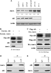
FIGURE 1.
FIGURE 1.
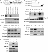
Aurora-A phosphorylates and interacts with AR. A, expression of Aurora-A in prostate cancer cell lines. The indicated cells were immunoblotted with the indicated antibodies. Expression of Aurora-A was quantified (top). Notably, a higher level of Aurora-A was detected in androgen-independent LNCaP-RF than in parent LNCaP cells. B, AR is phosphorylated by Aurora-A in vitro. In vitro kinase was performed by incubation of recombinant AR with and without recombinant Aurora-A (top). Panels 2 and 3 are immunoblots showing the proteins used for in vitro kinase assay. C, Aurora-A phosphorylates AR in vivo. HEK293 cells were transfected with FLAG-AR together with and without HA-Aurora-A. After 36 h of transfection, cells were labeled with [32P]Pi (0.5 mCi/ml) in phenol red-free minimum Eagle's medium without phosphate and serum for 4 h. FLAG-AR was immunoprecipitated, separated on SDS-PAGE, and exposed (top). Expression of the transfected plasmids is shown in panels 2 and 3. Actin was used as a loading control (bottom). D and E, Aurora-A interacts with AR. LNCaP-RF cells were immunoprecipitated (IP) with anti-AR and detected with anti-Aurora-A antibody (D) and vice versa (E). NS, nonspecific band. Each experiment was repeated three times.
FIGURE 2.
FIGURE 2.
Identification of Aurora-A phosphorylation of AR at Thr282 and Ser293. A, domain structure and location of three putative Aurora-A phosphorylation motifs of AR (top). In vitro Aurora-A kinase was carried out by incubation of recombinant Aurora-A with each GST-fused wild-type and Thr/Ser → Ala-mutated AR motif as substrate (middle). Panels 3 and 4 are Western blots showing the proteins used for the kinase assay. B and C, further definition of the phosphorylation sites with truncated and point-mutated AR. HEK293 cells were transfected with the indicated truncation (B) and point mutation (C) of full-length AR and then immunoprecipitated with anti-FLAG antibody. The immunoprecipitates were used as substrates for in vitro Aurora-A kinase assay (top panels). Bottom panels are immunoblotting with anti-FLAG antibody showing expression of transfected plasmids. Note that the size of the nonspecific band (NS) is comparable with FLAG-AR-N3 (B). D, Aurora-A phosphorylates AR-Thr282/Ser293 in vivo. HEK293 cells were transfected with the indicated plasmids and labeled with [32P]Pi (0.5 mCi/ml). FLAG-AR was immunoprecipitated, separated in SDS-PAGE, and exposed (top). Panels 2 and 3 show expression of the transfected AR and Aurora-A. E, knockdown of Aurora-A reduces phosphorylation of AR. PC3 cells were transfected with FLAG-AR and shRNA of Aurora-A, labeled with [32P]Pi (0.5 mCi/ml), and analyzed as described in D (top). Panels 2–4 are Western blots hybridized with the indicated antibodies.
FIGURE 3.
FIGURE 3.
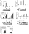
Aurora-A induces AR activity and potentiates androgen action in AR. A, basal level of AR activity is higher in LNCaP-RF than LNCaP cells. The indicated cells were transfected with ARR3-Luc and β-galactosidase. Following 48 h of incubation, luciferase activity was measured and normalized to β-galactosidase. Results are the mean ± S.E. (error bars) of three independent experiments performed in triplicate. B, wild-type Aurora-A stimulates and DN-Aurora-A inhibits AR activity. LNCaP cells were transfected with indicated plasmids and assayed for luciferase activity as described above. C, androgen-induced AR activity is enhanced by wild-type Aurora-A and inhibited by DN-Aurora-A. LNCaP cells were transfected with the indicated plasmids. After 36 h of incubation, cells were treated with and without R1881 and then subjected to a luciferase assay. D, expression of Aurora-A in AR-negative HEK293 cells had no effect on ARR3-Luc activity; however, co-expression of AR and Aurora-A stimulates the reporter activity. HEK293 cells were transfected with the indicated plasmids and assayed for luciferase activity. The bottom panels of A–D show the expression of transfected plasmids. E, knockdown of Aurora-A reduces AR transactivation activity. LNCaP cells were transfected with Aurora-A shRNA and control shRNA. After treatment with and without R1881, cells were subjected to a luciferase assay. Each experiment was repeated three times in triplicate. *, p < 0.05.
FIGURE 4.
FIGURE 4.
Aurora-A activation of AR depends on phosphorylation of Thr282 and Ser393. A and B, reporter assay. PC3 (A) and HEK293 (B) cells were transfected with the indicated plasmids, treated with and without R1881. After 36 h of incubation, cells were assayed for luciferase activity. The bottom panels of A–D show the expression of transfected plasmids. All of the experiments were repeated three times in triplicate. *, p < 0.05. Error bars, S.E.
FIGURE 5.
FIGURE 5.
Ectopic expression of Aurora-A in LNCaP cells induces a PSA- and androgen-independent phenotype. A and B, effect of Aurora-A and/or androgen on PSA expression. LNCaP cells were stably transfected with HA-Aurora-A or pHM6 vector and immunoblotted with the indicated antibody (A). Aurora-A- and pHM6-transfected cells were treated with and without R1881 and subjected to RT-PCR (top panels) and immunoblotting (bottom panels) analysis (B). C and D, overexpression of Aurora-A renders LNCaP cells androgen-independent. Aurora-A- and pHM6-transfected LNCaP cells were cultured in 24-well plates with phenol red-free RPMI 1640 medium containing 10% charcoal-stripped serum in the absence or the presence of androgen. The cell number was counted in the indicated time. *, p < 0.05 (C). The same cells were cultured in either regular FBS or charcoal-stripped serum (CS) for 3 days and examined for apoptosis using a Cell Death Detection ELISAPLUS kit (D). Error bars, S.E.
FIGURE 6.
FIGURE 6.
Knockdown of Aurora-A in LNCaP-RF cells reduces PSA expression and androgen-independent phenotype; Aurora-A-phosphorylated AR induces cell growth in the presence and absence of androgen. A, knockdown of Aurora-A decreases PSA expression. LNCaP-RF cells were transfected with shRNA-Aurora-A and scramble shRNA and detected for expression of Aurora-A (top two panels) and PSA (panels 3 and 5) as described in the legend to Fig. 5. Actin was used as a control. B and C, knockdown of Aurora-A reduces LNCaP-RF androgen-independent phenotype. Aurora-A/shRNA- and scramble shRNA-treated LNCaP-RF cells were cultured in phenol red-free RPMI 1640 medium containing 10% charcoal-stripped serum and then assayed for cell growth (B) and apoptosis (C). D and E, AR-2D but not AR-2A induces cell proliferation in the absence and presence of R1881. PC3 (D) and LNCaP (E) cells were seeded in a 6-well plate (105 cells/well) and then transfected with the indicated plasmids (bottom panels). Following incubation for 4 days in phenol red-free RPMI 1640 medium containing 10% charcoal-stripped serum in the absence or the presence of R1881, cell number was counted. The experiments were repeated three times in triplicate. *, p < 0.05. Error bars, S.E.
FIGURE 7.
FIGURE 7.
Aurora-A induces AR DNA binding activity and is frequently up-regulated in anti-androgen-resistant prostate cancer. A, ectopic expression of Aurora-A induces and knockdown of Aurora-A decreases AR DNA binding activity. LNCaP and LNCaP-RF cells were transfected with the indicated plasmids and shRNAs. After 48 h of incubation, a ChIP assay was performed as described under “Experimental Procedures.” B, effect of Aurora-A on AR DNA binding activity depends on phosphorylation of Thr282 and Ser393. HEK293 cells were transfected with the indicated plasmids and then subjected to a ChIP assay (top). Panels 3–5 show immunoblotting with indicated antibodies. C and D, frequent up-regulation of Aurora-A in anti-androgen-resistant/AR-positive prostate cancer. Seven anti-androgen-resistant/AR-positive prostate tumors were immunoblotted with the indicated antibodies (C). A representative anti-androgen-resistant tumor and prostatic intraepithelial neoplasia (PIN) were immunostained with anti-Aurora-A antibody (D).

Similar articles

Cited by

References

    1. Heinlein C. A., Chang C. (2004) Endocr. Rev. 25, 276–308 - PubMed
    1. Wang Q., Li W., Zhang Y., Yuan X., Xu K., Yu J., Chen Z., Beroukhim R., Wang H., Lupien M., Wu T., Regan M. M., Meyer C. A., Carroll J. S., Manrai A. K., Jänne O. A., Balk S. P., Mehra R., Han B., Chinnaiyan A. M., Rubin M. A., True L., Fiorentino M., Fiore C., Loda M., Kantoff P. W., Liu X. S., Brown M. (2009) Cell 138, 245–256 - PMC - PubMed
    1. Chen C. D., Welsbie D. S., Tran C., Baek S. H., Chen R., Vessella R., Rosenfeld M. G., Sawyers C. L. (2004) Nat. Med. 10, 33–39 - PubMed
    1. Taplin M. E., Bubley G. J., Shuster T. D., Frantz M. E., Spooner A. E., Ogata G. K., Keer H. N., Balk S. P. (1995) N. Engl. J. Med. 332, 1393–1398 - PubMed
    1. Culig Z. (2004) Growth Factors 22, 179–184 - PubMed

Publication types

MeSH terms