

The association of dynamin with synaptophysin regulates quantal size and duration of exocytotic events in chromaffin cells
- PMID: 20702699
- PMCID: PMC6634707
- DOI: 10.1523/JNEUROSCI.5210-09.2010
The association of dynamin with synaptophysin regulates quantal size and duration of exocytotic events in chromaffin cells
Abstract
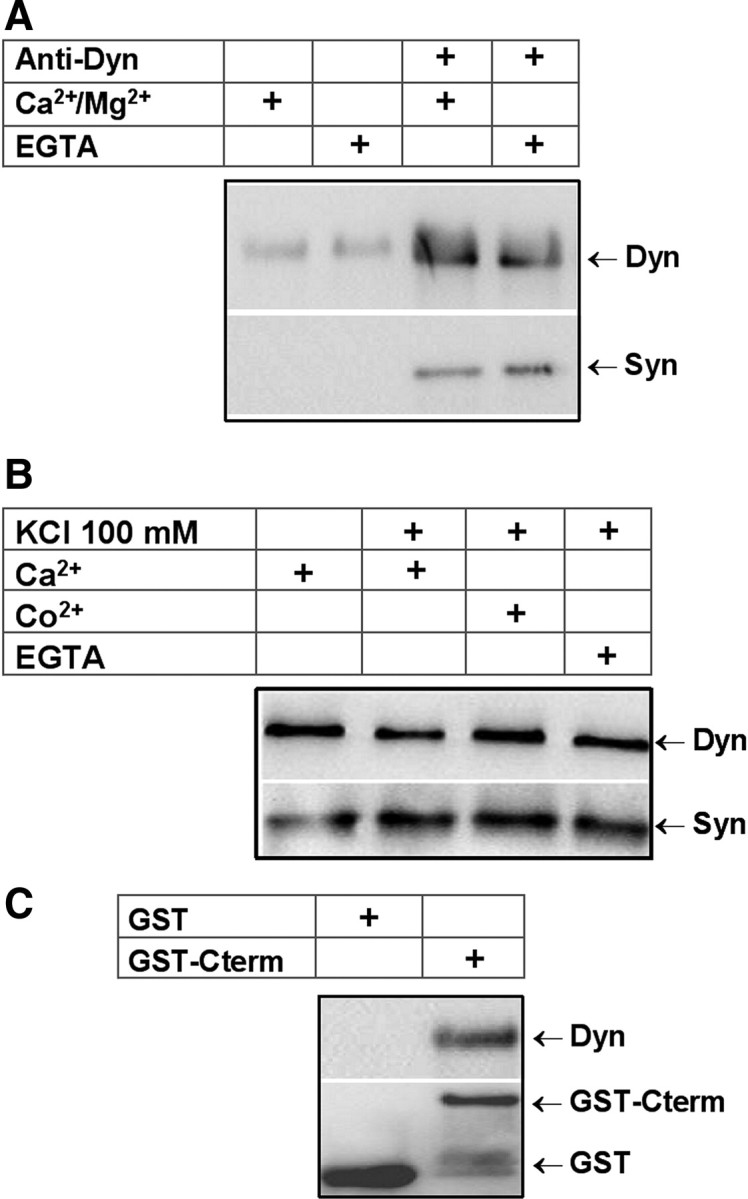
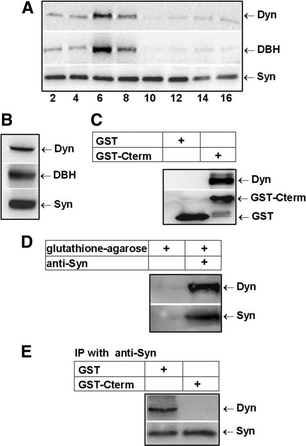
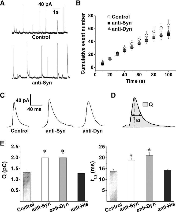
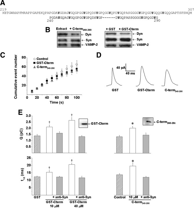
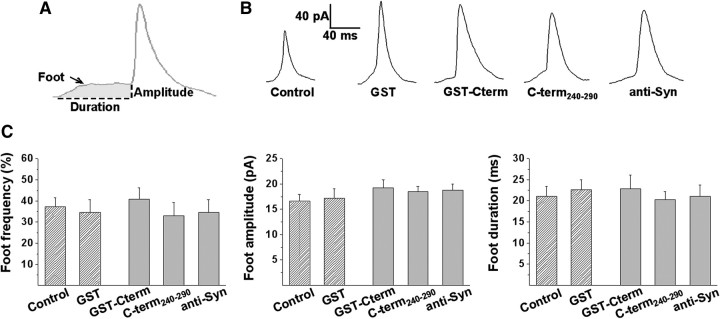
Although synaptophysin is one of the most abundant integral proteins of synaptic vesicle membranes, its contribution to neurotransmitter release remains unclear. One possibility is that through its association with dynamin it controls the fine tuning of transmitter release. To test this hypothesis, we took advantage of amperometric measurements of quantal catecholamine release from chromaffin cells. First, we showed that synaptophysin and dynamin interact in chromaffin granule-rich fractions and that this interaction relies on the C terminal of synaptophysin. Experimental maneuvers that are predicted to disrupt the association between these two proteins, such as injection of antibodies against dynamin or synaptophysin, or peptides homologous to the C terminal of synaptophysin, increased the quantal size and duration of amperometric spikes. In contrast, the amperometric current that precedes the spike remained unchanged, indicating that synaptophysin/dynamin association does not regulate the initial fusion pore, but it appears to target a later step of exocytosis to control the amount of catecholamines released during a single vesicle fusion event.
Figures
Similar articles
-
Synaptophysin Regulates Fusion Pores and Exocytosis Mode in Chromaffin Cells.J Neurosci. 2021 Apr 21;41(16):3563-3578. doi: 10.1523/JNEUROSCI.2833-20.2021. Epub 2021 Mar 4. J Neurosci. 2021. PMID: 33664131 Free PMC article.
-
Small molecules demonstrate the role of dynamin as a bi-directional regulator of the exocytosis fusion pore and vesicle release.Mol Psychiatry. 2015 Jul;20(7):810-9. doi: 10.1038/mp.2015.56. Epub 2015 May 5. Mol Psychiatry. 2015. PMID: 25939402
-
The exocytotic event in chromaffin cells revealed by patch amperometry.Nature. 1997 Oct 2;389(6650):509-12. doi: 10.1038/39081. Nature. 1997. PMID: 9333242
-
Dynamin and myosin regulate differential exocytosis from mouse adrenal chromaffin cells.Cell Mol Neurobiol. 2010 Nov;30(8):1351-7. doi: 10.1007/s10571-010-9591-z. Cell Mol Neurobiol. 2010. PMID: 21061163 Free PMC article. Review.
-
Relationship between amperometric pre-spike feet and secretion granule composition in chromaffin cells: an overview.Biophys Chem. 2007 Sep;129(2-3):181-9. doi: 10.1016/j.bpc.2007.05.018. Epub 2007 Jun 6. Biophys Chem. 2007. PMID: 17587484 Review.
Cited by
-
A new role for the dynamin GTPase in the regulation of fusion pore expansion.Mol Biol Cell. 2011 Jun 1;22(11):1907-18. doi: 10.1091/mbc.E11-02-0101. Epub 2011 Apr 1. Mol Biol Cell. 2011. PMID: 21460182 Free PMC article.
-
Concentration of stimulant regulates initial exocytotic molecular plasticity at single cells.Chem Sci. 2022 Jan 20;13(6):1815-1822. doi: 10.1039/d1sc05278k. eCollection 2022 Feb 9. Chem Sci. 2022. PMID: 35282618 Free PMC article.
-
Gain-of-Function Dynamin-2 Mutations Linked to Centronuclear Myopathy Impair Ca2+-Induced Exocytosis in Human Myoblasts.Int J Mol Sci. 2022 Sep 8;23(18):10363. doi: 10.3390/ijms231810363. Int J Mol Sci. 2022. PMID: 36142275 Free PMC article.
-
Pannexin 1 channels: new actors in the regulation of catecholamine release from adrenal chromaffin cells.Front Cell Neurosci. 2014 Sep 4;8:270. doi: 10.3389/fncel.2014.00270. eCollection 2014. Front Cell Neurosci. 2014. PMID: 25237296 Free PMC article.
-
Lumenal protein within secretory granules affects fusion pore expansion.Biophys J. 2014 Jul 1;107(1):26-33. doi: 10.1016/j.bpj.2014.04.064. Biophys J. 2014. PMID: 24988338 Free PMC article.
References
-
- Albillos A, Dernick G, Horstmann H, Almers W, Alvarez de Toledo G, Lindau M. The exocytotic event in chromaffin cells revealed by patch amperometry. Nature. 1997;389:509–512. - PubMed
-
- Alder J, Lu B, Valtorta F, Greengard P, Poo MM. Calcium-dependent transmitter secretion reconstituted in Xenopus oocytes: requirement for synaptophysin. Science. 1992a;257:657–661. - PubMed
-
- Alder J, Xie ZP, Valtorta F, Greengard P, Poo M. Antibodies to synaptophysin interfere with transmitter secretion at neuromuscular synapses. Neuron. 1992b;9:759–768. - PubMed
-
- Alés E, Tabares L, Poyato JM, Valero V, Lindau M, Alvarez de Toledo G. High calcium concentrations shift the mode of exocytosis to the kiss-and-run mechanism. Nat Cell Biol. 1999;1:40–44. - PubMed
-
- Annaert WG, Llona I, Backer AC, Jacob WA, De Potter WP. Catecholamines are present in a synaptic-like microvesicle-enriched fraction from bovine adrenal medulla. J Neurochem. 1993;60:1746–1754. - PubMed
Publication types
MeSH terms
Substances
LinkOut - more resources
Full Text Sources
Molecular Biology Databases