. 2010 Oct 1;21(19):3421-32.
doi: 10.1091/mbc.E10-01-0065. Epub 2010 Aug 11.

Cdk phosphorylation of a nucleoporin controls localization of active genes through the cell cycle

Affiliations

Cdk phosphorylation of a nucleoporin controls localization of active genes through the cell cycle

Donna Garvey Brickner et al. Mol Biol Cell. .

Abstract

Many inducible genes in yeast are targeted to the nuclear pore complex when active. We find that the peripheral localization of the INO1 and GAL1 genes is regulated through the cell cycle. Active INO1 and GAL1 localized at the nuclear periphery during G1, became nucleoplasmic during S-phase, and then returned to the nuclear periphery during G2/M. Loss of peripheral targeting followed the initiation of DNA replication and was lost in cells lacking a cyclin-dependent kinase (Cdk) inhibitor. Furthermore, the Cdk1 kinase and two Cdk phosphorylation sites in the nucleoporin Nup1 were required for peripheral targeting of INO1 and GAL1. Introduction of aspartic acid residues in place of either of these two sites in Nup1 bypassed the requirement for Cdk1 and resulted in targeting of INO1 and GAL1 to the nuclear periphery during S-phase. Thus, phosphorylation of a nuclear pore component by cyclin dependent kinase controls the localization of active genes to the nuclear periphery through the cell cycle.


Figures
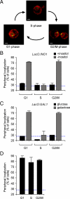
Figure 1.
Figure 1.
Gene localization through the cell cycle. (A) Representative images of unbudded, small-, and large-budded cells stained for GFP-Lac I (green) and Sec63-myc (red). Scale bar, 1 μm. The localization of the INO1 gene (B) and the GAL1 gene (C) was quantified under either repressing (■) or activating (▩) conditions in unbudded (G1), small- (S), and large-budded (G2/M) cells from an asynchronous culture. (D) Localization of artificially tethered INO1 through the cell cycle. Localization of tethered INO1 was performed as in B and C. In B–D, the blue, hatched line represents the level of colocalization of the lac repressor spot with the nuclear envelope predicted by chance (Brickner and Walter, 2004).
Figure 2.
Figure 2.
Loss of peripheral localization during S-phase. (A and B) Localization of INO1 (A) or GAL1 (B) arrested during mitosis. Wild-type (control) or cdc20-1 cells were grown under repressing conditions (■), activating conditions (dark gray bars) or activating conditions at 37°C (2 h; light gray bars) before scoring for localization. The right-most bar is the wild-type strain grown under activating conditions treated with nocodazole (+noc) for 2 h (dark gray bars). (C) Nocodazole-arrested cells having the lac operator array at INO1 were washed into fresh medium, and the localization of INO1 was scored at the indicated times. (D and E) Localization of INO1 (D) or GAL1 (E) arrested at the G1/S transition. Wild-type (control) or cdc6-1 cells were grown under activating conditions (dark gray bars) or shifted to the restrictive temperature of 37°C for 2 h (light gray bars) before scoring for localization. (F) cdc6-1 mutant cells arrested at the restrictive temperature were washed into fresh medium at 25°C and the localization of INO1 was scored at the indicated times. (G and H) Localization of INO1 (G) and GAL1 (H) in cells arrested during S-phase. Cells were grown under repressing conditions (■) or activating conditions (dark gray bars) with or without 0.1 M hydroxyurea (+HU) for 2 h and scored for localization. (I) Cells arrested with hydroxyurea were washed into activating medium without drug and the localization of INO1 was scored at the indicated times. The blue, hatched line represents the level of colocalization of the lac repressor spot with the nuclear envelope predicted by chance (Brickner and Walter, 2004).
Figure 3.
Figure 3.
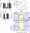
Cdk inhibitors negatively regulate gene recruitment to the nuclear periphery. (A) Wild-type (■) and sic1Δ mutant (▩) cells grown under activating conditions were scored for INO1 localization in cells with no buds (G1), small buds (S) or large buds (G2/M) as in Figure 1. (B) Newly born wild-type and sic1Δ mutant cells grown under activating conditions were recovered by centrifugal elutriation, and the localization of INO1 was scored at the indicated times. (C) Wild-type and ste2Δ mutant cells were grown under activating conditions in the presence (▩) or absence (■) of alpha factor. The population was scored for INO1 localization with respect to the nuclear periphery. (D) Alpha factor–arrested cells were washed into fresh activating medium lacking alpha factor at time 0 (red arrow) and INO1 localization (bottom panel) and budding index (top panel) were scored at the indicated times. The blue, hatched line represents the level of colocalization of the lac repressor spot with the nuclear envelope predicted by chance (Brickner and Walter, 2004).
Figure 4.
Figure 4.
Cdk1 promotes gene recruitment to the nuclear periphery. (A) cdc28-1 mutant cells having the lac operator array at INO1 (■) or at GAL1 (▩) were grown under activating conditions at 22°C or shifted to 37°C for 2 h. (B) cdc28-1 mutant cells having the lac operator array at INO1 (■) or at GAL1 (▩) were grown under activating conditions at 22°C. Cells were then treated with nocodazole for 2 h and then either maintained at 22°C or shifted to 37°C for 30 min before quantifying gene localization. (C) Complementation of the cdc28-1 mutation by cloned CDC28. The cdc28-1 mutant was transformed with the indicated plasmids and tested for growth at either 22 or 37°C. (D) The cdc28-1 mutant transformed with CDC28 was tested for localization as in B. In A, B, and D, the blue, hatched line represents the level of colocalization of the lac repressor spot with the nuclear envelope predicted by chance (Brickner and Walter, 2004).
Figure 5.
Figure 5.
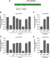
Phosphorylation of Nup1 at two Cdk consensus sites is required for gene recruitment to the nuclear periphery. (A) Schematic of the Nup1 protein. Two Cdk consensus sites with the phospho-acceptor sites corresponding to amino acids 161 and 344 are highlighted, and the FG repeats are indicated as gray bars. (B and D) Effect of loss of phospho-acceptor sites on INO1 (B) or GAL1 (D) localization at the nuclear periphery in an asynchronous population grown under activating conditions. nup1Δ cells were transformed with integrating plasmids expressing the indicated alleles of NUP1. ΔP1 = S161C, ΔP2 = T344C, PM1 = S161D, and PM2 = T344D. (C and E) Effect of phosphomimetic NUP1 mutations on localization of INO1 (C) or GAL1 (E) in small-budded cells grown under activating conditions. Cells expressing either wild-type NUP1, NUP1-PM1, or NUP1-PM2 were scored for localization of INO1 (C) or GAL1 (E) in small-budded cells. The blue, hatched line represents the level of colocalization of the lac repressor spot with the nuclear envelope predicted by chance (Brickner and Walter, 2004).
Figure 6.
Figure 6.
Phosphomimetic mutations in Nup1 bypass the requirement for Cdk1 in gene targeting to the nuclear periphery. cdc28-1 cells having the lac repressor array integrated at INO1 (A) or GAL1 (B) were transformed with integrating plasmids expressing NUP1-PM1 or NUP1-PM2. Cells were grown under activating conditions at 22°C and treated with nocodazole for 2 h as in Figure 4. Cells were then either maintained at 22°C or shifted to 37°C for 30 min before localization of INO1 (A) or GAL1 (B) was quantified. For comparison, untransformed cells were also scored (control). The blue, hatched line represents the level of colocalization of the lac repressor spot with the nuclear envelope predicted by chance (Brickner and Walter, 2004).
Figure 7.
Figure 7.
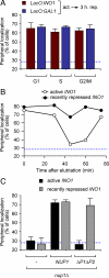
Recently repressed genes localize to the nuclear periphery throughout the cell cycle. (A) Strains having the lac operator array integrated either at INO1 or GAL1 were shifted from activating conditions to repressing conditions. After 3 h, cells were harvested, fixed, and processed for immunofluorescence. Cells were classified according to their bud morphology as in Figure 1 and, within each class, the percentage of cells in which the gene localized at the nuclear periphery was determined. (B) Newly born daughter cells having the lac operator array integrated at INO1 were purified by centrifugal elutriation under activating conditions or after 3 h of repression. Cells were harvested at the indicated times after elutriation, fixed, and processed for immunofluorescence. (C) nup1Δ cells having the lac operator array integrated at INO1 were transformed with either a vector, wild-type NUP1 or nup1ΔP1ΔP2. Cells were harvested either under activating conditions or after 3 h of repression, fixed, and processed for immunofluorescence. The blue, hatched line represents the level of colocalization of the lac repressor spot with the nuclear envelope predicted by chance (Brickner and Walter, 2004).

Similar articles

Cited by

References

    1. Ahmed S., Brickner D. G., Light W. H., McDonough M., Froyshteter A. B., Volpe T., Brickner J. H. DNA zip codes control an ancient mechanism for targeting genes to the nuclear periphery. Nat. Cell Biol. 2010;12:111–118. - PMC - PubMed
    1. Albuquerque C. P., Smolka M. B., Payne S. H., Bafna V., Eng J., Zhou H. A multidimensional chromatography technology for in-depth phosphoproteome analysis. Mol. Cell Proteom. 2008;7:1389–1396. - PMC - PubMed
    1. Azvolinsky A., Giresi P. G., Lieb J. D., Zakian V. A. Highly transcribed RNA polymerase II genes are impediments to replication fork progression in Saccharomyces cerevisiae. Mol. Cell. 2009;34:722–734. - PMC - PubMed
    1. Berger A. B., Cabal G. G., Fabre E., Duong T., Buc H., Nehrbass U., Olivo-Marin J. C., Gadal O., Zimmer C. High-resolution statistical mapping reveals gene territories in live yeast. Nat. Methods. 2008;5:1031–1037. - PubMed
    1. Blobel G. Gene gating: a hypothesis. Proc. Natl. Acad. Sci. USA. 1985;82:8527–8529. - PMC - PubMed

Publication types

MeSH terms