. 2010 Oct 1;21(19):3396-408.
doi: 10.1091/mbc.E10-06-0512. Epub 2010 Aug 11.

Coordination of substrate binding and ATP hydrolysis in Vps4-mediated ESCRT-III disassembly

Affiliations

Coordination of substrate binding and ATP hydrolysis in Vps4-mediated ESCRT-III disassembly

Brian A Davies et al. Mol Biol Cell. .

Abstract

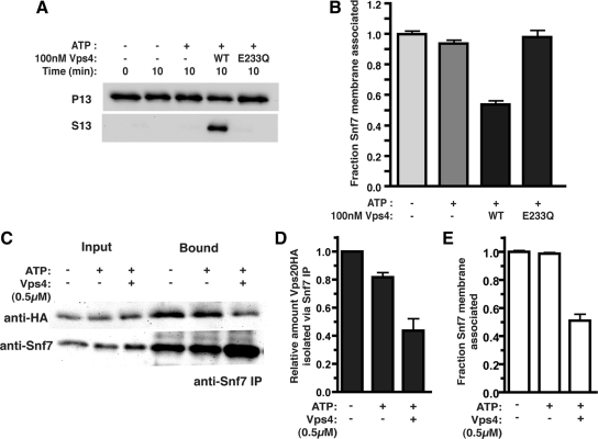
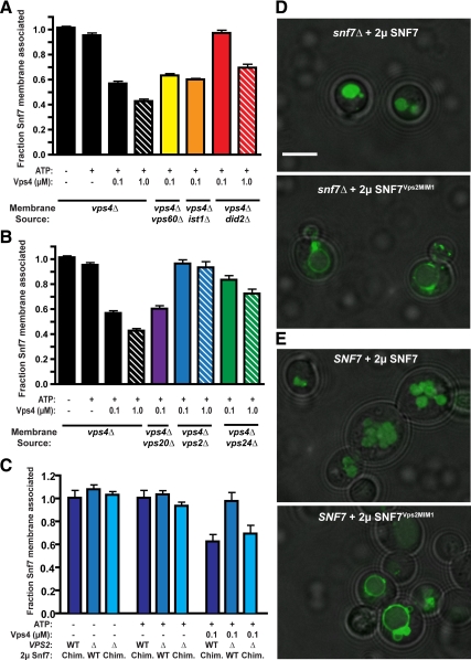
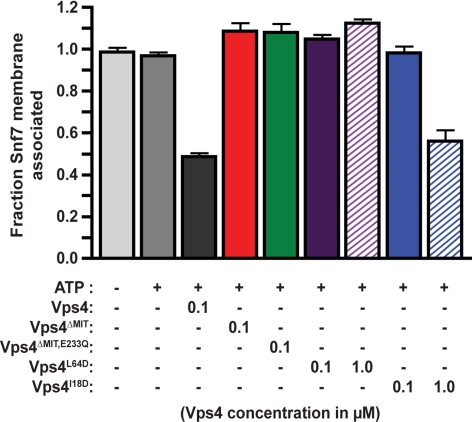
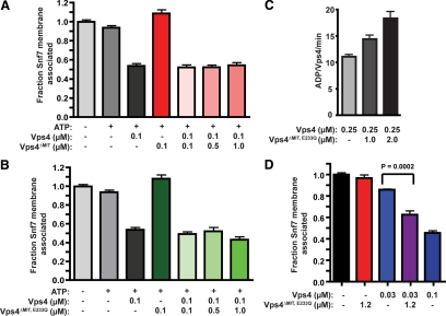
ESCRT-III undergoes dynamic assembly and disassembly to facilitate membrane exvagination processes including multivesicular body (MVB) formation, enveloped virus budding, and membrane abscission during cytokinesis. The AAA-ATPase Vps4 is required for ESCRT-III disassembly, however the coordination of Vps4 ATP hydrolysis with ESCRT-III binding and disassembly is not understood. Vps4 ATP hydrolysis has been proposed to execute ESCRT-III disassembly as either a stable oligomer or an unstable oligomer whose dissociation drives ESCRT-III disassembly. An in vitro ESCRT-III disassembly assay was developed to analyze Vps4 function during this process. The studies presented here support a model in which Vps4 acts as a stable oligomer during ATP hydrolysis and ESCRT-III disassembly. Moreover, Vps4 oligomer binding to ESCRT-III induces coordination of ATP hydrolysis at the level of individual Vps4 subunits. These results suggest that Vps4 functions as a stable oligomer that acts upon individual ESCRT-III subunits to facilitate ESCRT-III disassembly.


Figures

Figure 1.
Figure 1.
In vitro ESCRT-III release and disassembly. (A) ESCRT-III release reactions were initiated with resuspended P13 membranes (0.5 OD600 equivalents) generated from vps4Δ cells, in the presence (+) or absence (−) of 1 mM ATP and an ATP regeneration system (ATP), 100 nM Vps4 (WT), or 100 nM Vps4E233Q (E233Q). After a 10 min incubation at 30°C, the membrane-associated (P13) and soluble (S13) materials were separated. Western blotting with Snf7 antiserum was performed. (B) Quantitation of ESCRT-III release reaction. Data are presented as the fraction of Snf7 remaining membrane associated and represent the mean of at least three independent experiments with reactions performed in triplicate within each experiment. (C) Vps20HA-containing membranes (10 OD600 equivalents) were incubated for 30 min with 500 nM Vps4 and ATP or ATP alone. Snf7 immunoprecipitations were then performed after TritonX-100 extraction of the total reaction. Vps20HA was detected with the HA.11 mAb and Snf7 with the polyclonal antiserum. (D) Quantitation of Vps20HA isolation from the Snf7 immunoprecipitation. (E) Quantitation of the fraction of Snf7 remaining membrane associated under the reaction conditions used in C.
Figure 2.
Figure 2.
ESCRT-III subunit contributions to Vps4 disassembly of ESCRT-III. (A) ESCRT-III–containing membranes were generated from yeast lacking the accessory ESCRT-III subunits Did2 (red), Ist1 (orange), and Vps60 (red). Membranes (0.5 OD600 equivalent) were incubated with ATP and 100 nM or 1 μM Vps4, and the fraction of Snf7 remaining membrane associated was determined. (B) ESCRT-III membrane release assays performed with membranes generated from yeast lacking the core ESCRT-III subunits Vps20 (purple), Vps24 (green), and Vps2 (blue). (C) ESCRT-III membrane release assays performed with membranes generated from yeast lacking Snf7 and Vps2 (Δ) or Snf7 alone (WT) and expressing either Snf7 or the Snf7Vps2MIM1 chimera. (D and E) Distribution of the MVB sorting reporter GFP-CPS in snf7Δ (D) or wild-type yeast (E) expressing Snf7 or the Snf7Vps2MIM1 chimera. Bar, 5 μm.
Figure 3.
Figure 3.
ESCRT-III disassembly depends on Vps4 MIT associations with MIM1 and MIM2. ESCRT-III disassembly reactions were performed with inclusion of 100 nM Vps4 (black), 100 nM Vps4ΔMIT (red), 100 nM Vps4ΔMIT,E233Q (green), 100 nM and 1 μM Vps4L64D (purple, MIM1 binding surface), or 100 nM and 1 μM Vps4I18D (blue, MIM2 binding surface).
Figure 4.
Figure 4.
Vps4 oligomer MIT domain composition for ESCRT-III disassembly. (A) ESCRT-III disassembly reactions were performed with vps4Δ membranes (0.5 OD600 equivalents) and 100 nM Vps4, 100 nM Vps4ΔMIT, or 100 nM Vps4 with increasing concentrations of Vps4ΔMIT (100 nM, 500 nM, and 1 μM). (B) ESCRT-III release reactions were performed as in A but with increasing concentrations of Vps4ΔMIT,E233Q (100 nM, 500 nM, and 1 μM). (C) ATPase activity of 250 nM Vps4 alone or with addition of 1 μM or 2 μM Vps4ΔMIT,E233Q. The initial ATP concentration was 1 mM. Activity is presented as ADP generated per wild-type Vps4 per minute. (D) ESCRT-III disassembly reactions were performed as in A but with 30 nM Vps4 alone, 1.2 μM Vps4ΔMIT,E233Q alone, 30 nM Vps4 and 1.2 μM Vps4ΔMIT,E233Q, or 100 nM Vps4. The addition of Vps4ΔMIT,E233Q enhanced 30 nM Vps4-mediated ESCRT-III disassembly (p < 0.0002).
Figure 5.
Figure 5.
Vps4ΔMIT,E233Q colocalization with Did2 is Vps4-dependent. (A) In yeast lacking Vps4 (vps4Δ), Cherry-Did2 exhibits colocalization with the core ESCRT-III subunit Vps24-GFP. Dashed white lines represent the borders of individual yeast cells. Bar, 15 μm. (B) Vps4E233Q-GFP exhibits punctate distribution and colocalizes extensively with Cherry-Did2 in vps4Δ vta1Δ cells. (C) GFP-Vps4ΔMIT,E233Q exhibits a diffuse distribution and little colocalization with Cherry-Did2 in vps4Δ vta1Δ cells. (D) In the presence of Vps4 (vta1Δ cells), GFP-Vps4ΔMIT,E233Q displays some punctate association that colocalizes with Cherry-Did2 (indicated by arrows). Additional micrographs demonstrating colocalization of GFP-Vps4ΔMIT,E233Q with Cherry-Did2 in the presence of wild-type Vps4 are presented in Supplemental Figure 4.
Figure 6.
Figure 6.
Vps4E233Q inhibition of ESCRT-III disassembly. (A) ESCRT-III disassembly reactions were performed with vps4Δ membranes (0.5 OD600 equivalents) and 100 nM Vps4, 100 nM Vps4E233Q, or 100 nM Vps4 with decreasing concentrations of Vps4E233Q (100 nM, 50 nM, and 20 nM). (B) ATP hydrolysis reaction were performed with 250 nM Vps4 and increasing concentrations of Vps4E233Q. The initial ATP concentration was 1 mM. Rates are presented as ADP generated per wild-type Vps4 per minute. (C) ATP hydrolysis reactions were initiated with 2 μM Vps4E233Q (red), 4 μM Vps4E233Q (black), or Vps4 alone (blue) with Vps4 concentrations from 100 nM to 2 μM. Rates are presented as ADP generated per wild-type Vps4 per minute. (D and E) Distribution of the MVB sorting reporter GFP-CPS in wild-type yeast expressing Cherry-Vps4E233Q (D) or Cherry-Vps4ΔMIT,E233Q (E). Dashed white lines represent the borders of individual yeast cells. Bar, 5 μm.
Figure 7.
Figure 7.
The Vps4 oligomer is functionally stable in vitro. (A) ATP hydrolysis reactions were initiated with 500 nM Vps4 and 1 mM ATP. After 30 min, reactions were diluted 10-fold in the absence of additional ATP (black, red) or with ATP maintained at 1 mM (blue). After an additional 30 min, ATP restoration (to 1 mM, red) was performed. The ADP generated per Vps4 was determined at 5-min intervals and plotted versus time. (B) The activities of ATP hydrolysis (ADP generated/Vps4/min) relative to the initial rate (I) are presented. (C) ESCRT-III release reactions were initiated with 30 nM Vps4 (red, blue) or with 30 nM Vps4 and 100 nM Vps4E233Q (black). After a 5-min incubation, Vps4 (30 nM, red) or Vps4E233Q (100 nM, blue) were added to the reactions initiated with Vps4 only. The fraction of Snf7 released into the soluble material was assessed at both 5 and 20 min.
Figure 8.
Figure 8.
Coordination of Vps4 ESCRT-III engagement and ATP hydrolysis at the subunit level. (A) Vps4 ATP hydrolysis cycle in the presence or absence of the substrate ESCRT-III as suggested by in vitro analyses. Vps4 oligomerizes in an ATP-dependent manner, suggested to form a two-ring dodecamer (Yu et al., 2008; Landsberg et al., 2009). Neither concerted hydrolysis nor dissociation appear requisite for ATPase activity (upper row) or ESCRT-III disassembly (lower row), suggesting that individual subunits within the oligomer may hydrolyze and reload ATP. The presence of the substrate ESCRT-III can enhance Vps4 ATPase activity in an MIT-dependent manner, suggesting that engagement via the MIT domain potentiates ATP hydrolysis at the Vps4 subunit level to remodel and release ESCRT-III subunits (lower row). [Please note that the orientation of the asymmetric Vps4 oligomer relative to substrate is depicted with artistic license rather than adherence to supportive biochemical observations.] (B) If the engaged Vps4 subunit cannot hydrolyze ATP (Vps4E233Q), then ESCRT-III disassembly is arrested. However, Vps4E233Q does not disrupt substrate-independent ATPase activity (upper row in A). (C) The inhibition of ESCRT-III disassembly by Vps4E233Q is dependent upon the MIT domain, suggesting that coordination between ESCRT-III engagement via the MIT domain and ATP hydrolysis occurs at the subunit level within the Vps4 oligomer.

Similar articles

Cited by

References

    1. Agromayor M., Carlton J. G., Phelan J. P., Matthews D. R., Carlin L. M., Ameer-Beg S., Bowers K., Martin-Serrano J. Essential role of hIST1 in cytokinesis. Mol. Biol. Cell. 2009;20:1374–1387. - PMC - PubMed
    1. Azmi I., Davies B., Dimaano C., Payne J., Eckert D., Babst M., Katzmann D. J. Recycling of ESCRTs by the AAA-ATPase Vps4 is regulated by a conserved VSL region in Vta1. J. Cell Biol. 2006;172:705–717. - PMC - PubMed
    1. Azmi I. F., Davies B. A., Xiao J., Babst M., Xu Z., Katzmann D. J. ESCRT-III family members stimulate Vps4 ATPase activity directly or via Vta1. Dev. Cell. 2008;14:50–61. - PubMed
    1. Babst M., Katzmann D. J., Estepa-Sabal E. J., Meerloo T., Emr S. D. Escrt-III: an endosome-associated heterooligomeric protein complex required for mvb sorting. Dev. Cell. 2002a;3:271–282. - PubMed
    1. Babst M., Katzmann D. J., Snyder W. B., Wendland B., Emr S. D. Endosome-associated complex, ESCRT-II, recruits transport machinery for protein sorting at the multivesicular body. Dev. Cell. 2002b;3:283–289. - PubMed

Publication types

MeSH terms

Substances

LinkOut - more resources