

T-cell receptor complex is essential for Fas signal transduction
- PMID: 20696918
- PMCID: PMC2930531
- DOI: 10.1073/pnas.1005419107
T-cell receptor complex is essential for Fas signal transduction
Abstract
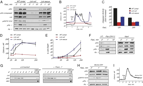
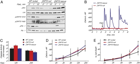
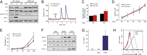
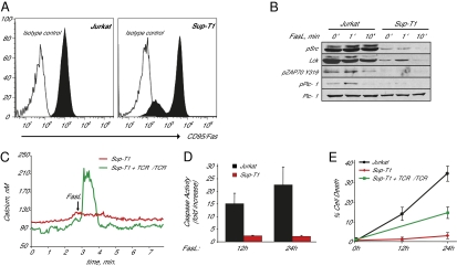
The Fas receptor (also known as CD95 and APO-1) is a member of the tumor necrosis factor alpha-family of death receptors that mediate T-cell responses. Here, we show that Fas receptor signaling requires a functional T-cell receptor (TCR) complex. Fas receptor directly binds to and activates TCR components in a stimulus-dependent manner. Fas receptor stimulation does not activate canonical downstream TCR pathways, but instead the TCR complex is required specifically for Fas-mediated calcium release. Importantly, null mutations in Lck, ZAP70, and the TCR alpha- and beta-chains abrogate Fas signaling. Our results reveal a direct role for the TCR complex in mediating Fas-specific signaling events critical for T-cell homeostasis.
Conflict of interest statement
The authors declare no conflict of interest.
Figures
Similar articles
-
Regulation of T cell development by c-Cbl: essential role of Lck.Int Immunol. 2015 May;27(5):245-51. doi: 10.1093/intimm/dxu105. Epub 2014 Dec 4. Int Immunol. 2015. PMID: 25477210 Free PMC article.
-
p56Lck tyrosine kinase enhances the assembly of death-inducing signaling complex during Fas-mediated apoptosis.J Biol Chem. 2007 Dec 7;282(49):36048-56. doi: 10.1074/jbc.M706007200. Epub 2007 Oct 11. J Biol Chem. 2007. PMID: 17932036
-
Lck-dependent tyrosine phosphorylation of diacylglycerol kinase alpha regulates its membrane association in T cells.J Immunol. 2008 May 1;180(9):5805-15. doi: 10.4049/jimmunol.180.9.5805. J Immunol. 2008. PMID: 18424699
-
Two receptors, two kinases, and T cell lineage determination.Sci Signal. 2010 Mar 23;3(114):pe11. doi: 10.1126/scisignal.3114pe11. Sci Signal. 2010. PMID: 20332426 Review.
-
The role of competing mechanisms on Lck regulation.Immunol Res. 2020 Oct;68(5):289-295. doi: 10.1007/s12026-020-09148-2. Epub 2020 Aug 14. Immunol Res. 2020. PMID: 32794043 Review.
Cited by
-
Ca2+-dependent protein acyltransferase DHHC21 controls activation of CD4+ T cells.J Cell Sci. 2022 Mar 1;135(5):jcs258186. doi: 10.1242/jcs.258186. Epub 2021 Jun 3. J Cell Sci. 2022. PMID: 34080635 Free PMC article.
-
An integrative computational systems biology approach identifies differentially regulated dynamic transcriptome signatures which drive the initiation of human T helper cell differentiation.BMC Genomics. 2012 Oct 30;13:572. doi: 10.1186/1471-2164-13-572. BMC Genomics. 2012. PMID: 23110343 Free PMC article.
-
S-acylation of Orai1 regulates store-operated Ca2+ entry.J Cell Sci. 2022 Mar 1;135(5):jcs258579. doi: 10.1242/jcs.258579. Epub 2021 Jun 22. J Cell Sci. 2022. PMID: 34156466 Free PMC article.
-
Relationship between infiltrating lymphocytes in cancerous ascites and dysfunction of Cajal mesenchymal cells in the small intestine.Int J Clin Exp Pathol. 2018 Apr 1;11(4):2201-2213. eCollection 2018. Int J Clin Exp Pathol. 2018. PMID: 31938332 Free PMC article.
-
Fas/FasL Signaling Regulates CD8 Expression During Exposure to Self-Antigens.Front Immunol. 2021 Mar 24;12:635862. doi: 10.3389/fimmu.2021.635862. eCollection 2021. Front Immunol. 2021. PMID: 33841416 Free PMC article.
References
-
- Green DR, Droin N, Pinkoski M. Activation-induced cell death in T cells. Immunol Rev. 2003;193:70–81. - PubMed
-
- Takahashi T, et al. Generalized lymphoproliferative disease in mice, caused by a point mutation in the Fas ligand. Cell. 1994;76:969–976. - PubMed
-
- Fisher GH, et al. Dominant interfering Fas gene mutations impair apoptosis in a human autoimmune lymphoproliferative syndrome. Cell. 1995;81:935–946. - PubMed
-
- Siegel RM, Muppidi J, Roberts M, Porter M, Wu Z. Death receptor signaling and autoimmunity. Immunol Res. 2003;27:499–512. - PubMed
Publication types
MeSH terms
Substances
Associated data
- Actions
Grants and funding
LinkOut - more resources
Full Text Sources
Other Literature Sources
Molecular Biology Databases
Research Materials
Miscellaneous
