. 2010 Oct 22;285(43):33307-33314.
doi: 10.1074/jbc.M110.153940. Epub 2010 Aug 6.

Palmitoylation of the S0-S1 linker regulates cell surface expression of voltage- and calcium-activated potassium (BK) channels

Affiliations

Palmitoylation of the S0-S1 linker regulates cell surface expression of voltage- and calcium-activated potassium (BK) channels

Owen Jeffries et al. J Biol Chem. .

Abstract

S-palmitoylation is rapidly emerging as an important post-translational mechanism to regulate ion channels. We have previously demonstrated that large conductance calcium- and voltage-activated potassium (BK) channels are palmitoylated within an alternatively spliced (STREX) insert. However, these studies also revealed that additional site(s) for palmitoylation must exist outside of the STREX insert, although the identity or the functional significance of these palmitoylated cysteine residues are unknown. Here, we demonstrate that BK channels are palmitoylated at a cluster of evolutionary conserved cysteine residues (Cys-53, Cys-54, and Cys-56) within the intracellular linker between the S0 and S1 transmembrane domains. Mutation of Cys-53, Cys-54, and Cys-56 completely abolished palmitoylation of BK channels lacking the STREX insert (ZERO variant). Palmitoylation allows the S0-S1 linker to associate with the plasma membrane but has no effect on single channel conductance or the calcium/voltage sensitivity. Rather, S0-S1 linker palmitoylation is a critical determinant of cell surface expression of BK channels, as steady state surface expression levels are reduced by ∼55% in the C53:54:56A mutant. STREX variant channels that could not be palmitoylated in the S0-S1 linker also displayed significantly reduced cell surface expression even though STREX insert palmitoylation was unaffected. Thus our work reveals the functional independence of two distinct palmitoylation-dependent membrane interaction domains within the same channel protein and demonstrates the critical role of S0-S1 linker palmitoylation in the control of BK channel cell surface expression.


Figures
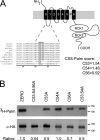
FIGURE 1.
FIGURE 1.
The S0-S1 linker is palmitoylated in BK channels. A, schematic illustrating the topology of the BK channel pore-forming α-subunit. Sequence alignments of cysteine residues in the S0-S1 linker indicate evolutionary conservation (gray box) across vertebrates, Drosophila and C. elegans. Murine sequence numbered starting from the initiation methionine (MDALI), accession number: AF156674. CSS-Palm prediction scores were determined with the CSS-Palm v2.0 platform. B, representative fluorographs (upper) and Western blots (lower) of full-length ZERO-HA channels and ZERO channels with mutation of key cysteine residues in the S0-S1 linker, expressed in HEK293 cells. Constructs were labeled with [3H]palmitate for 4 h and immunoprecipitated (IP) by using α-HA magnetic microbeads and detected by fluorography. Ratios (normalized to the wild-type ZERO channel) of [3H]palmitate detection in comparison to total protein expression are included.
FIGURE 2.
FIGURE 2.
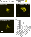
Palmitoylation targets the S0-S1 linker to the plasma membrane. A schematic of the short S0-S1 linker YFP fusion construct (S0-S1-YFP), that encodes the 70 amino acid intracellular S0-S1 linker between amino acids Arg-44 and Arg-11 fused in-frame with YFP, and relative position of the palmitoylated cysteine residues. A, representative single cell confocal images of the S0-S1-YFP linker, S0-S1 C53:54:56A-YFP linker, and the S0-S1-YFP linker fusion proteins after incubation with the palmitoylation inhibitor 2BP (100 μm) for 24 h, expressed in HEK293 cells (scale bars: 10 μm). B, summary bar graph illustrates the effect of site-directed mutagenesis of cysteine residues in the S0-S1 linker on localization of the respective S0-S1-YFP fusion protein at the plasma membrane expressed as a percentage of the wild-type S0-S1-YFP fusion protein (where wild-type S0-S1-YFP membrane expression is normalized to 100%). (For all S0-S1 linker constructs, N>3, n>330 cells analyzed). **, p < 0.01; ***, p < 0.001 compared with wild-type S0-S1 (ANOVA with Tukey post hoc test).
FIGURE 3.
FIGURE 3.
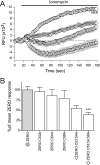
Ionomycin-driven activation of BK channels is attenuated in S0-S1 mutant channels. A, representative time course plots of mean change in relative fluorescence units (RFU) of the FLIPR-blue membrane potential dye in HEK293 cells expressing ZERO (closed gray circles), ZERO C53:54A (inverted triangles,▽), ZERO C53:54:56A (diamonds,♢), and mock-transfected HEK293 (open circles,○), in response to calcium influx induced by 1 μm ionomycin. B, summary bar chart of the membrane potential change for each construct expressed as a percentage of the maximal hyperpolarization, following subtraction of the HEK293 response, in the ZERO (gray) variant (where the ZERO response is normalized to 100%). Data were determined at the maximum hyperpolarizing response in the wild-type ZERO channel (t = 70 s) in the time course plots in A. All data are means ± S.E. (n = 3, n>24), ***, p < 0.001, compared with ZERO (ANOVA with Tukey post hoc test).
FIGURE 4.
FIGURE 4.
The intrinsic channel properties are un-affected in de-palmitoylated BK channels. A, representative single channel conductance recordings of excised inside-out patches at +40 and −40 mV in 0.33 μm Ca2+. B, current (pA) voltage (V) plot for ZERO channels (closed gray circles) and ZERO C53:54:56A (diamonds,♢), showing that single channel conductance is unaffected in 0.33 μm Ca2+ (n = 3). C, representative macropatch recordings traces showing BK currents in response to a depolarizing voltage step protocol (−120 mV to +120 mV) from a holding potential of −80mV from excised inside-out patch recordings in equimolar potassium gradients and 1 μm free Ca2+ (scale bars: 1 pA/25 ms). D, G/GMAX conductance curves show no change in channel activation at 1 μm free Ca2+ between ZERO (closed gray circles) and ZERO C53:54:56A (diamonds,♢). E, summary bar graph illustrates no significant changes in V0.5max across the physiological calcium range 0.33–10 μm free calcium. All data are means ± S.E. (n >3).
FIGURE 5.
FIGURE 5.
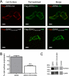
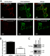
Palmitoylation of the S0-S1 linker regulates cell surface expression of BK channels. A, representative confocal images of HEK293 cells expressing Flag-ZERO-HA (top panels), and Flag-ZERO C53:54:56A-HA (bottom panels). The extracellular Flag epitope was labeled (red) under non-permeabilized conditions (cell surface) with the C-terminal HA epitope tag (green) labeled following cell permeabilization. Flag and HA labeling from the same cell are then overlaid (merge) (scale bars: 10 μm). B, quantification of cell surface expression between ZERO (gray bars) and ZERO C53:54:56A (white bars). Data are means ± S.E. (n >3). **, p < 0.01, ANOVA with post hoc Tukey test compared with Flag-ZERO-HA construct. C, representative Western blots of HA immunoreactivity from cell surface biotinylation assays (top panels) and corresponding whole cell lysates (bottom panels) in HEK293 cells expressing ZERO-HA and ZERO C53:54:56A-HA channels and control mock transfected cells.
FIGURE 6.
FIGURE 6.
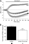
Disruption of the S0-S1 linker palmitoylation site in STREX splice variant channels also attenuates the ionomycin-driven channel activation. A, representative time course plots of mean change in relative fluorescence units (RFU) of the FLIPR-blue membrane potential dye in HEK293 cells expressing STREX (closed black circles, ●), STREX C53:54:56A (upright triangles, △), and mock-transfected HEK293 (open circles, ○), in response to calcium influx induced by 1 μm ionomycin. B, summary bar chart of the membrane potential change for each construct expressed as a percentage of the maximal hyperpolarization, following subtraction of the HEK293 response, in the STREX (black) variant (where the STREX response is normalized to 100%). Data were determined at the maximum hyperpolarizing response in STREX (t = 70 s) in the time course plots in A. All data are means ± S.E. (n = 3, n > 24), ***, p < 0.001, compared with STREX (ANOVA with Tukey post hoc test).
FIGURE 7.
FIGURE 7.
S0-S1 palmitoylation site functions independently of additional STREX splice variant palmitoylation site to control BK channel surface expression. A, representative confocal images of HEK293 cells expressing Flag-STREX-HA (top panels), and Flag- STREX C53:54:56A -HA (bottom panels). The extracellular Flag epitope was labeled (red) under non-permeabilized conditions (cell surface) with the C-terminal HA epitope tag (green) labeled following cell permeabilization. Flag and HA labeling from the same cell are then overlaid (merge) (scale bars: 10 μm). B, quantification of cell surface expression between STREX (black bars) and STREX C53:54:56A (white bars). Data are means ± S.E. (n >3). *, p < 0.05, ANOVA with post hoc Tukey test compared with Flag-STREX-HA construct. C, representative fluorographs (upper) and Western blots (lower) of full-length STREX-HA channels and STREX C53:54:56A channels expressed in HEK293 cells. Constructs were labeled with [3H]palmitate for 4 h and immunoprecipitated (IP) by using α-HA magnetic microbeads and detected by fluorography. In this particular experiment protein expression of the two constructs was not equivalent; however, the C53:54:56A mutations do not compromise STREX expression per se. These data reveal the residual palmitoylation of the channel mediated via the STREX insert in contrast to the ZERO variant (see Fig. 1). Ratios (normalized to the wild-type STREX channel) of palmitate incorporation relative to total protein expression are included.

Similar articles

Cited by

References

    1. Raffaelli G., Saviane C., Mohajerani M. H., Pedarzani P., Cherubini E. (2004) J. Physiol. 557, 147–157 - PMC - PubMed
    1. Sausbier M., Hu H., Arntz C., Feil S., Kamm S., Adelsberger H., Sausbier U., Sailer C. A., Feil R., Hofmann F., Korth M., Shipston M. J., Knaus H. G., Wolfer D. P., Pedroarena C. M., Storm J. F., Ruth P. (2004) Proc. Natl. Acad. Sci. U.S.A. 101, 9474–9478 - PMC - PubMed
    1. Brenner R., Peréz G. J., Bonev A. D., Eckman D. M., Kosek J. C., Wiler S. W., Patterson A. J., Nelson M. T., Aldrich R. W. (2000) Nature 407, 870–876 - PubMed
    1. Sausbier M., Arntz C., Bucurenciu I., Zhao H., Zhou X. B., Sausbier U., Feil S., Kamm S., Essin K., Sailer C. A., Abdullah U., Krippeit-Drews P., Feil R., Hofmann F., Knaus H. G., Kenyon C., Shipston M. J., Storm J. F., Neuhuber W., Korth M., Schubert R., Gollasch M., Ruth P. (2005) Circulation 112, 60–68 - PubMed
    1. Brenner R., Chen Q. H., Vilaythong A., Toney G. M., Noebels J. L., Aldrich R. W. (2005) Nat. Neurosci. 8, 1752–1759 - PubMed

Publication types

MeSH terms

Substances

LinkOut - more resources