COL9A2 and COL9A3 mutations in canine autosomal recessive oculoskeletal dysplasia
- PMID: 20686772
- PMCID: PMC2954766
- DOI: 10.1007/s00335-010-9276-4
COL9A2 and COL9A3 mutations in canine autosomal recessive oculoskeletal dysplasia
Abstract
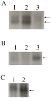
Oculoskeletal dysplasia segregates as an autosomal recessive trait in the Labrador retriever and Samoyed canine breeds, in which the causative loci have been termed drd1 and drd2, respectively. Affected dogs exhibit short-limbed dwarfism and severe ocular defects. The disease phenotype resembles human hereditary arthro-ophthalmopathies such as Stickler and Marshall syndromes, although these disorders are usually dominant. Linkage studies mapped drd1 to canine chromosome 24 and drd2 to canine chromosome 15. Positional candidate gene analysis then led to the identification of a 1-base insertional mutation in exon 1 of COL9A3 that cosegregates with drd1 and a 1,267-bp deletion mutation in the 5' end of COL9A2 that cosegregates with drd2. Both mutations affect the COL3 domain of the respective gene. Northern analysis showed that RNA expression of the respective genes was reduced in affected retinas. These models offer potential for studies such as protein-protein interactions between different members of the collagen gene family, regulation and expression of these genes in retina and cartilage, and even opportunities for gene therapy.
Figures


Similar articles
-
Clinical, histopathological and genetic characterisation of oculoskeletal dysplasia in the Northern Inuit Dog.PLoS One. 2019 Aug 15;14(8):e0220761. doi: 10.1371/journal.pone.0220761. eCollection 2019. PLoS One. 2019. PMID: 31415586 Free PMC article.
-
A loss of function mutation in the COL9A2 gene causes autosomal recessive Stickler syndrome.Am J Med Genet A. 2011 Jul;155A(7):1668-72. doi: 10.1002/ajmg.a.34071. Epub 2011 Jun 10. Am J Med Genet A. 2011. PMID: 21671392
-
Autosomal recessive Stickler syndrome associated with homozygous mutations in the COL9A2 gene.Ophthalmic Genet. 2021 Apr;42(2):161-169. doi: 10.1080/13816810.2020.1861309. Epub 2020 Dec 27. Ophthalmic Genet. 2021. PMID: 33356723
-
Autosomal recessive Stickler syndrome resulting from a COL9A3 mutation.Am J Med Genet A. 2018 Dec;176(12):2887-2891. doi: 10.1002/ajmg.a.40647. Epub 2018 Nov 18. Am J Med Genet A. 2018. PMID: 30450842 Free PMC article. Review.
-
Hearing Loss in Stickler Syndrome: An Update.Genes (Basel). 2022 Sep 1;13(9):1571. doi: 10.3390/genes13091571. Genes (Basel). 2022. PMID: 36140739 Free PMC article. Review.
Cited by
-
IQCB1 and PDE6B mutations cause similar early onset retinal degenerations in two closely related terrier dog breeds.Invest Ophthalmol Vis Sci. 2013 Oct 25;54(10):7005-19. doi: 10.1167/iovs.13-12915. Invest Ophthalmol Vis Sci. 2013. PMID: 24045995 Free PMC article.
-
FGF4 retrogene on CFA12 is responsible for chondrodystrophy and intervertebral disc disease in dogs.Proc Natl Acad Sci U S A. 2017 Oct 24;114(43):11476-11481. doi: 10.1073/pnas.1709082114. Epub 2017 Oct 11. Proc Natl Acad Sci U S A. 2017. PMID: 29073074 Free PMC article.
-
The genetics of inherited retinal disorders in dogs: implications for diagnosis and management.Vet Med (Auckl). 2016 Mar 15;7:41-51. doi: 10.2147/VMRR.S63537. eCollection 2016. Vet Med (Auckl). 2016. PMID: 30050836 Free PMC article. Review.
-
Canine chondrodysplasia caused by a truncating mutation in collagen-binding integrin alpha subunit 10.PLoS One. 2013 Sep 25;8(9):e75621. doi: 10.1371/journal.pone.0075621. eCollection 2013. PLoS One. 2013. PMID: 24086591 Free PMC article.
-
A COL11A2 mutation in Labrador retrievers with mild disproportionate dwarfism.PLoS One. 2013;8(3):e60149. doi: 10.1371/journal.pone.0060149. Epub 2013 Mar 20. PLoS One. 2013. PMID: 23527306 Free PMC article.
References
-
- Acland GM, Aguirre GD. Oculoskeletal dysplasia in samoyed and Labrador retriever dogs: 2 nonallelic disorders akin to Stickler-like syndromes affecting humans. Presented at the 2nd International DOGMAP meeting; Cambridge, Great Britain. 1995.
-
- Acland G, Ray K, Mellersh C, Langston A, Rine J, Ostrander E, Aguirre G. A Novel Retinal Degeneration Locus Identified by Linkage and Comparative Mapping of Canine Early Retinal Degeneration. Genomics. 1999;59(2):134–142. - PubMed
-
- Acland GM, Ray K, Mellersh CS, Gu W, Langston AA, Rine J, Ostrander EA, Aguirre GD. Linkage analysis and comparative mapping of canine progressive rod-cone degeneration (prcd) establishes potential locus homology with retinitis pigmentosa (RP17) in humans. Proc Nat Acad Sc USA. 1998;95(6):3048–53. - PMC - PubMed
-
- Annunen S, Korkko J, Czarny M, Warman ML, Brunner HG, Kaariainen H, Mulliken JB, Tranebjaerg L, Brooks DG, Cox GF, Cruysberg JR, Curtis MA, Davenport SL, Friedrich CA, Kaitila I, Krawczynski MR, Latos-Bielenska A, Mukai S, Olsen BR, Shinno N, Somer M, Vikkula M, Zlotogora J, Prockop DJ, Ala-Kokko L. Splicing mutations of 54-bp exons in the COL11A1 gene cause Marshall syndrome but other mutations cause overlapping Marshall/Stickler phenotypes. Am J Hum Genet. 1999 Oct;65(4):974–83. - PMC - PubMed
Publication types
MeSH terms
Substances
Supplementary concepts
Associated data
- Actions
- Actions
- Actions
- Actions
Grants and funding
LinkOut - more resources
Full Text Sources
Other Literature Sources
Medical

