. 2010 Oct;299(4):E665-74.
doi: 10.1152/ajpendo.00349.2010. Epub 2010 Aug 3.

Activation and repression of glucose-stimulated ChREBP requires the concerted action of multiple domains within the MondoA conserved region

Affiliations

Activation and repression of glucose-stimulated ChREBP requires the concerted action of multiple domains within the MondoA conserved region

Michael N Davies et al. Am J Physiol Endocrinol Metab. 2010 Oct.

Abstract

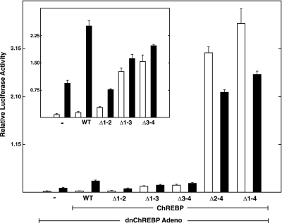
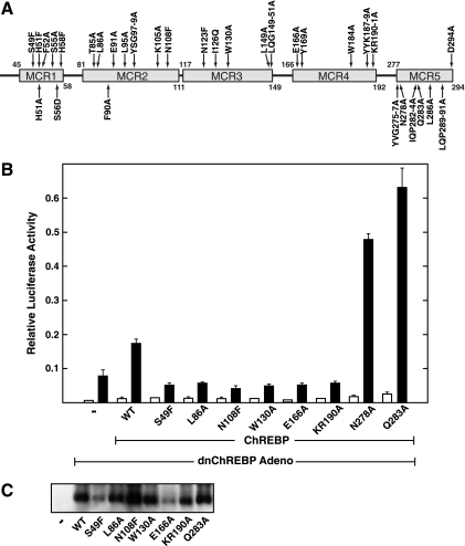
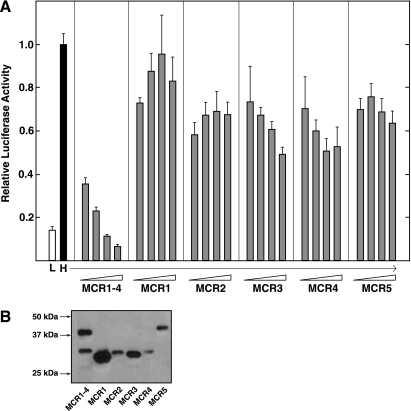
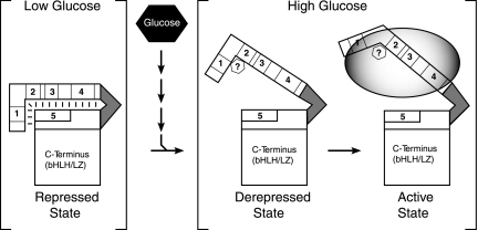
Carbohydrate response element-binding protein (ChREBP) is a glucose-dependent transcription factor that stimulates the expression of glycolytic and lipogenic genes in mammals. Glucose regulation of ChREBP has been mapped to its conserved NH(2)-terminal region of 300 amino acids, designated the MondoA conserved region (MCR). Within the MCR, five domains (MCR1-5) have a particularly high level of conservation and are likely to be important for glucose regulation. We carried out a large-scale deletion and substitution mutational analysis of the MCR domain of ChREBP. This analysis revealed that MCRs 1-4 function in a concerted fashion to repress ChREBP activity in basal (nonstimulatory) conditions. Deletion of the entire MCR1-4 segment or the combination of four specific point mutations located across this region leads to a highly active, glucose-independent form of ChREBP. However, deletion of any individual MCR domain and the majority of point mutations throughout MCR1-4 rendered ChREBP inactive. These observations suggest that the MCR1-4 region interacts with an additional coregulatory factor required for activation. This possibility is supported by the observation that the MCR1-4 region can compete for activity with wild-type ChREBP in stimulatory conditions. In contrast, mutations in the MCR5 domain result in increased activity, suggesting that this domain may be the target of intramolecular repression in basal conditions. Thus, the MCR domains act in a complex and coordinated manner to regulate ChREBP activity in response to glucose.


Figures

Fig. 1.
Fig. 1.
Combining activating mutations in the NH2-terminal segment of carbohydrate response element (ChoRE)-binding protein (ChREBP). A: diagram of the functional domains of ChREBP. MCR, MondoA conserved region, the region required for glucose sensing; pro-rich, a proline-rich segment that has been implicated in transcriptional activation; bHLH/LZ, basic helix-loop-helix/leucine zipper basic region DNA-binding domain; Mlx, Max-like factor X, a segment with homology to Mlx that is involved in dimerization. Numbers indicate the amino acid residue boundaries of each functional domain for mouse ChREBP (ζ-isoform, 864 residues). BD: INS-1 cells were transduced with a dominant negative (dn)ChREBP-expressing adenovirus for 2 h. After transduction, cells were cotransfected with an expression plasmid containing either Flag-tagged wild-type ChREBP or various ChREBP mutants and a reporter gene containing 2 copies of the acetyl-CoA carboxylase (ACC) ChoRE fused to the firefly luciferase gene. Transfection was performed overnight in RPMI medium containing 11 mM glucose. Cells were then treated with medium containing low (2.5 mM; open bars) or high glucose (25 mM; filled bars) for the following 24 h. After 24 h, extracts were prepared and firefly luciferase levels measured. Renilla luciferase was included in the cotransfection and used as an internal control. Values are shown as relative light units (firefly/Renilla) and represent the means (+ SD) for triplicate samples. B: activity of single mutations with increased activity compared with wild-type ChREBP. C: activity of mutants with various combinations of activating mutants. D: Western blotting with an anti-Flag antibody performed to detect expression of Flag-tag wild-type ChREBP and various ChREBP mutants.
Fig. 2.
Fig. 2.
Deletion of multiple MCR domains results in constitutive activation. Function of deletion mutants was tested using the rescue assay described in the legend to Fig. 1. Cells were treated with medium containing low (2.5 mM; open bars) or high glucose (25 mM; filled bars) for 24 h. Values are shown as relative light units (firefly/Renilla) and represent the means (+ SD) for triplicate samples. Inset is a replot of data omitting ΔMCR2–4 and ΔMCR1–4. Western blotting demonstrated that all mutants were expressed comparably with wild-type (WT) ChREBP, except for ΔMCR1–3 and ΔMCR1–4. These latter mutants were expressed at lower levels but were active in the functional assay.
Fig. 3.
Fig. 3.
Deletions of individual MCRs result in inactivation of ChREBP. A: function of deletion mutants of individual MCR domains was tested using the rescue assay described in the legend to Fig. 1. Cells were treated with medium containing low (2.5 mM; open bars) or high glucose (25 mM; filled bars) for 24 h. Values are shown as relative light units (firefly/Renilla) and represent the means (+ SD) for triplicate samples. B: electrophoretic mobility shift assay (EMSA) was performed using extracts of human embryonic kidney (HEK)-293 cells transfected with various ChREBP deletion mutants and an ACC ChoRE probe. The band adjacent to the asterisk represents a background band. The band adjacent to the right-pointing arrow represents the gel shift observed with WT ChREBP and the ChREBP deletion mutants. C: Western blotting of the extracts used for the EMSA was performed using an anti-Flag antibody.
Fig. 4.
Fig. 4.
Effect of amino acid substitutions spanning MCR1–5 on the functional activity of ChREBP. A: schematic of the amino acid substitutions of the NH2-terminal segment of ChREBP. Upward arrows represent mutants that have increased activity in low and high glucose compared with WT ChREBP. Downward arrows represent inactive mutants. The function of mutants was tested in INS-1 cells using the rescue assay described in the legend to Fig. 1. B: a representative functional assay of single mutants from each MCR domain. Cells were treated with medium containing low (2.5 mM; open bars) or high glucose (25 mM; filled bars) for 24 h. Values are shown as relative light units (firefly/Renilla) and represent the means (+ SD) for triplicate samples. C: expression of the Flag-tagged mutants in B from HEK-293 transfected cells was measured by Western blotting as described in the legend to Fig. 1.
Fig. 5.
Fig. 5.
Increasing amounts of the MCR1–4 fragment decrease ChREBP activity in a dose-dependent manner. INS-1 cells were cotransfected with an expression plasmid containing Gal4-ChREBP and a reporter gene plasmid consisting of the firefly luciferase gene fused to a promoter region containing 5 copies of the Gal4 response element. INS-1 cells were also transfected with increasing amounts of yellow fluorescent protein (YFP)-MCR1–4 competitor fragment. Total DNA concentrations were kept constant for each test point by balancing total DNA with a plasmid expressing only YFP. Cells were transfected overnight in RPMI medium containing 11 mM glucose. After ∼18 h, cells were treated with low or high glucose for 24 h. ○Low glucose, ■high glucose. Values are shown as relative light units (firefly/Renilla) normalized to the activity of cells treated with no competitor and high glucose and represent the means (+ SE) for 8–12 samples from 4 separate experiments.
Fig. 6.
Fig. 6.
Individual MCR fragments do not effectively compete for ChREBP activity. A: function of Gal4-ChREBP in the presence of increasing amounts of various competitor fragments was measured as described in the legend to Fig. 5. Individual MCR fragments consisting of MCR1, -2, -3, -4, and -5 were added in increasing amounts, and ChREBP activity was measured. The open bar and black bar represent Gal4-ChREBP activity in low- (L) and high-glucose (H) conditions, respectively, without competitor. B: Western blot of YFP competing fragments expressed in HEK-293 cells, as described in the legend to Fig. 1, except that an antibody to YFP was used. The specific regions used for each YFP fragment are stated in methods. The migration for each band represents the appropriate size of each fragment.
Fig. 7.
Fig. 7.
The MCR1–4 fragment competes with the Quad mutant for activity in both low- and high-glucose conditions. Activity of the Gal4-Quad mutant in the presence of the MCR1–4 competitor fragment was measured as described in the legend to Fig. 5. The bars with L and H below them represent Gal4-Quad mutant activity in low- and high-glucose conditions, respectively, without competitor. The bars that have an MCR1–4-labeled gradient depicted below represent Gal4-Quad mutant activity in the presence of 100, 200, or 300 ng of plasmid. Open bars represent low glucose, and black bars represent high glucose.
Fig. 8.
Fig. 8.
Model of glucose-stimulated ChREBP activation. In low-glucose conditions, ChREBP is repressed by an intramolecular interaction that involves MCR1–5 of the NH2-terminal segment of ChREBP. MCRs 1–4 are depicted as the repressor, and MCR5 is the target of repression. The resulting conformation is incapable of binding to DNA and stimulating transcription. When glucose metabolism increases (depicted by the small arrows emanating from glucose), ChREBP is targeted and modified in a manner that relieves the repressive intramolecular interaction. One possibility is that a glucose metabolite binds directly to the MCR1–4 region, as depicted by the hexagonal structure containing a question mark, representing this derepressed state. When repression of ChREBP is relieved, the conformation that results allows for MCR1–4 to interact with a coregulatory protein required for activation. This protein is represented by the ellipse in the active state conformation.

Similar articles

Cited by

References

    1. Billin AN, Eilers AL, Coulter KL, Logan JS, Ayer DE. MondoA, a novel basic helix-loop-helix-leucine zipper transcriptional activator that constitutes a positive branch of a max-like network. Mol Cell Biol 20: 8845–8854, 2000 - PMC - PubMed
    1. Billin AN, Eilers AL, Queva C, Ayer DE. Mlx, a novel Max-like BHLHZip protein that interacts with the Max network of transcription factors. J Biol Chem 274: 36344–36350, 1999 - PubMed
    1. Collier JJ, Zhang P, Pedersen KB, Burke SJ, Haycock JW, Scott DK. c-Myc and ChREBP regulate glucose-mediated expression of the L-type pyruvate kinase gene in INS-1-derived 832/13 cells. Am J Physiol Endocrinol Metab 293: E48–E56, 2007 - PubMed
    1. da Silva Xavier G, Rutter GA, Diraison F, Andreolas C, Leclerc I. ChREBP binding to fatty acid synthase and L-type pyruvate kinase genes is stimulated by glucose in pancreatic beta-cells. J Lipid Res 47: 2482–2491, 2006 - PubMed
    1. Davies MN, O'Callaghan BL, Towle HC. Glucose activates ChREBP by increasing its rate of nuclear entry and relieving repression of its transcriptional activity. J Biol Chem 283: 24029–24038, 2008 - PMC - PubMed

Publication types

MeSH terms

Substances

LinkOut - more resources