Review
. 2010 Dec;67(24):4109-34.
doi: 10.1007/s00018-010-0464-x. Epub 2010 Jul 31.

Deciphering the complexity of Toll-like receptor signaling

Affiliations
Review

Deciphering the complexity of Toll-like receptor signaling

Renato Ostuni et al. Cell Mol Life Sci. 2010 Dec.

Abstract

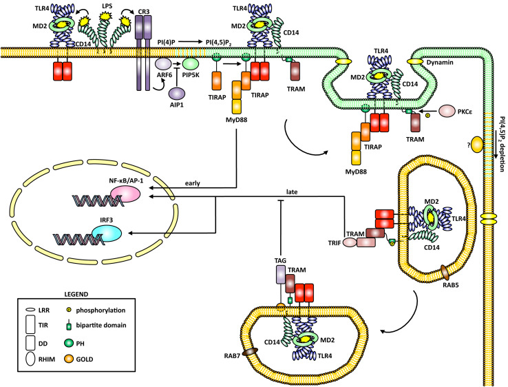
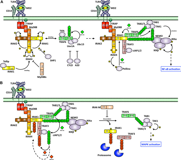
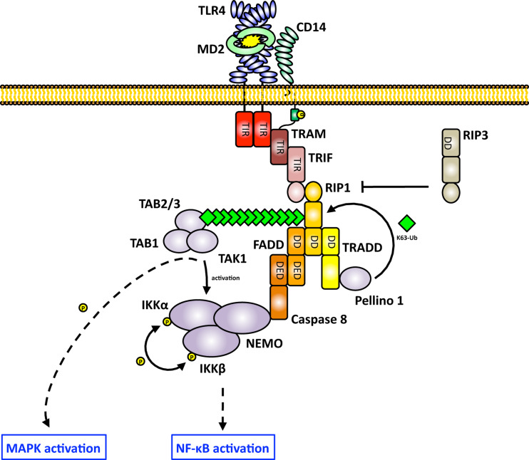
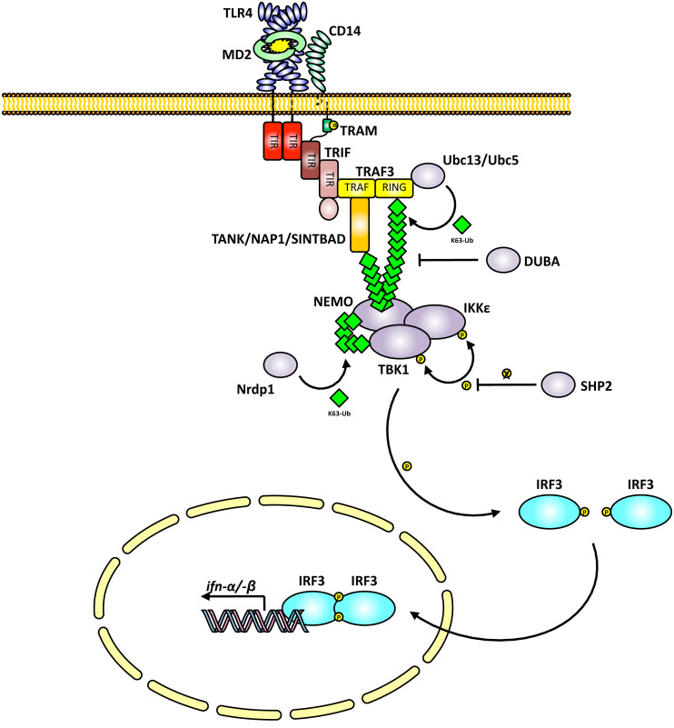
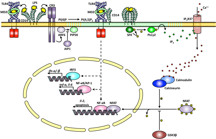
Toll-like receptors (TLRs) are essential players in the innate immune response to invading pathogens. Although extensive research efforts have provided a considerable wealth of information on how TLRs function, substantial gaps in our knowledge still prevent the definition of a complete picture of TLR signaling. However, several recent studies describe additional layers of complexity in the regulation of TLR ligand recognition, adaptor recruitment, posttranslational modifications of signaling proteins, and the newly described, autonomous role of the TLR4 co-receptor CD14. In this review, by using it as model system for the whole TLR family, we attempt to provide a complete description of the signal transduction pathways triggered by TLR4, with a particular emphasis on the molecular and cell biological aspects regulating its function. Finally, we discuss a recently reported model of CD14-dependent signaling and highlight its biological implications.


Figures

Fig. 1
Fig. 1
Cell biology of the Toll-like receptor (TLR)4 adaptor recruitment. Upon lipopolysaccharide (LPS) recognition, the TLR4 co-receptor CD14 mediates endotoxin transfer to TLR4 and promotes its homodimerization. CD14 also triggers CR3 activation by LPS, resulting in a localized ARF6-dependent synthesis of PI(4,5)P2 through phosphorylation of PI(4)P by PIP5K. Activation of the latter by ARF6 is negatively regulated by AIP1. Newly generated PI(4,5)P2 allows TIRAP recruitment and consequent sorting of MyD88 to the activated TLR4:CD14 complex, which signals early NF-κB/AP-1 activation from the plasma membrane. LPS recognition by TLR4:CD14 also promotes receptor endocytosis through a dynamin-dependent process that is initially promoted by PI(4,5)P2 synthesis but which requires PI(4,5)P2 consumption for its termination. As a consequence of the drop in PI(4,5)P2 concentration, whose regulation is currently unknown, TIRAP:MyD88 detach from PI(4,5)P2-depleted Rab5+ early endosomes, thereby freeing TLR4 for subsequent interaction with TRAM:TRIF. Due to its bipartite localization domain, TRAM resides both in early endosomes that fuse with TLR4:CD14-containing vesicles upon recycling and is coendocytosed with the receptor complex. This allows TRAM to sort TRIF to TLR4:CD14, resulting in the TRIF-dependent late wave of signaling to NF-κB/AP-1 and IRF3. As Rab5+ early endosomes mature, TAG, which resides in Rab7+ late endosomes, displaces TRIF from TRAM and thereby inhibits TRIF-dependent signaling
Fig. 2
Fig. 2
MyD88-dependent NF-κB and AP-1 activation. a MyD88 mediates recruitment to TLR4 of IRAK4, IRAK1 (through Toll interacting protein, Tollip), and IRAK2 by means of a homotypic death domain (DD) interaction. This results in the activation of IRAK4 (by autophosphorylation), IRAK1, and IRAK2, which are initially phosphorylated by IRAK4 and then undergo additional autophosphorylation. Recruitment of IRAK4 and IRAK1 phosphorylation by IRAK4 are negatively regulated by MyD88s and SHP1, respectively. Phosphorylated IRAK1 shows an increased binding affinity for TRAF6, which is in turn recruited to the receptor. TIFA-dependent oligomerization of TRAF6 stimulates its E3 ubiquitin ligase activity and, in coordination with the E2 complex Uev1A:Ubc13, TRAF6 catalyzes the attachment of K63-linked polyubiquitin chains on a number of substrates, including itself, in a process that is inhibited by CYLD and A20. Ubiquitinated TRAF6 interacts with TAK1 via TAB 2/3. TRAF6 also promotes K63-linked polyubiquitination of IRAK1 and TAK1, which directly recruit NEMO to the receptor complex. Ubiquitination of IRAK1 may also be operated by Pellino, which is in turn activated by IRAK4 and IRAK1 itself (not shown). Upon co-recruitment at the receptor complex, TAK1 promotes IKKα/β activation through a process that is independent of TAK1 kinase activity and which occurs at the plasma membrane. This ultimately results in MyD88-dependent activation of NF-κB. Even if not shown, it has to be noted that IRAK2 is likely to behave similarly to IRAK1 in promoting sustained NF-κB activation after IRAK1 degradation (see below). b In addition to TRAF6, IRAK1 probably mediates the recruitment of TRAF3 and cIAP1/2 to the receptor complex where TRAF6 catalyzes K63-linked polyubiquitination of cIAP1/2. K63-linked polyubiquitinated cIAP1/2 is enzymatically active as an E3 ligase that promotes degradative K48-linked polyubiquitination of TRAF3 and possibly IRAK1. Upon subsequent proteasomal degradation of TRAF3 and IRAK1, the TRAF6-nucleated complex containing TAK1 dissociates from the receptor and is released into the cytosol in a process that is inhibited by IRAK-M. Once in the cytoplasm (the cellular compartment where TAK1 substrates are located), TAK1 triggers effective MAPK activation by initiating a cascade of phosphorylating events. The kinase activity of TAK1 is therefore absolutely required for cytosolic MAPK activation
Fig. 3
Fig. 3
TRIF-dependent NF-κB/AP-1 activation. TRAM and TRIF are recruited to endosomal TLR4 after TIRAP:MyD88 dissociation from the internalized receptor. Through its RHIM, TRIF mediates direct recruitment of RIP1, which acts as a scaffold for the DD-containing proteins FADD and TRADD. Inactive RIP3 negatively regulates this process by competing with RIP1 for binding TRIF. TRADD binds Pellino1, an E3 ligase that catalyzes K63-linked polyubiquitination of RIP1. NF-κB/AP-1 activation is operated through the concomitant recruitment to the receptor complex of TAK1, which interacts with modified RIP1 via TAB 2/3, and IKKs. IKK recruitment is thought to occur by means of uncleaved caspase 8, which bridges FADD and NEMO. TAK1 in turn mediates IKK activation independently of its kinase activity, which is instead required for downstream MAPK activation
Fig. 4
Fig. 4
TRIF-dependent IRF3 activation. TRIF mediates direct recruitment of TRAF3 to endosomal TLR4 and promotes its consequent oligomerization, resulting in the Ubc13/Ubc5-dependent K63-linked polyubiquitination of TRAF3 and possibly of the downstream adaptors TANK/NAP1/SINTBAD. K63-linked polyubiquitination of TRAF3 is negatively regulated by the deubiquitinase DUBA. TRAF3 and/or one of the TRAF3-interacting adaptors recruit TBK1 and IKKε via the ubiquitin-binding domain of NEMO, thereby linking upstream signaling with IRF3 activation. The interaction of TBK1 with NEMO is also favored by the Nrdp1-dependent K63-linked polyubiquitination of TBK1. Upon transautophosphorylation, TBK1/IKKε are activated and phosphorylate IRF3 monomers, which in turn dimerize and translocate into the nucleus to promote type I IFN gene expression
Fig. 5
Fig. 5
CD14-dependent and TLR4-independent NFAT activation in dendritic cells. In addition to its role in LPS recognition and presentation to TLR4 and CR3 (see Fig. 1), CD14 has autonomous signaling functions in dendritic cells (DCs). Upon LPS-induced clusterization, CD14 transiently recruits and activates a Src family kinase (SKF) member through an ill-defined mechanism that relies on the CD14 GPI anchor and on its residency in lipid rafts. Active SFK then phosphorylates PLCγ2, which in turn catalyzes the hydrolysis of PI(4,5)P2 into the second messengers diacylglycerol (DAG) and IP3. Whereas the biological role of DAG in this system has not been investigated, it is likely to contribute to NF-κB activation through PKCs (not shown). On the other side, IP3 directly triggers Ca++ influx by acting on the plasma membrane Ca++ channel receptor (IP3R3?). The increased [Ca++]I stimulates activation of calcineurin, which dephosphorylates NFAT and promotes its nuclear translocation. Active NFAT cooperates with NF-κB to drive the expression of the genes coding for IL-2 as well as several proapoptotic proteins. It has to be noted that, although LPS-induced activation of NFAT in DCs is TLR4 independent, no change in gene expression is observed in the absence of TLR4, which is therefore required for full transcriptional activity of NFAT through activation of NF-κB

Similar articles

Cited by

References

    1. Janeway CA., Jr Approaching the asymptote? Evolution and revolution in immunology. Cold Spring Harb Symp Quant Biol. 1989;54:1–13. - PubMed
    1. West AP, Koblansky AA, Ghosh S. Recognition and signaling by toll-like receptors. Annu Rev Cell Dev Biol. 2006;22:409–437. - PubMed
    1. Gay NJ, Gangloff M. Structure and function of Toll receptors and their ligands. Annu Rev Biochem. 2007;76:141–165. - PubMed
    1. Beutler B, Jiang Z, Georgel P, Crozat K, Croker B, Rutschmann S, Du X, Hoebe K. Genetic analysis of host resistance: Toll-like receptor signaling and immunity at large. Annu Rev Immunol. 2006;24:353–389. - PubMed
    1. Barton GM, Kagan JC. A cell biological view of Toll-like receptor function: regulation through compartmentalization. Nat Rev Immunol. 2009;8:535–542. - PMC - PubMed

MeSH terms