

IKAP/hELP1 down-regulation in neuroblastoma cells causes enhanced cell adhesion mediated by contactin overexpression
- PMID: 20671422
- PMCID: PMC3011265
- DOI: 10.4161/cam.4.4.12923
IKAP/hELP1 down-regulation in neuroblastoma cells causes enhanced cell adhesion mediated by contactin overexpression
Abstract
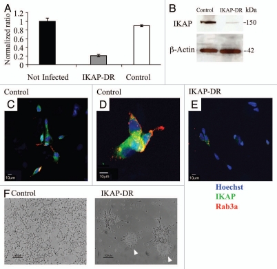
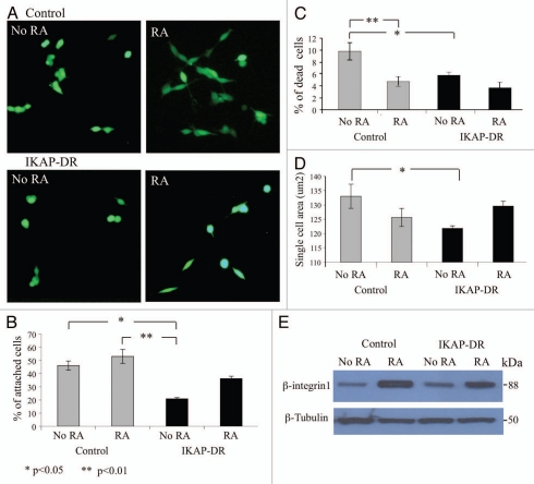
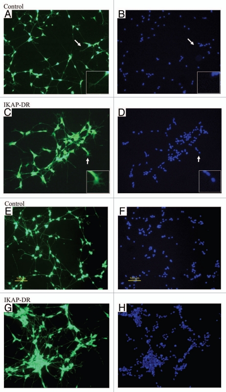
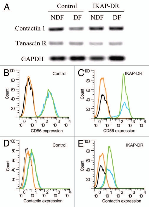
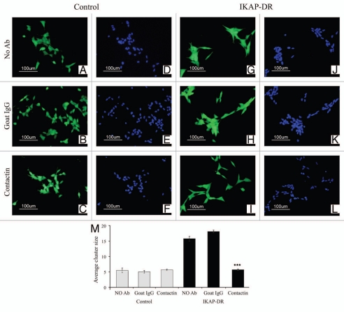
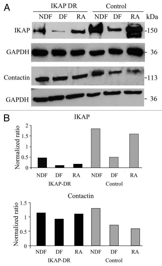
A splicing mutation in the IKBKAP gene encoding the IKAP/hELP1 (IKAP) protein was found to be the major cause of Familial Dysautonomia (FD). This mutation affects both the normal development and survival of sensory and sympathetic neurons of the peripheral nervous system (PNS). To understand the FD phenotype it is important to study the specific role played by IKAP in developing and mature PNS neurons. We used the neuroblastoma SHSY5Y cell line, originated from neural crest adrenal tumor, and simulated the FD phenotype by reducing IKAP expression with retroviral constructs. We observed that IKAP – down - regulated cells formed cell clusters compared to control cells under regular culture conditions. We examined the ability of these cells to differentiate into mature neurons in the presence of laminin, an essential extracellular matrix for developing PNS neurons. We found that the cells showed reduced attachment to laminin, morphological changes and increased cell-to-cell adhesion resulting in cell aggregates. We identified Contactin as the adhesion molecule responsible for this phenotype. We show that Contactin expression is related to IKAP expression, suggesting that IKAP regulates Contactin levels for appropriate cell-cell adhesion that could modulate neuronal growth of PNS neurons during development.
Figures
Similar articles
-
Effects of IKAP/hELP1 deficiency on gene expression in differentiating neuroblastoma cells: implications for familial dysautonomia.PLoS One. 2011 Apr 29;6(4):e19147. doi: 10.1371/journal.pone.0019147. PLoS One. 2011. PMID: 21559466 Free PMC article.
-
Familial Dysautonomia (FD) Human Embryonic Stem Cell Derived PNS Neurons Reveal that Synaptic Vesicular and Neuronal Transport Genes Are Directly or Indirectly Affected by IKBKAP Downregulation.PLoS One. 2015 Oct 5;10(10):e0138807. doi: 10.1371/journal.pone.0138807. eCollection 2015. PLoS One. 2015. PMID: 26437462 Free PMC article.
-
IKAP/Elp1 is required in vivo for neurogenesis and neuronal survival, but not for neural crest migration.PLoS One. 2012;7(2):e32050. doi: 10.1371/journal.pone.0032050. Epub 2012 Feb 23. PLoS One. 2012. PMID: 22384137 Free PMC article.
-
Familial dysautonomia.Curr Opin Genet Dev. 2002 Jun;12(3):307-11. doi: 10.1016/s0959-437x(02)00303-9. Curr Opin Genet Dev. 2002. PMID: 12076674 Review.
-
The molecular basis of familial dysautonomia: overview, new discoveries and implications for directed therapies.Neuromolecular Med. 2008;10(3):148-56. doi: 10.1007/s12017-007-8019-5. Epub 2007 Nov 6. Neuromolecular Med. 2008. PMID: 17985250 Review.
Cited by
-
The Interaction between hERG1 and β1 Integrins Modulates hERG1 Current in Different Pathological Cell Models.Membranes (Basel). 2022 Nov 18;12(11):1162. doi: 10.3390/membranes12111162. Membranes (Basel). 2022. PMID: 36422154 Free PMC article.
-
Effects of IKAP/hELP1 deficiency on gene expression in differentiating neuroblastoma cells: implications for familial dysautonomia.PLoS One. 2011 Apr 29;6(4):e19147. doi: 10.1371/journal.pone.0019147. PLoS One. 2011. PMID: 21559466 Free PMC article.
-
Elongator promotes neuritogenesis via regulation of tau stability through acly activity.Front Cell Dev Biol. 2022 Oct 26;10:1015125. doi: 10.3389/fcell.2022.1015125. eCollection 2022. Front Cell Dev Biol. 2022. PMID: 36393857 Free PMC article.
References
-
- Slaugenhaupt SA, Gusella JF. Familial dysautonomia. Curr Opin Genet Dev. 2002;12:307–311. - PubMed
-
- Axelrod FB. Familial dysautonomia. Muscle Nerve. 2004;29:352–363. - PubMed
-
- Neer EJ, Schmidt CJ, Nambudripad R, Smith TF. The ancient regulatory-protein family of WD-repeat proteins. Nature. 1994;371:297–300. - PubMed
Publication types
MeSH terms
Substances
LinkOut - more resources
Full Text Sources
Medical