. 2010 Aug 15;123(Pt 16):2743-9.
doi: 10.1242/jcs.073098. Epub 2010 Jul 27.

The cyclin A centrosomal localization sequence recruits MCM5 and Orc1 to regulate centrosome reduplication

Affiliations

The cyclin A centrosomal localization sequence recruits MCM5 and Orc1 to regulate centrosome reduplication

Rebecca L Ferguson et al. J Cell Sci. .

Abstract

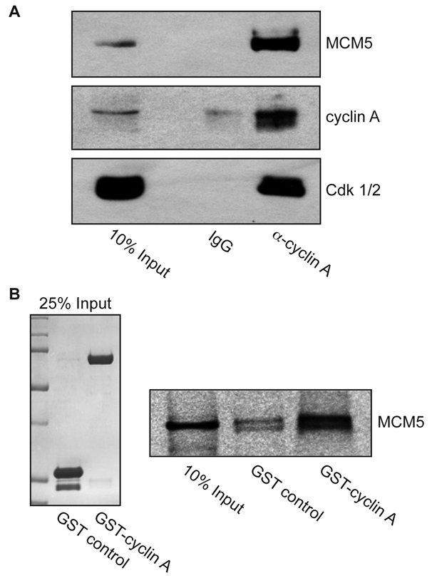
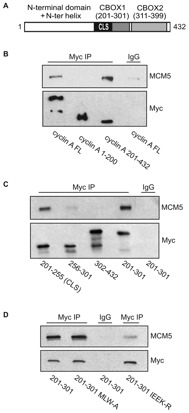
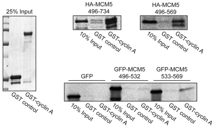
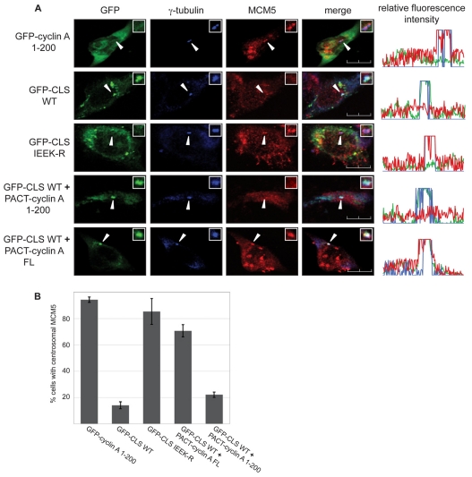
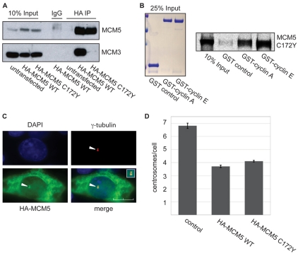
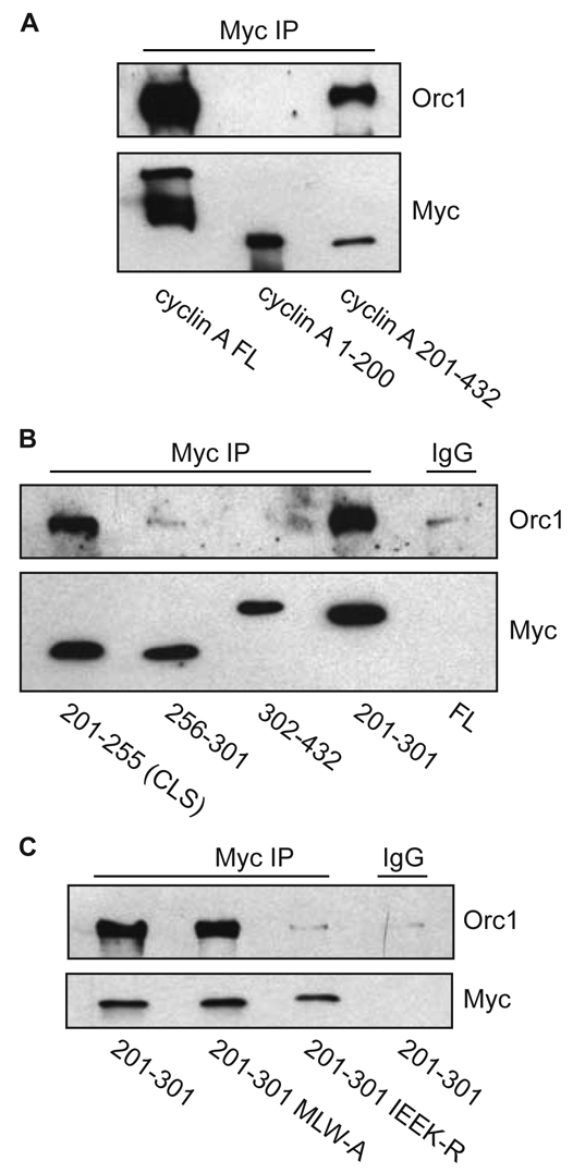
Centrosomes are the major microtubule-organizing centers in animal cells and regulate formation of a bipolar mitotic spindle. Aberrant centrosome number causes chromosome mis-segregation, and has been implicated in genomic instability and tumor development. Previous studies have demonstrated a role for the DNA replication factors MCM5 and Orc1 in preventing centrosome reduplication. Cyclin A-Cdk2 localizes on centrosomes by means of a modular centrosomal localization sequence (CLS) that is distinct from that of cyclin E. Here, we show that cyclin A interacts with both MCM5 and Orc1 in a CLS-dependent but Cdk-independent manner. Although the MRAIL hydrophobic patch is contained within the cyclin A CLS, binding of both MCM5 and Orc1 to cyclin A does not require a wild-type hydrophobic patch. The same domain in MCM5 that mediates interaction with cyclin E also binds cyclin A, resulting in centrosomal localization of MCM5. Finally, unlike its function in DNA synthesis, MCM5-mediated inhibition of centrosome reduplication in S-phase-arrested CHO cells does not require binding to other MCM family members. These results suggest that cyclins E and A sequentially prevent centrosome reduplication throughout interphase by recruitment of DNA replication factors such as MCM5 and Orc1.


Figures

Fig. 1.
Fig. 1.
Cyclin A interacts with MCM5. (A) Endogenous cyclin A co-immunoprecipitates MCM5. Lysates from asynchronous HeLa S3 cells were subjected to immunoprecipitation with antibodies to either cyclin A or control IgG, as indicated. Immunoprecipitates were analyzed by western blot using antibodies to MCM5 (upper), cyclin A (middle) and PSTAIR (lower), which recognizes both Cdk2 and Cdk1. (B) Cyclin A directly interacts with MCM5. MCM5 protein was produced and radiolabeled with [35S]-methionine in an in vitro TNT-coupled system, and incubated with glutathione-agarose beads bound to GST or to GST-cyclin A. Beads were washed, eluted with SDS-PAGE sample buffer, electrophoresed on 10% gels and analyzed by autoradiography. Left: Coomassie-stained gel of 25% input of GST and GST-cyclin A. Right: autoradiograph of MCM5.
Fig. 2.
Fig. 2.
MCM5 interaction with cyclin A is CLS dependent. (A) Schematic representation of human cyclin A. The N-terminal domain (amino acids 1-200) is shown in white. The C-terminal region (amino acids 201-432) contains the cyclin domain, which can be divided into two cyclin box folds (CBOX1 and CBOX2), represented in gray. The CLS motif is highlighted in black. (B) MCM5 interacts with the C-terminus of cyclin A. CHO-K1 cells were transiently co-transfected with the indicated constructs of Myc-tagged cyclin A (FL, full length) and wild-type MCM5. Approximately 20 hours after transfection, cell lysates were prepared and subjected to immunoprecipitation (IP) with control IgG or antibody to Myc. The immunoprecipitates were separated on 10% SDS-PAGE gels and analyzed by western blotting for MCM5 and Myc-cyclin A. (C) MCM5 interacts with the CLS region of cyclin A. CHO-K1 cells were transiently co-transfected with the indicated constructs of Myc-tagged cyclin A. Analysis of MCM5 binding was carried out as in B. (D) Mutation of the cyclin A CLS disrupts MCM5 interaction. CHO-K1 cells were transiently co-transfected with wild-type MCM5 and Myc-tagged cyclin A amino acids 201-301 containing a wild-type CLS, the hydrophobic patch mutant CLS (MLW-A) or mutant CLS (IEEK-R), as indicated. Analysis of MCM5 co-immunoprecipitation was carried out as in B.
Fig. 3.
Fig. 3.
Identification of the cyclin A-binding domain of MCM5. HA-tagged MCM5 (top panels) and GFP-tagged MCM5 (bottom panel) constructs were radiolabeled with [35S]-methionine in an in vitro TNT reaction and subjected to pulldown analysis with GST or GST-tagged cyclin A as in Fig. 1B. Right: autoradiographs of MCM5. Left: Coomassie-stained gel of 25% input of GST and GST-tagged cyclin A.
Fig. 4.
Fig. 4.
Centrosomal localization of MCM5 is dependent on cyclin A. (A) Localization of endogenous MCM5 was analyzed in CHO-K1 cells expressing either wild-type or mutant GFP-CLS, or co-expressing wild-type GFP-CLS and the indicated PACT-fused cyclin A constructs. Cells were stained with antibody to γ-tubulin (blue) and MCM5 (red). Expression and localization of GFP-tagged constructs were observed by direct fluorescence (green). Line scans measuring centrosome-associated relative fluorescence intensity are shown on the right, with the green, blue and red lines representing the GFP-, γ-tubulin- and MCM5-associated fluorescence, respectively. Arrows indicate the position of centrosomes. Insets: magnified image of the centrosomal region. Scale bars: 10 μm. (B) Graphical analysis of the centrosomal localization of MCM5. Several experiments similar to the one in A were performed with the indicated constructs. Over 100 cells were analyzed for each condition in each experiment. Error bars indicate mean ± s.d. (n=3).
Fig. 5.
Fig. 5.
MCM5 centrosomal function is independent of other MCMs. (A) MCM5 C172Y does not interact with MCM3. CHO-K1 cells were transiently transfected with either HA-tagged wild-type MCM5 or HA-tagged MCM5 C172Y mutant. Approximately 20 hours after transfection, cell lysates were subjected to immunoprecipitation with antibody to the HA tag. The immunoprecipitates were separated on 10% SDS-PAGE gels and analyzed by western blotting with antibodies to MCM5 and MCM3. (B) MCM5 C172Y interacts with both cyclin A and cyclin E. HA-tagged MCM5 C172Y protein was produced and radiolabeled with [35S]-methionine in an in vitro TNT-coupled system. The MCM5 C172Y protein was then incubated with glutathione-agarose beads bound to GST, GST-tagged cyclin A or GST-tagged cyclin E. Beads were washed, eluted with SDS-PAGE sample buffer, electrophoresed on 10% gels and analyzed by autoradiography. Left: Coomassie-stained gel of 25% input of GST, GST-cyclin A and GST-cyclin E. Right: autoradiograph of MCM5 C172Y. (C) MCM5 C172Y is centrosomally localized. CHO-K1 cells were transiently transfected with HA-tagged MCM5 C172Y. Approximately 20 hours after transfection, cells were methanol fixed and stained with antibodies to γ-tubulin and HA, as indicated. Arrowheads mark the centrosome. Inset: magnified image of the centrosomal region of the merged image. Scale bar: 10 μm. (D) Expression of MCM5 C172Y inhibits centrosome reduplication. CHO-K1 cells arrested in S phase by HU were transiently transfected with either HA-tagged wild-type MCM5 or HA-tagged MCM5 C172Y. The number of γ-tubulin-staining foci (centrosomes) was monitored by immunofluorescence in HA-expressing cells and compared with control non-expressing cells. The error bar indicates mean ± s.d. (n=3). Over 150 cells were counted for each condition in each experiment.
Fig. 6.
Fig. 6.
Cyclin A interaction with Orc1 is CLS dependent, but MRAIL independent. (A) Orc1 interacts with the C-terminus of cyclin A. CHO-K1 cells were transiently co-transfected with wild-type Orc1 and full-length (FL) Myc-tagged cyclin A, Myc-tagged cyclin A N-terminus (amino acids 1-200) or Myc-tagged cyclin A C-terminus (amino acids 201-432), as indicated. Approximately 20 hours after transfection, cell lysates were prepared and subjected to immunoprecipitation with anti-Myc antibody. The immunoprecipitates were separated on 10% SDS-PAGE gels and analyzed by western blotting for Orc1 and Myc. Non-immune rabbit IgG was used as a control. (B) Orc1 specifically interacts with the CLS of cyclin A. CHO-K1 cells were transiently co-transfected with indicated constructs of Myc-tagged cyclin A and wild-type Orc1. Analysis of Orc1 binding to cyclin A was carried out by immunoprecipitation analysis as in A. Amino acids 201-255 form the cyclin A CLS. (C) Mutation of the cyclin A CLS but not of the MRAIL hydrophobic pocket (MLW-A) disrupts Orc1 interaction with cyclin A. CHO-K1 cells were transiently co-transfected with indicated constructs of Myc-tagged cyclin A and wild-type Orc1. Analysis of Orc1 binding to cyclin A was carried out by immunoprecipitation analysis as in A.

Similar articles

Cited by

References

    1. Balczon R., Bao L., Zimmer W. E., Brown K., Zinkowski R. P., Brinkley B. R. (1995). Dissociation of centrosome replication events from cycles of DNA synthesis and mitotic division in hydroxyurea-arrested Chinese hamster ovary cells. J. Cell Biol. 130, 105-115 - PMC - PubMed
    1. Berthet C., Aleem E., Coppola V., Tessarollo L., Kaldis P. (2003). Cdk2 knockout mice are viable. Curr. Biol. 13, 1775-1785 - PubMed
    1. Bettencourt-Dias M., Glover D. M. (2007). Centrosome biogenesis and function: centrosomics brings new understanding. Nat. Rev. Mol. Cell Biol. 8, 451-463 - PubMed
    1. Bochman M. L., Bell S. P., Schwacha A. (2008). Subunit organization of Mcm2-7 and the unequal role of active sites in ATP hydrolysis and viability. Mol. Cell. Biol. 28, 5865-5873 - PMC - PubMed
    1. Chen Z., Indjeian V. B., McManus M., Wang L., Dynlacht B. D. (2002). CP110, a cell cycle-dependent CDK substrate, regulates centrosome duplication in human cells. Dev. Cell 3, 339-350 - PubMed

Publication types

MeSH terms