

Expression and cell localization of brain-derived neurotrophic factor and TrkB during zebrafish retinal development
- PMID: 20649707
- PMCID: PMC2972535
- DOI: 10.1111/j.1469-7580.2010.01268.x
Expression and cell localization of brain-derived neurotrophic factor and TrkB during zebrafish retinal development
Abstract
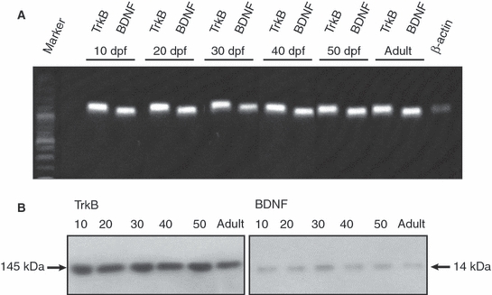
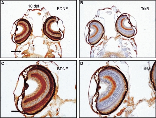
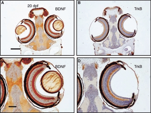
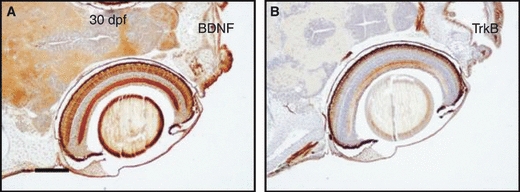
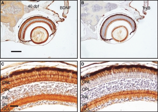
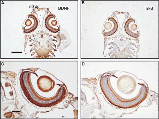
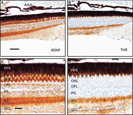
Brain-derived neurotrophic factor (BDNF) signaling through TrkB regulates different aspects of neuronal development, including survival, axonal and dendritic growth, and synapse formation. Despite recent advances in our understanding of the functional significance of BDNF and TrkB in the retina, the cell types in the retina that express BDNF and TrkB, and the variations in their levels of expression during development, remain poorly defined. The goal of the present study is to determine the age-dependent changes in the levels of expression and localization of BDNF and TrkB in the zebrafish retina. Zebrafish retinas from 10 days post-fertilization (dpf) to 180 dpf were used to perform PCR, Western blot and immunohistochemistry. Both BDNF and TrkB mRNAs, and BDNF and full-length TrkB proteins were detected at all ages sampled. The localization of these proteins in the retina was very similar at all time points studied. BDNF immunoreactivity was found in the outer nuclear layer, the outer plexiform layer and the inner plexiform layer, whereas TrkB immunoreactivity was observed in the inner plexiform layer and, to a lesser extent, in the ganglion cell layer. These results demonstrate that the pattern of expression of BDNF and TrkB in the retina of zebrafish remains unchanged during postembryonic development and adult life. Because TrkB expression in retina did not change with age, cells expressing TrkB may potentially be able to respond during the entire lifespan of zebrafish to BDNF either exogenously administered or endogenously produced, acting through paracrine mechanisms.
Figures
Similar articles
-
Expression of brain-derived neurotrophic factor and TrkB in the lateral line system of zebrafish during development.Cell Mol Neurobiol. 2010 Jul;30(5):787-93. doi: 10.1007/s10571-010-9506-z. Epub 2010 Feb 17. Cell Mol Neurobiol. 2010. PMID: 20162349
-
Light regulates the expression of the BDNF/TrkB system in the adult zebrafish retina.Microsc Res Tech. 2013 Jan;76(1):42-9. doi: 10.1002/jemt.22133. Epub 2012 Oct 16. Microsc Res Tech. 2013. PMID: 23070877
-
Expression and Localization of BDNF/TrkB System in the Zebrafish Inner Ear.Int J Mol Sci. 2020 Aug 12;21(16):5787. doi: 10.3390/ijms21165787. Int J Mol Sci. 2020. PMID: 32806650 Free PMC article.
-
Neuroanatomical distribution and functions of brain-derived neurotrophic factor in zebrafish (Danio rerio) brain.J Neurosci Res. 2020 May;98(5):754-763. doi: 10.1002/jnr.24536. Epub 2019 Sep 18. J Neurosci Res. 2020. PMID: 31532010 Review.
-
Brain-derived neurotrophic factor in gastroenterology oncology - short review of current literature.Ann Agric Environ Med. 2021 Sep 16;28(3):367-371. doi: 10.26444/aaem/122628. Epub 2020 Jul 8. Ann Agric Environ Med. 2021. PMID: 34558255 Review.
Cited by
-
Characterization of Progenitor Cells during Canine Retinal Development.Stem Cells Int. 2012;2012:675805. doi: 10.1155/2012/675805. Epub 2012 Feb 27. Stem Cells Int. 2012. PMID: 22567026 Free PMC article.
-
Potential Neuroprotective Role of Calretinin-N18 and Calbindin-D28k in the Retina of Adult Zebrafish Exposed to Different Wavelength Lights.Int J Mol Sci. 2023 Jan 6;24(2):1087. doi: 10.3390/ijms24021087. Int J Mol Sci. 2023. PMID: 36674603 Free PMC article.
-
Localization of BDNF expression in the developing brain of zebrafish.J Anat. 2014 May;224(5):564-74. doi: 10.1111/joa.12168. Epub 2014 Mar 4. J Anat. 2014. PMID: 24588510 Free PMC article.
-
A Brain-Derived Neurotrophic Factor Mimetic Is Sufficient to Restore Cone Photoreceptor Visual Function in an Inherited Blindness Model.Sci Rep. 2017 Sep 12;7(1):11320. doi: 10.1038/s41598-017-11513-5. Sci Rep. 2017. PMID: 28900183 Free PMC article.
-
Proliferative role of BDNF/TrkB signaling is associated with anoikis resistance in cervical cancer.Oncol Rep. 2018 Aug;40(2):621-634. doi: 10.3892/or.2018.6515. Epub 2018 Jun 20. Oncol Rep. 2018. PMID: 29989647 Free PMC article.
References
-
- Arango-González B, Cellerino A, Kohler K. Exogenous brain-derived neurotrophic factor (BDNF) reverts phenotypic changes in the retinas of transgenic mice lacking the BDNF gene. Invest Ophthalmol Vis Sci. 2009;50:1416–1422. - PubMed
-
- Bai Y, Xu J, Brahimi F, et al. An agonistic anti-TrkB mAb, but not BDNF, causes sustained TrkB activation, delays RGC death, and protects the retinal structure in optic nerve axotomy and in glaucoma. Invest Ophthalmol Vis Sci. 2010 in press (Published in PubMed. - PubMed
-
- Bessero AC, Clarke PG. Neuroprotection for optic nerve disorders. Curr Opin Neurol. 2010;23:10–15. - PubMed
-
- Bilotta J, Saszik S. The zebrafish as a model visual system. Int J Dev Neurosci. 2001;19:621–629. - PubMed
-
- Caminos E, Becker E, Martín-Zanca D, et al. Neurotrophins and their receptors in the tench retina during optic nerve regeneration. J Comp Neurol. 1999;404:321–331. - PubMed
MeSH terms
Substances
LinkOut - more resources
Full Text Sources
Molecular Biology Databases
