. 2010 Aug;137(16):2763-72.
doi: 10.1242/dev.048108. Epub 2010 Jul 14.

Drosophila PAT1 is required for Kinesin-1 to transport cargo and to maximize its motility

Affiliations

Drosophila PAT1 is required for Kinesin-1 to transport cargo and to maximize its motility

Philippe Loiseau et al. Development. 2010 Aug.

Abstract

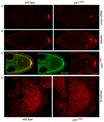
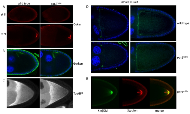
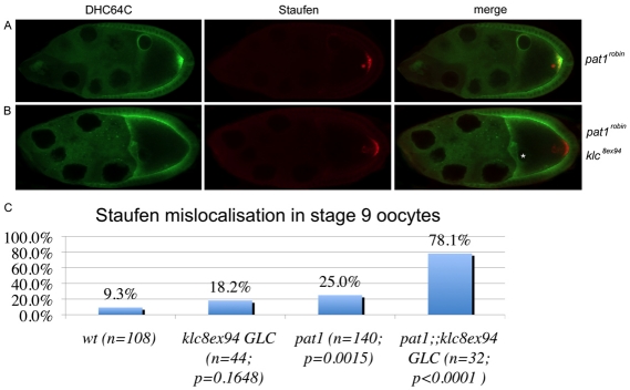
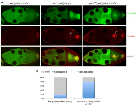
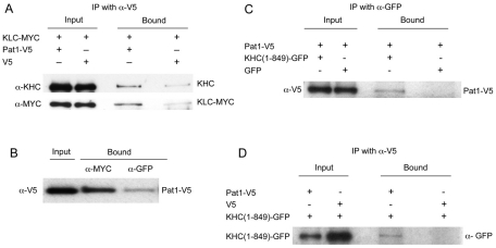
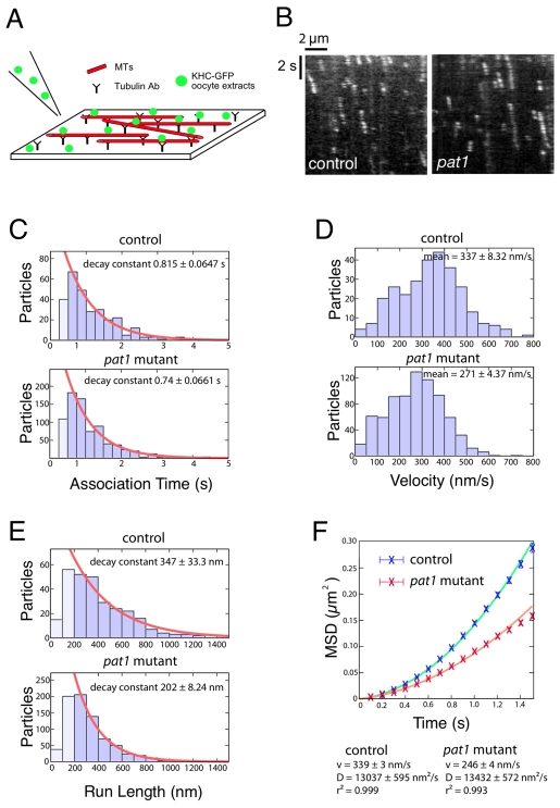
Kinesin heavy chain (KHC), the force-generating component of Kinesin-1, is required for the localization of oskar mRNA and the anchoring of the nucleus in the Drosophila oocyte. These events are crucial for the establishment of the anterior-posterior and dorsal-ventral axes. KHC is also essential for the localization of Dynein and for all ooplasmic flows. Interestingly, oocytes without Kinesin light chain show no major defects in these KHC-dependent processes, suggesting that KHC binds its cargoes and is activated by a novel mechanism. Here, we shed new light on the molecular mechanism of Kinesin function in the germline. Using a combination of genetic, biochemical and motor-tracking studies, we show that PAT1, an APP-binding protein, interacts with Kinesin-1, functions in the transport of oskar mRNA and Dynein and is required for the efficient motility of KHC along microtubules. This work suggests that the role of PAT1 in cargo transport in the cell is linked to PAT1 function as a positive regulator of Kinesin motility.


Figures

Fig. 1.
Fig. 1.
PAT1 is required for oskar mRNA localization, but not for Dynein transport or cytoplasmic flows. (A) In situ hybridization for oskar mRNA at stage 9 of oogenesis in wild-type (left) and Pat1 mutant (right) Drosophila ovaries. In wild-type egg chambers, oskar mRNA localizes to the posterior pole of the oocyte, where it remains anchored throughout oogenesis. This localization is impaired in Pat1 mutant ovaries. Ectopic dots of oskar mRNA are found in 22% of Pat1 stage 9 oocytes (n=40), but in only 7% of wild-type oocytes (n=40). (B) Staufen protein (a marker for oskar mRNA) in wild-type (left) and Pat1 mutant (right) stage 9 egg chambers. As with oskar mRNA, Staufen is found in an ectopic region in 25% of the Pat1 oocytes (n=140; see Fig. 3). (C) Dynein heavy chain (green) and Staufen (red) in stage 9 Pat1 oocytes. Unlike Staufen, Dynein is not found as a dot in Pat1 mutants. (D) Particles that reflect 568 nm light (`red' particles) in wild-type (left) and Pat1 mutant (right) egg chambers. Each image results from a continuous confocal scan, averaged eight times using the Kalman function. Insets show representative streaks of dots, which are used as a qualitative measure of cytoplasmic streaming.
Fig. 2.
Fig. 2.
PAT1 is not required for microtubule organization or regulation of Oskar translation. (A) Oskar protein (red) in stage 8 (top) and stage 9 (bottom) wild-type (left) and Pat1 (right) Drosophila egg chambers. In wild-type and Pat1 ovaries, Oskar protein is not detected at stage 8 of oogenesis, but starts accumulating at stage 9. (B) Gurken protein localizes normally to the anterior-dorsal corner in stage 9 wild-type (left) and Pat1 mutant (right) oocytes. (C) Tau-GFP labeling of microtubules in living wild-type (left) and Pat1 mutant (right) stage 9 oocytes. The overall organization of the microtubule cytoskeleton appears normal in Pat1 oocytes. (D) In situ hybridization for bicoid mRNA in wild-type (top) and Pat1 mutant (bottom), stage 9 (left) and stage 10b (right) oocytes. bicoid mRNA localization to the anterior pole is not affected by Pat1 loss of function. (E) Kinesin-β-galactosidase (KinβGal, green) and Staufen (red) in Pat1 mutant oocytes. Unlike Staufen, Kinesin-β-galactosidase localizes only to the posterior pole of Pat1 oocytes.
Fig. 3.
Fig. 3.
PAT1 and KLC interact genetically. (A) Dynein (green) and Staufen (red) in stage 9 Pat1 egg chambers. Unlike Staufen, Dynein is not mislocalized in Pat1 mutant egg chambers. (B) Dynein (green) and Staufen (red) in stage 9 Pat1, Klc oocytes. In these double mutants, Dynein is mislocalized at the anterior/lateral cortex. The Pat1, Klc double-mutant oocytes were obtained by inducing Klc8ex94 germline clones in a Pat1robin mutant background. Note that the oocyte nucleus (asterisk) is not mislocalized in this double-mutant background, and it only seems so because of the orientation of the egg chamber, with its anterior-dorsal corner towards the observer. (C) Quantification of the Staufen mislocalization phenotype in stage 9 wild-type egg chambers, Klc germline clones (Klc8ex94 GLC), Pat1 mutants and Pat1, Klc (Pat1;;Klc8ex94 GLC) double mutants. Although Staufen mislocalization is not statistically different between Klc and wild-type egg chambers (P=0.16, Fisher's exact test), it is significantly higher in Pat1 mutants compared with wild type (P=0.0015, Fisher's exact test). The penetrance of the Staufen mislocalization phenotype is dramatically increased (by more than threefold) in Pat1, Klc double mutants compared with single Pat1 or Klc mutants, which demonstrates that Pat1 and Klc interact genetically (P<0.0001, Fisher's exact test). The mislocalization of Staufen to a dot in the center of the oocyte was never found in wild-type flies, although dots closely attached to the posterior crescent were occasionally observed and included in the quantification.
Fig. 4.
Fig. 4.
Loss of function of Pat1 enhances the phenotype of a mutant form of KHC. (A) Staufen (red) in Drosophila egg chambers that express one copy of KHC(1-975)-GFP (left), one copy of KHC(1-849)-GFP (middle), or one copy of KHC(1-849)-GFP but are also mutant for Pat1 (right). Top panels show the localization of the various KHC-GFP fusions. In contrast to oocytes expressing the full-length KHC, Staufen (red) is mislocalized in 18% of the KHC(1-849)-GFP-expressing oocytes (see B). This mislocalization is more severe and four times more penetrant in Pat1robin;;KHC(1-849)-GFP oocytes than in the KHC(1-849)-GFP-expressing egg chambers. Arrows point to the region where Staufen is mislocalized. (B) Quantification of the Staufen mislocalization phenotype in stage 9 egg chambers that express one copy of tailless KHC fused to GFP, and that are either wild-type [KHC(1-849)-GFP/+] or mutant for Pat1 [Pat1;;KHC(1-849)-GFP/+].
Fig. 5.
Fig. 5.
PAT1 interacts with Kinesin-1. (A) Drosophila S2 cells co-expressing KLC-MYC and either PAT1-V5 or empty V5 were immunoprecipitated with an anti-V5 antibody (α-V5). Endogenous KHC (top) and KLC-MYC (bottom) were specifically co-purified with PAT1. (B) Drosophila S2 cells expressing PAT1-V5 and KLC-MYC were immunoprecipitated using an anti-MYC antibody (α-MYC) or an unrelated antibody (α-GFP) as a control. PAT1 is specifically co-immunoprecipitated with KLC-MYC by the anti-MYC antibody. (C) Drosophila S2 cells expressing PAT1-V5 and either KHC(1-849)-GFP or GFP alone were immunoprecipitated using anti-GFP antibody. PAT1-V5 is specifically pulled down by the anti-GFP antibody when KHC(1-849)GFP is present, indicating that PAT1 is in a complex with tailless KHC. (D) Drosophila S2 cells expressing KHC(1-849)-GFP and either PAT1-V5 or V5 alone were immunoprecipitated using anti-V5 antibody. KHC(1-849)-GFP is specifically pulled down by the anti-V5 antibody when PAT1-V5 is present, indicating that tailless KHC is in a complex with PAT1. Note that the expression of KHC(1-849)-GFP in cells transformed with the empty V5 plasmid is higher than in cells expressing PAT1-V5 (the amount of extract loaded in each input was the same). Each immunoprecipitation was performed in triplicate. Representative blots are shown.
Fig. 6.
Fig. 6.
The velocity and run length of Kinesin are reduced in the absence of Pat1. (A) The in vitro assay used to study the motility of KHC in control and Pat1 ovarian extracts. KHC-GFP-expressing ovaries are dissected and added to immobilized Rhodamine-labeled microtubules in the presence of ATP. The motility of KHC is analyzed by TIRF microscopy, which allows single fluorophores close to the surface to be tracked when moving along an immobilized microtubule. (B) Representative kymographs of KHC in extracts that express tailless KHC-GFP, and that are either wild-type or mutant for Pat1. (C-F) The velocity and run length of KHC are reduced in Pat1 mutants. (C-E) Bar charts of association time (C), velocity (D) and run length (E) of the individual KHC-GFP particles moving along microtubules in control and Pat1 extracts (n=285 and n=771, respectively). Orange curves (C,E) represent fitting of the data (bins in darker blue) to exponential decays. (F) Mean square displacements (MSDs) calculated from the consolidated data were plotted against time intervals (t) and fitted with quadratic curves MSD=v2t2+2Dt (v, velocity; D, diffusion coefficient). The velocity and run length of KHC are reduced by 20% and 42% in Pat1 extracts, respectively. A statistically significant difference between control and Pat1 extracts was seen in three independent experiments, although some variation occurred in the mean velocities. There was no significant difference in the diffusion coefficient (13,000±600 versus 13,400±600 nm2/second).

Similar articles

Cited by

References

    1. Adio S., Jaud J., Ebbing B., Rief M., Woehlke G. (2009). Dissection of kinesin's processivity. PLoS ONE 4, e4612 - PMC - PubMed
    1. Blasius T. L., Cai D., Jih G. T., Toret C. P., Verhey K. J. (2007). Two binding partners cooperate to activate the molecular motor Kinesin-1. J. Cell Biol. 176, 11-17 - PMC - PubMed
    1. Bowman A. B., Kamal A., Ritchings B. W., Philp A. V., McGrail M., Gindhart J. G., Goldstein L. S. (2000). Kinesin-dependent axonal transport is mediated by the sunday driver (SYD) protein. Cell 103, 583-594 - PubMed
    1. Brendza K. M., Rose D. J., Gilbert S. P., Saxton W. M. (1999). Lethal kinesin mutations reveal amino acids important for ATPase activation and structural coupling. J. Biol. Chem. 274, 31506-31514 - PMC - PubMed
    1. Brendza R. P., Serbus L. R., Duffy J. B., Saxton W. M. (2000a). A function for Kinesin I in the posterior transport of oskar mRNA and Staufen protein. Science 289, 2120-2122 - PMC - PubMed

Publication types

LinkOut - more resources