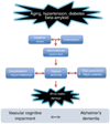
The overlap between neurodegenerative and vascular factors in the pathogenesis of dementia
- PMID: 20623294
- PMCID: PMC3001188
- DOI: 10.1007/s00401-010-0718-6
The overlap between neurodegenerative and vascular factors in the pathogenesis of dementia
Abstract
There is increasing evidence that cerebrovascular dysfunction plays a role not only in vascular causes of cognitive impairment but also in Alzheimer's disease (AD). Vascular risk factors and AD impair the structure and function of cerebral blood vessels and associated cells (neurovascular unit), effects mediated by vascular oxidative stress and inflammation. Injury to the neurovascular unit alters cerebral blood flow regulation, depletes vascular reserves, disrupts the blood-brain barrier, and reduces the brain's repair potential, effects that amplify the brain dysfunction and damage exerted by incident ischemia and coexisting neurodegeneration. Clinical-pathological studies support the notion that vascular lesions aggravate the deleterious effects of AD pathology by reducing the threshold for cognitive impairment and accelerating the pace of the dementia. In the absence of mechanism-based approaches to counteract cognitive dysfunction, targeting vascular risk factors and improving cerebrovascular health offers the opportunity to mitigate the impact of one of the most disabling human afflictions.
Figures


Similar articles
-
Vascular contributions to cognitive impairment, clinical Alzheimer's disease, and dementia in older persons.Biochim Biophys Acta. 2016 May;1862(5):878-86. doi: 10.1016/j.bbadis.2015.12.023. Epub 2016 Jan 5. Biochim Biophys Acta. 2016. PMID: 26769363 Free PMC article. Review.
-
Neurovascular dysfunction and neurodegeneration in dementia and Alzheimer's disease.Biochim Biophys Acta. 2016 May;1862(5):887-900. doi: 10.1016/j.bbadis.2015.12.016. Epub 2015 Dec 17. Biochim Biophys Acta. 2016. PMID: 26705676 Free PMC article. Review.
-
Evidence of endothelial dysfunction in the development of Alzheimer's disease: Is Alzheimer's a vascular disorder?Am J Cardiovasc Dis. 2013 Nov 1;3(4):197-226. Am J Cardiovasc Dis. 2013. PMID: 24224133 Free PMC article. Review.
-
A Perfect sTORm: The Role of the Mammalian Target of Rapamycin (mTOR) in Cerebrovascular Dysfunction of Alzheimer's Disease: A Mini-Review.Gerontology. 2018;64(3):205-211. doi: 10.1159/000485381. Epub 2018 Jan 11. Gerontology. 2018. PMID: 29320772 Free PMC article. Review.
-
Neurovascular mechanisms of Alzheimer's neurodegeneration.Trends Neurosci. 2005 Apr;28(4):202-8. doi: 10.1016/j.tins.2005.02.001. Trends Neurosci. 2005. PMID: 15808355 Review.
Cited by
-
Pathophysiology of the neurovascular unit: disease cause or consequence?J Cereb Blood Flow Metab. 2012 Jul;32(7):1207-21. doi: 10.1038/jcbfm.2012.25. Epub 2012 Mar 7. J Cereb Blood Flow Metab. 2012. PMID: 22395208 Free PMC article. Review.
-
SSAO/VAP-1 in Cerebrovascular Disorders: A Potential Therapeutic Target for Stroke and Alzheimer's Disease.Int J Mol Sci. 2021 Mar 25;22(7):3365. doi: 10.3390/ijms22073365. Int J Mol Sci. 2021. PMID: 33805974 Free PMC article. Review.
-
Capillary dysfunction: its detection and causative role in dementias and stroke.Curr Neurol Neurosci Rep. 2015 Jun;15(6):37. doi: 10.1007/s11910-015-0557-x. Curr Neurol Neurosci Rep. 2015. PMID: 25956993 Free PMC article. Review.
-
Untangling Neurons With Endothelial Nitric Oxide.Circ Res. 2016 Oct 28;119(10):1052-1054. doi: 10.1161/CIRCRESAHA.116.309927. Circ Res. 2016. PMID: 27789581 Free PMC article. No abstract available.
-
The role of NOTCH3 variants in Alzheimer's disease and subcortical vascular dementia in the Chinese population.CNS Neurosci Ther. 2021 Aug;27(8):930-940. doi: 10.1111/cns.13647. Epub 2021 May 4. CNS Neurosci Ther. 2021. PMID: 33942994 Free PMC article.
References
-
- Abbott NJ, Patabendige AAK, Dolman DEM, Yusof SR, Begley DJ. Structure and function of the blood-brain barrier. Neurobiol Dis. 2010;37:13–25. - PubMed
-
- Aho L, Jolkkonen J, Alafuzoff I. Beta-amyloid aggregation in human brains with cerebrovascular lesions. Stroke. 2006;37:2940–2945. - PubMed
-
- Andresen J, Shafi NI, Bryan RM., Jr Endothelial influences on cerebrovascular tone. J Appl Physiol. 2006;100:318–327. - PubMed
Publication types
MeSH terms
Grants and funding
LinkOut - more resources
Full Text Sources
Other Literature Sources
Medical

