Rational design of envelope identifies broadly neutralizing human monoclonal antibodies to HIV-1
- PMID: 20616233
- PMCID: PMC2965066
- DOI: 10.1126/science.1187659
Rational design of envelope identifies broadly neutralizing human monoclonal antibodies to HIV-1
Abstract
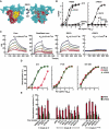
Cross-reactive neutralizing antibodies (NAbs) are found in the sera of many HIV-1-infected individuals, but the virologic basis of their neutralization remains poorly understood. We used knowledge of HIV-1 envelope structure to develop antigenically resurfaced glycoproteins specific for the structurally conserved site of initial CD4 receptor binding. These probes were used to identify sera with NAbs to the CD4-binding site (CD4bs) and to isolate individual B cells from such an HIV-1-infected donor. By expressing immunoglobulin genes from individual cells, we identified three monoclonal antibodies, including a pair of somatic variants that neutralized over 90% of circulating HIV-1 isolates. Exceptionally broad HIV-1 neutralization can be achieved with individual antibodies targeted to the functionally conserved CD4bs of glycoprotein 120, an important insight for future HIV-1 vaccine design.
Figures


Comment in
-
AIDS/HIV. A boost for HIV vaccine design.Science. 2010 Aug 13;329(5993):770-3. doi: 10.1126/science.1194693. Science. 2010. PMID: 20705840 No abstract available.
Similar articles
-
Isolation and characterization of a novel neutralizing antibody targeting the CD4-binding site of HIV-1 gp120.Antiviral Res. 2016 Aug;132:252-61. doi: 10.1016/j.antiviral.2016.06.013. Epub 2016 Jul 5. Antiviral Res. 2016. PMID: 27387828
-
AIDS/HIV. A boost for HIV vaccine design.Science. 2010 Aug 13;329(5993):770-3. doi: 10.1126/science.1194693. Science. 2010. PMID: 20705840 No abstract available.
-
Structure of an N276-Dependent HIV-1 Neutralizing Antibody Targeting a Rare V5 Glycan Hole Adjacent to the CD4 Binding Site.J Virol. 2016 Oct 28;90(22):10220-10235. doi: 10.1128/JVI.01357-16. Print 2016 Nov 15. J Virol. 2016. PMID: 27581986 Free PMC article.
-
Structural Features of Broadly Neutralizing Antibodies and Rational Design of Vaccine.Adv Exp Med Biol. 2018;1075:73-95. doi: 10.1007/978-981-13-0484-2_4. Adv Exp Med Biol. 2018. PMID: 30030790 Review.
-
GP120: target for neutralizing HIV-1 antibodies.Annu Rev Immunol. 2006;24:739-69. doi: 10.1146/annurev.immunol.24.021605.090557. Annu Rev Immunol. 2006. PMID: 16551265 Review.
Cited by
-
Architectural insight into inovirus-associated vectors (IAVs) and development of IAV-based vaccines inducing humoral and cellular responses: implications in HIV-1 vaccines.Viruses. 2014 Dec 17;6(12):5047-76. doi: 10.3390/v6125047. Viruses. 2014. PMID: 25525909 Free PMC article. Review.
-
Immunization with HIV-1 trimeric SOSIP.664 BG505 or founder virus C (FVCEnv) covalently complexed to two-domain CD4S60C elicits cross-clade neutralizing antibodies in New Zealand white rabbits.Vaccine X. 2022 Sep 30;12:100222. doi: 10.1016/j.jvacx.2022.100222. eCollection 2022 Dec. Vaccine X. 2022. PMID: 36262212 Free PMC article.
-
Structural Repertoire of HIV-1-Neutralizing Antibodies Targeting the CD4 Supersite in 14 Donors.Cell. 2015 Jun 4;161(6):1280-92. doi: 10.1016/j.cell.2015.05.007. Epub 2015 May 21. Cell. 2015. PMID: 26004070 Free PMC article.
-
Strategies to guide the antibody affinity maturation process.Curr Opin Virol. 2015 Apr;11:137-47. doi: 10.1016/j.coviro.2015.04.002. Epub 2015 Apr 24. Curr Opin Virol. 2015. PMID: 25913818 Free PMC article. Review.
-
Long trimer-immunization interval and appropriate adjuvant reduce immune responses to the soluble HIV-1-envelope trimer base.iScience. 2024 Jan 11;27(2):108877. doi: 10.1016/j.isci.2024.108877. eCollection 2024 Feb 16. iScience. 2024. PMID: 38318357 Free PMC article.
References
Publication types
MeSH terms
Substances
Associated data
- Actions
- Actions
- Actions
- Actions
- Actions
- Actions
Grants and funding
LinkOut - more resources
Full Text Sources
Other Literature Sources
Molecular Biology Databases
Research Materials
Miscellaneous

