. 2010 Jul 7;30(27):9228-40.
doi: 10.1523/JNEUROSCI.0418-10.2010.

Reduced Reelin expression accelerates amyloid-beta plaque formation and tau pathology in transgenic Alzheimer's disease mice

Affiliations

Reduced Reelin expression accelerates amyloid-beta plaque formation and tau pathology in transgenic Alzheimer's disease mice

Samira Kocherhans et al. J Neurosci. .

Abstract

In addition to the fundamental role of the extracellular glycoprotein Reelin in neuronal development and adult synaptic plasticity, alterations in Reelin-mediated signaling have been suggested to contribute to neuronal dysfunction associated with Alzheimer's disease (AD). In vitro data revealed a biochemical link between Reelin-mediated signaling, Tau phosphorylation, and amyloid precursor protein (APP) processing. To directly address the role of Reelin in amyloid-beta plaque and Tau pathology in vivo, we crossed heterozygous Reelin knock-out mice (reeler) with transgenic AD mice to investigate the temporal and spatial AD-like neuropathology. We demonstrate that a reduction in Reelin expression results in enhanced amyloidogenic APP processing, as indicated by the precocious production of amyloid-beta peptides, the significant increase in number and size of amyloid-beta plaques, as well as age-related aggravation of plaque pathology in double mutant compared with single AD mutant mice of both sexes. Numerous amyloid-beta plaques accumulate in the hippocampal formation and neocortex of double mutants, precisely in layers with strongest Reelin expression and highest accumulation of Reelin plaques in aged wild-type mice. Moreover, concentric accumulations of phosphorylated Tau-positive neurons around amyloid-beta plaques were evident in 15-month-old double versus single mutant mice. Silver stainings indicated the presence of neurofibrillary tangles, selectively associated with amyloid-beta plaques and dystrophic neurites in the entorhinal cortex and hippocampus. Our findings suggest that age-related Reelin aggregation and concomitant reduction in Reelin-mediated signaling play a proximal role in synaptic dysfunction associated with amyloid-beta deposition, sufficient to enhance Tau phosphorylation and tangle formation in the hippocampal formation in aged Reelin-deficient transgenic AD mice.


Figures

Figure 1.
Figure 1.
Early neuropathological changes in the hippocampus of heterozygous reeler (reln) mice. Immunoperoxidase labelings of hippocampal brain sections obtained from 3-month-old (A, B, E–H) and 9-month-old (C, D) wt and heterozygous reeler mice (reln). A, C, Aging-related reduction in Reelin immunoreactivity and concomitant appearance of Reelin-positive plaques (enlarged view in C) in the CA1 stratum radiatum, lacunosum-moleculare (slm), and outer molecular layer (ml) of the DG in 9-month-old compared with 3-month-old wt mice. The two boxes in A are enlarged in E and F to show the dense accumulation of Reelin-positive cells in the stratum oriens and the distinct neuropil staining in the slm and ml, respectively, the latter likely representing the extracellular Reelin secreted from perforant path axonal projections. B, Reelin-positive plaques are already detectable at 3 months of age in heterozygous reeler mice. Note the reduction in Reelin-immunoreactive cells in stratum oriens (box in B is enlarged in G) and selective enrichment in plaques in the slm (arrow; box in B is enlarged in H). D, Prominent accumulation of Reelin in amyloid-like plaques in the subiculum, slm, and outer ml at 9 months of age. Scale bars: D, 500 μm; H, 20 μm.
Figure 2.
Figure 2.
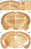
Early and accelerated amyloid-plaque deposition in reln/app double transgenic mice. Representative images of coronal brain sections taken from 6- (A–C), 9- (D), and 15-month-old single mutant (app) and double mutant (reln/app) mice (E) processed for anti-amyloid-β immunoperoxidase staining. A, No amyloid-β plaque deposition was detected in single transgenic AD mice at 6 months. B, In contrast, numerous amyloid-β plaques were evident in both hippocampus and cortex of reln/app mice at this age. C, Typical dense-core fibrillar plaque in the CA1 stratum oriens (so). C′, Granular Aβ plaque, potentially representing precursor amyloid deposits in the DG molecular layer. D, Representative images of half-brain sections of a single (left) and double mutant (right) mouse processed for immunoperoxidase staining using 6E10 monoclonal anti-Aβ antibody. Note the increase in plaque size and the prominent layer-specific localization of amyloid-β plaques in the CA1 lacunosum-moleculare and piriform cortex layer I (arrows) of reln/app mice. E, Representative brain sections of an app (left) and reln/app (right) mouse at 15 months showing the aggravated plaque load in reln/app mice compared with app subjects. Note the ventricular enlargement and reduced cortical thickness in reln/app brains, indicative of progressive neurodegeneration. Scale bars: A, D, E, 500 μm; C', 20 μm.
Figure 3.
Figure 3.
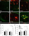
Increased inflammatory responses in reln/app double transgenic mice. Low-magnification images of double immunofluorescence staining of cortical (A–C) and hippocampal (D–F) brain sections obtained from 9-month-old single mutant app and double mutant reln/app mice. A, B, D, E, Pronounced increase in GFAP-positive astrocytes (red) selectively associated with amyloid-β plaques (anti-Aβ1-40/42 antibody; green) was found in reln/app compared with app subjects. C, F, A similar increase in reactive microglia was evident in the neocortex (C) and hippocampus (F) of 9 month reln/app mice. Note the selective accumulation of CD68-positive microglia (red) with Reelin-immunoreactive plaques (green). G, Quantification of the area fraction of GFAP-immunoreactive astrocytes revealed a main effect of genotype (F (3,20) = 4.4; p = 0.016) and significant differences between reln/app versus wt (p = 0.022), reln/app versus reln (p = 0.043), and reln/app versus app (p = 0.010) at 9 months of age. H, Similar differences emerged between reln/app versus wt (p = 0.039) and reln/app versus app (p = 0.046) subjects for the CD68 area fraction. Values are given as mean ± SEM. *p < 0.05 (comparison with reln/app subjects), statistical significance based on Fisher's LSD post hoc analysis. Scale bars: C, 50 μm; F, 10 μm.
Figure 4.
Figure 4.
Aggravation and segregation of Reelin and amyloid-β plaques pathology in the hippocampus and cortex of 9- and 15-month-old double mutant reln/app mice. A–I, Triple immunofluorescence stainings using Cy3-GFAP (red), anti-Aβ1-40/42 (green), and anti-Reelin (blue) antibodies on cortical (A–F) and hippocampal (G–I) brain sections of 9-month-old app (A–C) and reln/app mice (D–I). In the double mutants, amyloid-β plaques were densely surrounded and tightly associated with reactive astrocytes, whereas in app subjects only moderate astrogliosis was evident. Note the intense Reelin immunoreactivity associated with fibrillary amyloid-β plaques (B vs E, arrows) and their striking segregation from Aβ deposits (H, arrow) in the CA1 slm in reln/app compared with app subjects. J–O, Aggravation of the microgliosis and astrogliosis in reln/app mice at 15 months of age was particularly prominent in the entorhinal cortex (ECtx). Note the reduction in APP-expressing neurons in the aged double (N) versus single mutants (K). Scale bars: C, F, I, 10 μm; L, O, 20 μm.
Figure 5.
Figure 5.
Increase in amyloidogenic APP processing and insoluble Aβ levels in double mutant reln/app mice. A, Representative Western blots of the SDS-soluble supernatant (SN) derived from hippocampal brain lysates of 9-month-old mice using anti-Aβ (6E10) antibodies. B, Semiquantitative analysis involving densitometry of the immunoreactive β-cleaved C-terminal APP fragments (β-stubs), run in duplicate, corrected for nonspecific background and equal loading using β-actin as control, revealed a significant increase in reln/app compared with app (p = 0.049; n = 4). Soluble Aβ-levels were very low, but clearly detectable in samples of reln/app subjects. C, Representative Western blots using anti-Reelin (G10) antibody recognizing both full-length (FL-Reln) and the N-terminal 180 kDa fragment (N-Reln). The N-terminal-specific APP antibody (22C11) antibody recognized in addition to full-length APP (FL-APP) and the soluble α- or β-secretase-cleaved APP ectodomains (sAPP) short N-terminal fragments, presumably representing N-APP. D, Statistical analysis of the densitometrical measurements of FL-APP and sAPP-immunoreactive fragments revealed a significant difference between genotypes as indicated by the reduced FL-APP/sAPP ratio in reln/app compared with app subjects (p = 0.033; n = 4). In addition, a marked shift toward higher N-APP-immunoreactive fragments in 9-month-old reln/app mice was evident, as demonstrated by the significant reduction in the FL-Reelin/N-APP ratio (G) (p = 0.034; n = 4). E, Representative Western blots using anti-Aβ (6E10) antibodies of SDS-insoluble fractions (pellet) of hippocampal brain lysates, which was resuspended in formic acid (FA). F, Quantitative analysis using ELISA with Aβ40- and Aβ42-specific antibodies revealed a significant increase in Aβ peptides in the insoluble fractions of reln/app compared with app littermates in the hippocampus (Aβ40, p = 0.021; Aβ42, p = 0.046) and neocortex (Aβ40, p = 0.016; Aβ42, p = 0.016; n = 7–8). Values are expressed as fraction of total protein content and given as mean ± SEM. *p < 0.05 **p < 0.01, statistical significance based on Mann–Whitney U test.
Figure 6.
Figure 6.
Increase in Thioflavin S-positive amyloid-β plaques in reln/app mice in neocortex and hippocampal formation. Double immunofluorescence using anti-Aβ (6E10; red) and anti-α1-syntrophin (αSyn; blue) antibodies combined with Thioflavin S counterstaining (green) were performed to investigate the fibrillary amyloid-β plaque load in both neuropil and cerebral vasculature in 9-month-old app (A) and reln/app mice (B). Note the increase in Thioflavin S signals concomitantly with the anti-Aβ immunoreactivity in the outer layers of the neocortex. C, Densitometric analysis of the brightness and surface area covered by Thioflavin S-positive signals in the neocortical layers I–VI (Ctx), lateral entorhinal cortex (EC), and dorsal and ventral hippocampus (Hip). D, Quantification of the pixel overlap between the green Thioflavin S-positive signals and red anti-Aβ immunoreactive staining, averaged from measurements of eight images (512 × 512 pixel in size) per animal and brain region (n = 4 per genotype) acquired in the areas indicated. Values are given as mean ± SEM. *p < 0.05, statistical significance based on Mann–Whitney U test. Scale bar, 50 μm.
Figure 7.
Figure 7.
Selective association of anti-phospho-Tau immunoreactivity in plaque-dense areas within CA1 slm. A, B, Double immunofluorescence using anti-phospho-T205 (red) and anti-Aβ (6E10, blue) antibodies combined with Sytox Green nuclear counterstaining to investigate the relationship between amyloid-β plaque and Tau pathology in app (A) and reln/app (B) at 15 months of age. A, Representative amyloid-β plaque in the CA1 slm of app mice with a dense core and some diffuse anti-Aβ immunoreactivity surrounding it (A′). In line with the immunoperoxidase stainings, anti-phospho-Tau immunoreactivity in app subjects was readily detectable in the neuropil, but no evidence for any enrichment in cells surrounding the plaques was found (A″, enlarged view is shown in inset). B–B″, Anti-amyloid-β immunoreactivity covered large areas within the core and surrounding areas of plaques in the CA1 slm of reln/app mice. Note the selective fibrillary enrichment of anti-phospho-Tau signals in the cytoplasm (inset) and dendrites (arrow) of neurons selectively associated with amyloid plaques. The distinct spatial distribution of the amyloid-β and Tau pathology is also evident on the xz and yz view at the bottom and right side of the image. Scale bars: A, B, 30 μm.
Figure 8.
Figure 8.
Concentric accumulations of phospho-Tau-positive neurons and neurofibrillary tangles around amyloid-β plaques in 15-month-old reln/app mice. Representative images of brain sections obtained from app (A, E) and reln/app mice (B–D, F, G) processed for immunoperoxidase staining using anti-phospho-T205 antibody (A–D) or silver staining (E–G). A, In app subjects, uniform phospho-Tau immunoreactivity was evident in the cortical neuropil, indicative of axonal localization. No particular enrichment was seen in the vicinity of amyloid-β plaques, recognized by their lack of phospho-Tau immunoreactivity (asterisk). B, C, In reln/app mice, strongly stained phospho-Tau-positive neurons were evident around amyloid-β plaques in neocortical areas (insets). D, In the hippocampus of reln/app mice, a similar pattern was evident in close vicinity to plaques; some neurons appeared dystrophic, strongly suggestive of tangle-like intraneuronal accumulations (D, inset). E, Silver staining revealed some dark precipitates (arrowhead) in the vicinity of large plaques (asterisk) in the entorhinal cortex of app mice. F, In contrast, dense silver precipitates surrounding amyloid-β plaques, indicative of dystrophic neurites, were very prominent in reln/app mice. In addition, black-labeled intraneuronal aggregates, likely representing neurofibrillary tangles, were seen in neurons associated with these plaques (arrows; enlarged in the insets). G, Representative image of a large amyloid-β plaque in the entorhinal cortex of a reln/app mouse, showing a concentric pattern of silver-stained cells, likely representing degenerating neurons in the vicinity of plaques (arrowheads). Scale bars, 20 μm.

Similar articles

Cited by

References

    1. Beffert U, Morfini G, Bock HH, Reyna H, Brady ST, Herz J. Reelin-mediated signaling locally regulates protein kinase B/Akt and glycogen synthase kinase 3beta. J Biol Chem. 2002;277:49958–49964. - PubMed
    1. Beffert U, Weeber EJ, Morfini G, Ko J, Brady ST, Tsai LH, Sweatt JD, Herz J. Reelin and cyclin-dependent kinase 5-dependent signals cooperate in regulating neuronal migration and synaptic transmission. J Neurosci. 2004;24:1897–1906. - PMC - PubMed
    1. Beffert U, Weeber EJ, Durudas A, Qiu S, Masiulis I, Sweatt JD, Li WP, Adelmann G, Frotscher M, Hammer RE, Herz J. Modulation of synaptic plasticity and memory by Reelin involves differential splicing of the lipoprotein receptor Apoer2. Neuron. 2005;47:567–579. - PubMed
    1. Beffert U, Durudas A, Weeber EJ, Stolt PC, Giehl KM, Sweatt JD, Hammer RE, Herz J. Functional dissection of Reelin signaling by site-directed disruption of Disabled-1 adaptor binding to apolipoprotein E receptor 2: distinct roles in development and synaptic plasticity. J Neurosci. 2006a;26:2041–2052. - PMC - PubMed
    1. Beffert U, Nematollah Farsian F, Masiulis I, Hammer RE, Yoon SO, Giehl KM, Herz J. ApoE receptor 2 controls neuronal survival in the adult brain. Curr Biol. 2006b;16:2446–2452. - PubMed

Publication types

MeSH terms