. 2010 Jul 7;30(27):9103-16.
doi: 10.1523/JNEUROSCI.1049-10.2010.

Urocortin 3 modulates social discrimination abilities via corticotropin-releasing hormone receptor type 2

Affiliations

Urocortin 3 modulates social discrimination abilities via corticotropin-releasing hormone receptor type 2

Jan M Deussing et al. J Neurosci. .

Abstract

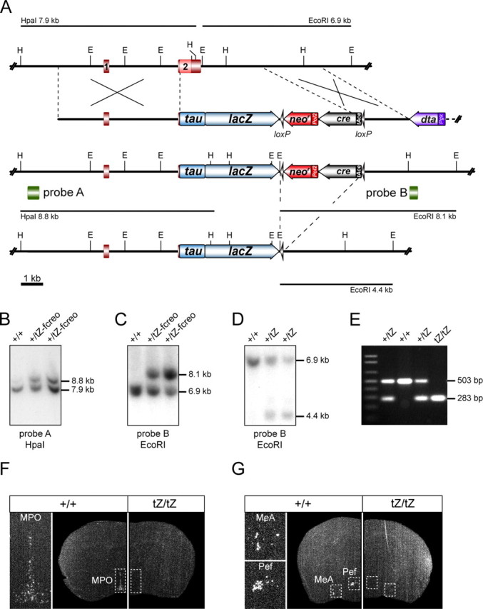
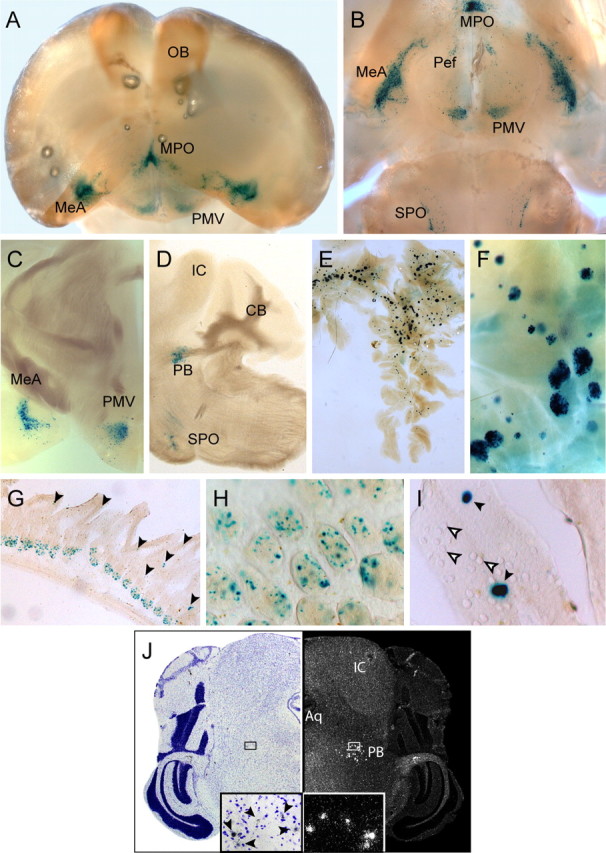
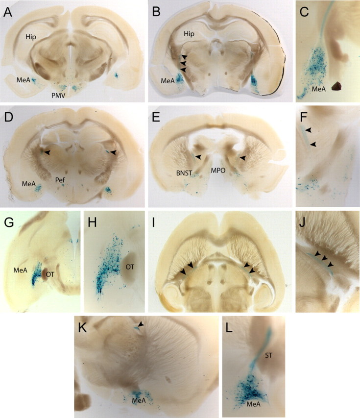
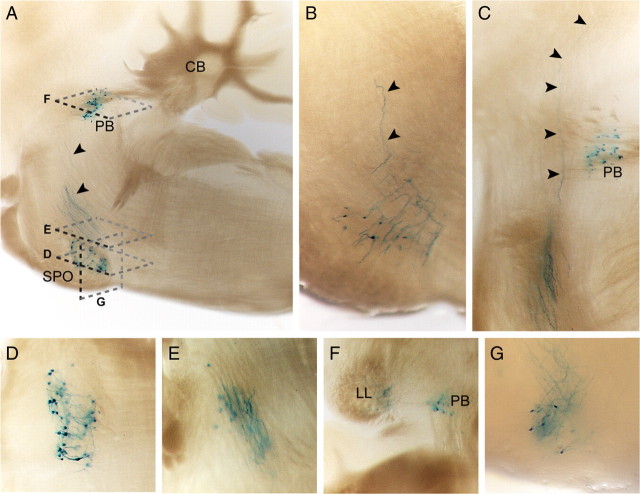
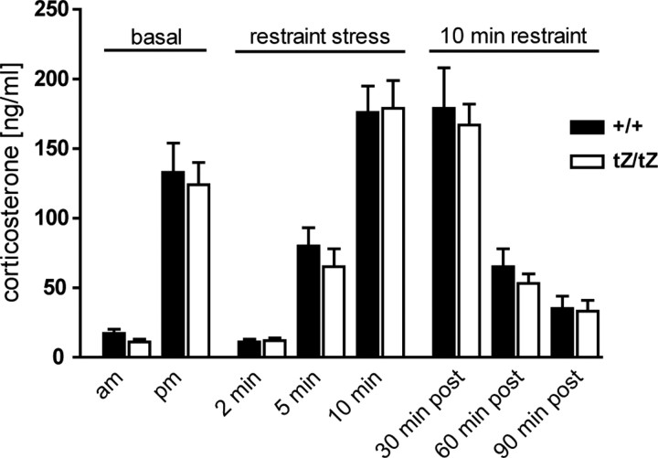
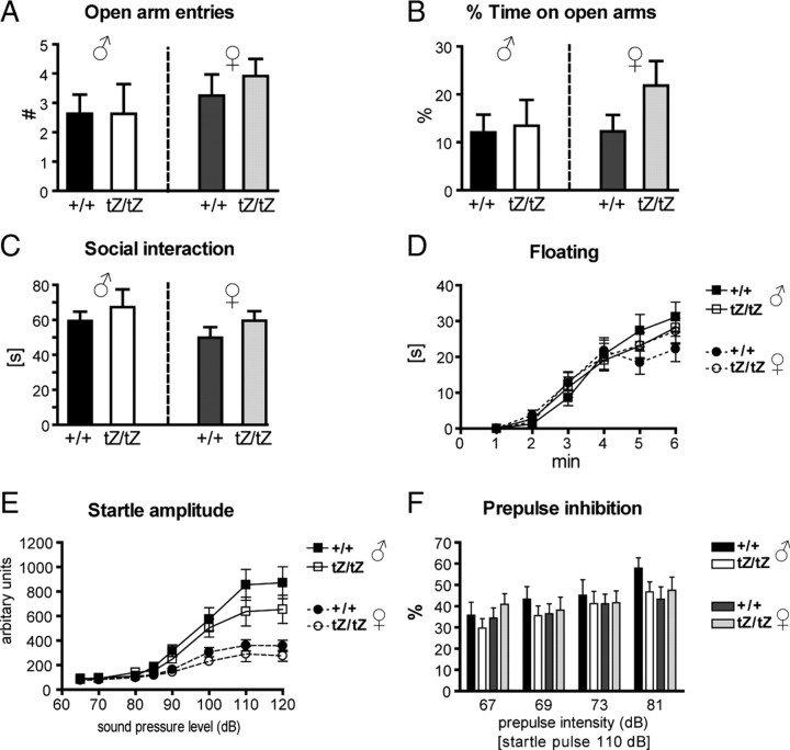
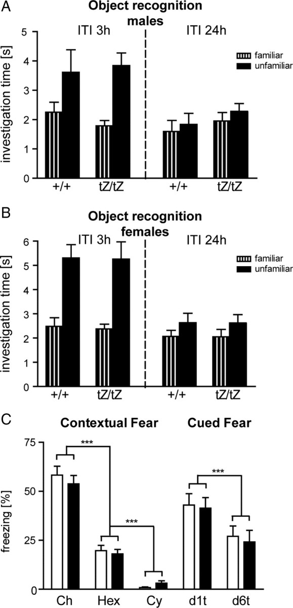
Urocortin 3 (UCN3) is strongly expressed in specific nuclei of the rodent brain, at sites distinct from those expressing urocortin 1 and urocortin 2, the other endogenous ligands of corticotropin-releasing hormone receptor type 2 (CRH-R2). To determine the physiological role of UCN3, we generated UCN3-deficient mice, in which the UCN3 open reading frame was replaced by a tau-lacZ reporter gene. By means of this reporter gene, the nucleus parabrachialis and the premammillary nucleus were identified as previously unknown sites of UCN3 expression. Additionally, the introduced reporter gene enabled the visualization of axonal projections of UCN3-expressing neurons from the superior paraolivary nucleus to the inferior colliculus and from the posterodorsal part of the medial amygdala to the principal nucleus of the bed nucleus of the stria terminalis, respectively. The examination of tau-lacZ reporter gene activity throughout the brain underscored a predominant expression of UCN3 in nuclei functionally connected to the accessory olfactory system. Male and female mice were comprehensively phenotyped but none of the applied tests provided indications for a role of UCN3 in the context of hypothalamic-pituitary-adrenocortical axis regulation, anxiety- or depression-related behavior. However, inspired by the prevalent expression throughout the accessory olfactory system, we identified alterations in social discrimination abilities of male and female UCN3 knock-out mice that were also present in male CRH-R2 knock-out mice. In conclusion, our results suggest a novel role for UCN3 and CRH-R2 related to the processing of social cues and to the establishment of social memories.


Figures

Figure 1.
Figure 1.
Generation of Ucn3tZ mice. A, Strategy for targeted disruption and substitution of Ucn3 by a tau-lacZ reporter gene (tZ). Partial restriction maps of wild-type Ucn3 locus, targeting vector, recombined Ucn3tZ-fcreo allele and UcntZ allele after cre-mediated self-excision of the floxed cre-neo (fcreo) cassette. E, EcoRI; H, HpaI; loxP sites are indicated as black arrowheads. B, Southern blot analysis of wild-type and targeted ES cell clones. The Ucn3 5′-probe A was hybridized to HpaI-digested genomic ES cell DNA. The targeted allele was indicated by the presence of an additional mutant 8.8 kb fragment. C, The Ucn3 3′-probe B was hybridized to EcoRI-digested DNA from the same ES cell clones, confirming homologous recombination by detection of an additional mutant fragment at 8.1 kb. D, Southern blot analysis of EcoRI-digested tail DNA of F1 progeny of germ line chimeras hybridized with the 3′-probe B. Self-excision of the fcreo cassette was indicated by the presence of an additional 4.4 kb fragment. E, Genotyping of F2 mice through PCR depicting wild-type, heterozygous, and homozygous Ucn3tZ mice. F, G, The loss of Ucn3 expression in Ucn3tZ/tZ mice was confirmed by radioactive in situ hybridization using a specific riboprobe for UCN3 exon 2. Shown are representative dark-field photomicrographs of coronal sections of Ucn3+/+ and Ucn3tZ/tZ mice at the level of the MPO (F) as well as at the level of the MeA and Pef (G).
Figure 2.
Figure 2.
Expression of the tau-LacZ reporter gene introduced into the Ucn3 locus fully recapitulates the central and peripheral sites of UCN3 expression. A, B, Frontal (A) and ventral (B) whole-mount view of a X-Gal-stained brain of an Ucn3+/tZ mouse after BABB clearing. Strong staining was visible in the MPO, MeA, Pef, PMV, and SPO. C, Coronal section depicting strong tau-LacZ/UCN3-expression in the MeA and PMV. D, Sagittal section depicting strong tau-LacZ/UCN3 expression in the SPO and in the PB. E, F, X-Gal staining reflected strong UCN3 expression in Langerhans islets of the pancreas. G, Tau-LacZ reporter gene expression in the small intestine. H, I, Higher magnification of X-Gal staining of cells within (H) intestinal crypts and in (I) a subset of goblet cells as indicated by black arrowheads. Tau-LacZ-negative goblet cells are indicated by white arrowheads. J, In situ hybridization of a coronal mouse brain sections displaying UCN3 expression in the PB. A bright-field picture of a cresyl violet-stained section (left) and corresponding dark-field photomicrograph (right) are shown. Enlargements of the indicated small boxes displaying UCN3-expressing cells within the PB are shown at the bottom. UCN3-positive cells in the bright-field enlargement are indicated by arrowheads. IC, Inferior colliculus; Aq, aqueduct.
Figure 3.
Figure 3.
UCN3-expressing neurons of the medial amygdala project via the stria terminalis. A–F, Coronal sections from caudal to rostral illustrating the full extension of UCN3-expressing neurons within the MeA. B, C, X-Gal-positive neurons of the posterodorsal part of the MeA send their axonal projections (black arrowheads) dorsally. D–F, X-Gal-stained axons continue horizontally at the level of the Pef (D) before they descend toward the BNST (E, F). G, H, Horizontal sections demonstrating the rostrocaudal distribution of UCN3-expressing neurons in the MeA. I, J, Horizontal view on axons originating in the MeA and projecting via the stria terminalis to the BNST. K, L, Sagittal view of the MeA at the level at which the axons turn rostrally (K) and more lateral view depicting the posterodorsal part of the MeA with originating axons (L).
Figure 4.
Figure 4.
UCN3-expressing neurons of the superior paraolivary nucleus project via the lateral lemniscus to the inferior colliculus. A–C, Sagittal sections illustrating UCN3-expressing neurons residing in the SPO and their ascending axons, as indicated by black arrowheads. D–F, Horizontal sections from ventral to dorsal (compare A) depicting UCN3-expressing neurons and their axons ascending via the LL. G, Coronal section demonstrating the origin and initial course of X-Gal-stained axons.
Figure 5.
Figure 5.
The HPA axis activity in male Ucn3tZ mice is normal. Male Ucn3tZ/tZ mice showed no differences in basal plasma corticosterone levels in the morning (am) or evening (pm). Restraint stress resulted in significantly elevated corticosterone levels in male Ucn3tZ/tZ mice and wild-type littermates. No difference was observed in negative-feedback regulation of HPA axis activity 30, 60, and 90 min after 10 min restraint stress. Error bars indicate SEM.
Figure 6.
Figure 6.
Anxiety- and depression-related behaviors are unchanged in male and female Ucn3tZ mice. A, B, In the elevated plus maze test, male and female Ucn3tZ/tZ and Ucn3+/+ mice did not show any differences when comparing open arm entries (A) and total time on open arms (B) (males, n = 8–11 mice per genotype; females, n = 12 mice per genotype). C, The active social interaction time was identical for male and female Ucn3tZ/tZ and Ucn3+/+ mice as assessed in the social interaction test (n = 5–7 mice per genotype). D, No difference in floating behavior in a 6 min forced-swim test was observed (n = 15 mice per genotype). E, F, The acoustic sensorimotor behavior of male and female Ucn3tZ/tZ and Ucn3+/+ littermate control mice is not significantly different. E, F, Intensity–response curves of acoustic startle amplitudes (E) and percentage of prepulse inhibition of the acoustic startle reflex at four prepulse intensities (startle pulse, 110 dB; n = 15 mice per genotype) (F) revealed no differences between male and female Ucn3tZ/tZ and Ucn3+/+ littermates. Error bars indicate SEM.
Figure 7.
Figure 7.
Ucn3tZ mice display altered social discrimination abilities. A–F, Investigation duration (in seconds) (mean + SEM) toward conspecific ovariectomized female. The white bars represent the duration during first exposure, and the hatched (same ovariectomized female) or black (novel ovariectomized female) bars, that of the second exposure (*p < 0.05, **p < 0.01, familiar vs unfamiliar conspecific). A–C, Social discrimination abilities of male Ucn3tZ/tZ and Ucn3+/+ mice after interexposure intervals of 2, 4, and 6 h (n = 12 mice per genotype). D, Female Ucn3tZ/tZ mice are able to discriminate between a familiar and an unfamiliar conspecific, whereas Ucn3+/+ mice cannot (n = 12 animals per genotype; interexposure interval, 2 h; *p < 0.05, familiar vs unfamiliar conspecific). E, Male Crhr2−/− mice are able to discriminate between a familiar and an unfamiliar conspecific, whereas Crhr2+/+ mice cannot (n = 12 mice per genotype; interexposure interval, 2 h). F, Both Ucn2tZ/tZ and Ucn2+/+ mice are unable to discriminate between a familiar and an unfamiliar conspecific (n = 12 mice per genotype; interexposure interval, 2 h). IEI, Interexposure interval. G, Investigation time of male Ucn3tZ/tZ and Ucn3+/+ littermate control mice at a neutral and a social odor stimulus in a discriminatory task (n = 19–21 mice per genotype). Both groups significantly discriminated between the neutral and the social stimulus (**p < 0.01, ***p < 0.001, neutral vs social odor). H, Fine olfactory discrimination with male Ucn3+/+ and Ucn3tZ/tZ mice. Ratios along the x-axis represent the concentration of [S+] in the binary mixture of [S+] and [S−]. The number of successful discriminations per session is expressed as mean percentage of first correct responses to the rewarded odorant [S+] of total trials per session [Mann–Whitney tests: 100% p = 0.6467, Mann–Whitney U (MWU) 48.00; 70% p = 0.2099, MWU 40.50; 55% p = 0.1579, MWU 34.50; 53% p = 0.5746, MWU 56.50; p = 0.2592, 38.50; 50% p = 0.3241, MWU 49.50]. I, Smell sensitivity test with male Ucn3+/+ and Ucn3tZ/tZ mice. Discrimination between a dish scented with [S+] in different steps of dilution and another dish with solvent (Mann–Whitney test: p = 0.0992, MWU 39.00).
Figure 8.
Figure 8.
Ucn3tZ mice display normal memory performance. A, B, Object recognition memory of male (n = 10–12) (A) and female (n = 12–13) (B) Ucn3tZ mice. Investigation time at a familiar and an unfamiliar object 3 h (left panel) and 24 h (right panel) after the last sample phase; shown are mean + SEM. ITI, Intertrial interval. C, Contextual and auditory-cued fear. Left panel, Freezing responses of Ucn3+/+ (open bars; n = 9) and Ucn3tZ/tZ mice (filled bars; n = 11) on (re-)exposure to the conditioning chamber (Ch), a hexagonal prism containing a grid floor (Hex), and a neutral cylindrical context (Cy) for 3 min each 1 d after conditioning. Right panel, Freezing responses of the same mice on exposure to 3 min tones in the neutral cylindrical context 1 d (d1t) and 6 d (d6t) after conditioning. Shown are mean + SEM. ***p < 0.001.

Similar articles

Cited by

References

    1. Bakshi VP, Smith-Roe S, Newman SM, Grigoriadis DE, Kalin NH. Reduction of stress-induced behavior by antagonism of corticotropin-releasing hormone 2 (CRH2) receptors in lateral septum or CRH1 receptors in amygdala. J Neurosci. 2002;22:2926–2935. - PMC - PubMed
    1. Bale TL, Vale WW. Increased depression-like behaviors in corticotropin-releasing factor receptor-2-deficient mice: sexually dichotomous responses. J Neurosci. 2003;23:5295–5301. - PMC - PubMed
    1. Bale TL, Contarino A, Smith GW, Chan R, Gold LH, Sawchenko PE, Koob GF, Vale WW, Lee KF. Mice deficient for corticotropin-releasing hormone receptor-2 display anxiety-like behaviour and are hypersensitive to stress. Nat Genet. 2000;24:410–414. - PubMed
    1. Bielsky IF, Hu SB, Szegda KL, Westphal H, Young LJ. Profound impairment in social recognition and reduction in anxiety-like behavior in vasopressin V1a receptor knockout mice. Neuropsychopharmacology. 2004;29:483–493. - PubMed
    1. Bunting M, Bernstein KE, Greer JM, Capecchi MR, Thomas KR. Targeting genes for self-excision in the germ line. Genes Dev. 1999;13:1524–1528. - PMC - PubMed

Publication types

MeSH terms