. 2010 Sep;84(18):9487-96.
doi: 10.1128/JVI.00763-10. Epub 2010 Jul 7.

HIV infection upregulates caveolin 1 expression to restrict virus production

Affiliations

HIV infection upregulates caveolin 1 expression to restrict virus production

Shanshan Lin et al. J Virol. 2010 Sep.

Abstract

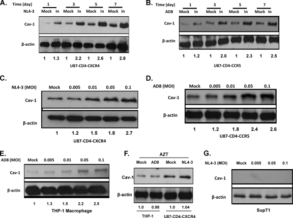
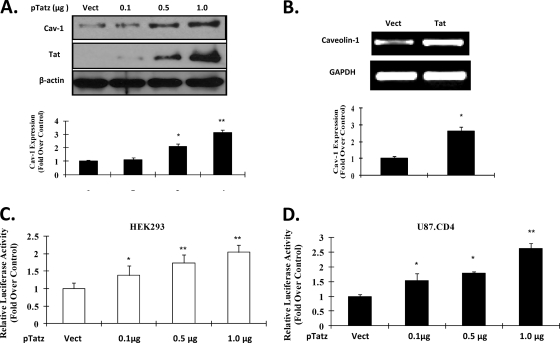
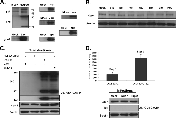
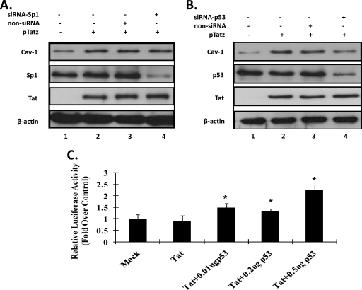
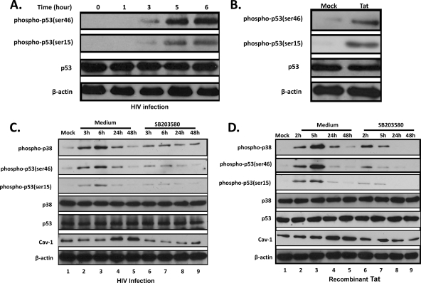
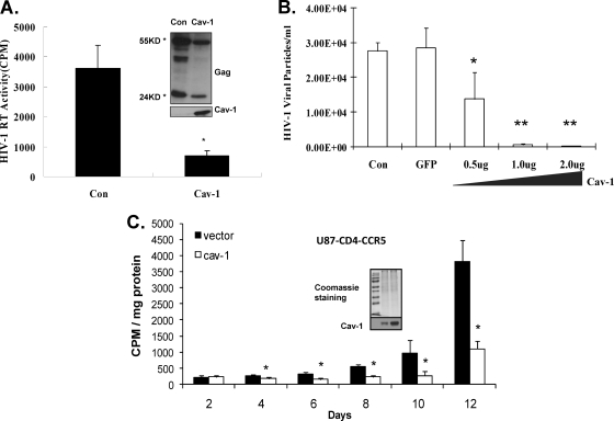
Caveolin 1 (Cav-1) is a major protein of a specific membrane lipid raft known as caveolae. Cav-1 interacts with the gp41 of the human immunodeficiency virus (HIV) envelope, but the role of Cav-1 in HIV replication and pathogenesis is not known. In this report, we demonstrate that HIV infection in primary human monocyte-derived macrophages (MDMs), THP-1 macrophages, and U87-CD4 cells results in a dramatic upregulation of Cav-1 expression mediated by HIV Tat. The activity of p53 is essential for Tat-induced Cav-1 expression, as our findings show enhanced phosphorylation of serine residues at amino acid positions 15 and 46 in the presence of Tat with a resulting Cav-1 upregulation. Furthermore, inhibition of p38 mitogen-activated protein kinase (MAPK) blocked phosphorylation of p53 in the presence of Tat. Infection studies of Cav-1-overexpressing cells reveal a significant reduction of HIV production. Taken together, these results suggest that HIV infection enhances the expression of Cav-1, which subsequently causes virus reduction, suggesting that Cav-1 may contribute to persistent infection in macrophages.


Figures

FIG. 1.
FIG. 1.
HIV infection upregulates the expression of caveolin 1 (Cav-1) in macrophages and U87 cells. (A) U87 cells expressing CD4 and CXCR4 or CCR5 were infected with NL4-3 or AD8 HIV cloned virus, respectively. (B) Primary monocyte-derived macrophages infected with AD8 HIV. (C). Human acute monocytic leukemia cells (THP-1), which were allowed to differentiate into macrophages by treatment with phorbol 12-myristate 13-acetate (PMA), infected with HIV-1 AD8 strain. (D) SupT1 cells infected with NL4-3 HIV. A multiplicity of infection (MOI) of 0.1 was used for infection for all cell types. Cell lysates were harvested 3 days after infection, and the expression levels of Cav-1 were detected by Western blotting. Mock denotes uninfected cells. The Cav-1 expression levels shown by the Western blot were quantified using densitometric analysis. β-Actin was used as a loading control. Blots were quantified by densitometry and normalized to the corresponding β-actin band. The relative values of Cav-1 protein expression are shown at the bottom. *, P < 0.05 compared to the control.
FIG. 2.
FIG. 2.
HIV infection increases Cav-1 expression in a time- and dose-dependent manner. U87-CD4-CXCR4 (A) and U87-CD4-CCR5 (B) cells were infected with HIV-1 NL4-3 and AD8, respectively, at an MOI of 0.005, and cell lysates were collected at 1, 3, 5 and 7 days postinfection. The numbers at the bottom of each blot are the relative values of Cav-1 upregulation in infected (In) cells compared to those in uninfected (Mock) cells. (C and D) Cav-1 expression in a dose-dependent infection of U87-CD4-CXCR4 or U87-CD4-CCR5 cells by NL4-3 or AD8, respectively. (E) Cav-1 expression in THP-1 macrophages infected with HIV AD8. (F) Cav-1 expression in HIV-infected THP-1 and U87-CD4-CXCR4 cells in the presence of viral inhibitor AZT. THP-1 cells were infected with the AD8 HIV strain and U87-CD4-CXCR4 with NL4-3. (G) SupT1 cells infected with NL4-3 HIV. Mock denotes uninfected cells. The expression of Cav-1 was normalized as a ratio of Cav-1 to β-actin.
FIG. 3.
FIG. 3.
Cav-1 upregulation by HIV infection is mediated by Tat. (A) U87-CD4-CXCR4 cells were transiently transfected with different doses of Tat expression vector (pTatz) or vector lacking the tat coding sequence (Vect). The Western blot is representative of results of three independent experiments. (B) Cav-1 expression as mRNA level in the presence of Tat. U87-CD4-CXCR4 cells were transfected with pTatz or vector lacking tat, and total RNA was extracted 24 h posttransfection and then subjected to RT-PCR using cav-1-specific primers. 293T (C) or U87-CD4-CXCR4 (D) cells were transfected with a plasmid construct (pGL3-cavFL) containing luciferase under the control of the cav-1 gene promoter along with different doses of pTatz. Cells were harvested 48 h posttransfection, and luciferase activities were measured. *, P < 0.05; **, P < 0.01 compared to the control.
FIG. 4.
FIG. 4.
Upregulation of Cav-1 by HIV is dependent on the Tat protein alone. (A) Constructs for expressing gag and pol (psPAX2), vif (pcDNA-Hvif), vpr (pEGFP-Vpr), vpu (pcDNA-Vphu), env (pCI-NL4-3-Env), rev (pCMV-rev), and nef (pcDNASF2Nef) were used to transfect U87-CD4-CXCR4, and expression of each of these proteins was analyzed by Western blotting using specific antibodies. (B) The influence of each of the HIV proteins on the level of Cav-1 expression. Each of the indicated viral gene expression cassettes was transfected into U87-CD4-CXCR4, and the level of Cav-1 was examined by Western blot analysis 72 h posttransfection. g-p refers to gag/pol. (C) Cav-1 expression in cells transfected with a tat-defective provirus DNA. Tat-defective provirus (pNL4-3-dTat) was transfected with Tat-expressing plasmid (pTatZ) or vector without tat (Vect) into U87-CD4-CXCR4 cells. The expressions of Cav-1 and viral protein Gag were determined in the presence or absence of Tat. (D) Cav-1 expression in U87-CD4-CXCR4 cells infected with virus harvested from 293T cells cotransfected with pNL4-3-dTat and pTatZ (pNL4-3-dTat+Tat) or pNL4-3-dTat and vector devoid of tat (pNL4-3-dTat). Virus production was assayed by reverse transcriptase activity, and supernatants (Sup) were used to infect U87-CD4-CXCR4. The levels of Tat and Cav-1 expression were examined by Western blot analysis.
FIG. 5.
FIG. 5.
p53 is necessary for HIV-induced Cav-1 expression. (A) Knockdown of Sp1 expression with specific siRNA has no impact on Cav-1 upregulation by Tat. U87-CD4-CXCR4 cells were pretreated with siRNA targeting Sp1 (lane 4) or with nonspecific siRNA (lane 3) overnight. Cells were then transfected with vector lacking tat (lane1) or Tat-expressing vector (lanes 2, 3, and 4) and cultured for an additional 48 h. (B) Knockdown of p53 expression with siRNA reduces Cav-1 upregulation by Tat. The experiments were performed as described for Sp1 in panel A. (C) cav-1 gene promoter-driven luciferase expression in cells lacking p53 in the presence or absence of Tat. The NCI-H1299 p53 null cell line was transfected with pGL3-cavFL alone or with pTatz or with pTatz and pEFp53. Cells were harvested 48 h posttransfection, and luciferase activity was measured. *, P < 0.05 compared to the control.
FIG. 6.
FIG. 6.
p38 MAPK-mediated p53 phosphorylation is required for Cav-1 upregulation by HIV infection or recombinant Tat. (A) HIV infection induces p53 phosphorylation. U87-CD4-CXCR4 cells were infected with HIV-1 NL4-3 at an MOI of 0.1 for 1, 3, 5, and 6 h. Cell lysates were collected and subjected to Western blotting to demonstrate the level of phosphorylation of p53 at serine residue 15 [phospho-p53(ser15)] and 46 [phospho-p53(ser46)] as well as detection of total p53. (B) Tat enhances p53 phosphorylation. U87-CD4-CXCR4 cells were treated with 0.5 μg/ml recombinant Tat protein for 6 h, and cell lysates were examined for phosphorylated as well as total p53 protein. Mock denotes samples from untreated cells. (C) p53 phosphorylation and Cav-1 expression in HIV-infected cells in the presence or absence of p38 MAPK activation inhibitor. U87-CD4-CXCR4 cells were treated with SB203580 (10 μM) and infected with NL4-3 while maintaining the SB203580 (10 μM) treatment during infection for 3, 6, 24, and 48 h. Control cells were also infected with NL4-3 in the absence of SB203580 for the same time points. Cell lysates were prepared at the indicated time points and subjected to Western blotting to monitor for the expression levels of p38, p53, Cav-1, phosphorylated p38, and phosphorylated p53. (D) The same experiments were performed as in panel C, with the exception that recombinant Tat (0.5 μg/ml) was used rather than HIV infection.
FIG. 7.
FIG. 7.
HIV replication in cells where Cav-1 is overexpressed or downregulated by siRNA treatment. (A) 293T cells, lacking Cav-1 expression as seen by Western blotting, were transfected with plasmid expressing Cav-1 (pCZ-cav-1) or vector without cav-1 sequence (pCZ) along with HIV provirus DNA (NL4-3). Cell supernatants for RT assays and cell lysates for Western blot analysis were harvested 3 days posttransfection to monitor Cav-1 expression and inhibition of virus production by Cav-1. (B) Dose-dependent inhibition of HIV replication by Cav-1. 293T cells were transfected with 0.5, 1, and 2 μg of pCZ-cav-1, with supernatants being harvested 3 days posttransfection and levels of virus production assayed by infecting TZM-bl indicator cells. Vector alone and a green fluorescent protein (GFP)-expressing construct were used as controls. (C) A stable cell line overexpressing Cav-1, U87-CD4-CCR5-Cav-1, was infected with AD8 at an MOI of 0.1. Supernatants were harvested at the indicated time intervals in triplicate and assayed for reverse transcriptase. The reverse transcriptase activity is shown in counts per minute (cpm)/mg protein concentration. A stable cell line containing vector without cav-1 sequence (U87-CD4-CCR5-vect) was used as a control. *, P < 0.05; **, P < 0.01 compared to the control.

Similar articles

Cited by

References

    1. Adachi, A., H. E. Gendelman, S. Koenig, T. Folks, R. Willey, A. Rabson, and M. A. Martin. 1986. Production of acquired immunodeficiency syndrome-associated retrovirus in human and nonhuman cells transfected with an infectious molecular clone. J. Virol. 59:284-291. - PMC - PubMed
    1. Anderson, R. G. 1993. Caveolae: where incoming and outgoing messengers meet. Proc. Natl. Acad. Sci. U. S. A. 90:10909-10913. - PMC - PubMed
    1. Ariumi, Y., A. Kaida, M. Hatanaka, and K. Shimotohno. 2001. Functional cross-talk of HIV-1 Tat with p53 through its C-terminal domain. Biochem. Biophys. Res. Commun. 287:556-561. - PubMed
    1. Bist, A., C. J. Fielding, and P. E. Fielding. 2000. p53 regulates caveolin gene transcription, cell cholesterol, and growth by a novel mechanism. Biochemistry 39:1966-1972. - PubMed
    1. Bist, A., P. E. Fielding, and C. J. Fielding. 1997. Two sterol regulatory element-like sequences mediate up-regulation of caveolin gene transcription in response to low density lipoprotein free cholesterol. Proc. Natl. Acad. Sci. U. S. A. 94:10693-10698. - PMC - PubMed

Publication types

MeSH terms

Substances