. 2010 Dec;80(3):445-52.
doi: 10.1016/j.mvr.2010.06.004. Epub 2010 Jun 22.

Tempol, a superoxide dismutase mimetic, prevents cerebral vessel remodeling in hypertensive rats

Affiliations

Tempol, a superoxide dismutase mimetic, prevents cerebral vessel remodeling in hypertensive rats

Paulo Wagner Pires et al. Microvasc Res. 2010 Dec.

Abstract

Increased reactive oxygen species (ROS) production is involved in the pathogenesis of hypertension and stroke. The effects of ROS on cerebral vessels from hypertensive rats have not been studied. We hypothesized that tempol, a superoxide dismutase mimetic, would prevent middle cerebral artery (MCA) remodeling in stroke-prone spontaneously hypertensive rats (SHRSP). Six-week-old male SHRSP were treated with tempol (1mM) for 6weeks. The MCA was then removed and mounted in a pressure myograph to study tone generation, vessel reactivity, and passive vessel structure. Data are shown as mean±SEM, tempol vs. control. Plasma thiobarbituric acid reactive substances (TBARS) were decreased by tempol treatment (14.15±1.46 vs. 20.55±1.25nM of malondialdehyde [MDA]/ml, p=0.008). Maximum serotonin-induced constriction was increased by tempol treatment, without changes in dilation to adenosine diphosphate or tone generation. At an intralumenal pressure of 80mmHg, tempol caused a dramatic increase in the MCA lumen diameter (246±5 vs. 207±3μm, p<0.001), outer diameter (281±5 vs. 241±3μm, p<0.001), lumen cross-sectional area, and vessel cross-sectional area. Collagen IV mRNA expressions were increased by 2.4-fold after tempol treatment. These results suggest that ROS are involved in the remodeling of the cerebral vasculature of SHRSP and that ROS scavenging can attenuate this process.


Figures

Fig 1
Fig 1
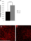
Antioxidant treatment with tempol caused a trend towards a reduction in superoxide accumulation in vascular smooth muscle cells from MCA wall observed as a reduction in mean fluorescence intensity after DHE staining in the MCA of tempol treated and untreated SHRSP (Tempol, n=10; Control, n=5) (A). Representative images of DHE staining in the MCA of untreated SHRSP (B) and tempol-treated SHRSP (C) (n=7 for both groups). The images were captured using a confocal microscope with a 40× objective.
Fig 2
Fig 2
Tempol treatment increased MCA reactivity to higher doses of 5-HT (A), but did not change endothelium-dependent vasodilation (B) or tone generation and 80 mmHg and 140 mmHg (C). *p<0.05, Tempol vs Control, ANOVA. Values are mean±SEM. The MCA was cannulated between two glass cannulas in a pressure arteriograph and kept in oxygenated warm PSS under no-flow conditions throughout the experiment (Tempol, n=10; Control, n=5).
Fig 2
Fig 2
Tempol treatment increased MCA reactivity to higher doses of 5-HT (A), but did not change endothelium-dependent vasodilation (B) or tone generation and 80 mmHg and 140 mmHg (C). *p<0.05, Tempol vs Control, ANOVA. Values are mean±SEM. The MCA was cannulated between two glass cannulas in a pressure arteriograph and kept in oxygenated warm PSS under no-flow conditions throughout the experiment (Tempol, n=10; Control, n=5).
Fig 3
Fig 3
Antioxidant treatment greatly improved MCA passive structure as observed by an increase in vessel outer and lumen diameter (A and B, respectively). *p<0.05, Tempol vs Control, ANOVA. Values are mean±SEM. The MCA was cannulated between two glass cannulas in a pressure arteriograph and kept in oxygenated warm calcium-free PSS under no-flow conditions throughout the experiment (Tempol, n=10; Control, n=5).
Fig 3
Fig 3
Antioxidant treatment greatly improved MCA passive structure as observed by an increase in vessel outer and lumen diameter (A and B, respectively). *p<0.05, Tempol vs Control, ANOVA. Values are mean±SEM. The MCA was cannulated between two glass cannulas in a pressure arteriograph and kept in oxygenated warm calcium-free PSS under no-flow conditions throughout the experiment (Tempol, n=10; Control, n=5).
Fig 4
Fig 4
Tempol treatment improved MCA passive structure as observed by an increase in vessel and lumen cross-sectional area (A and B, respectively). Vessel stress was increased in the MCA of tempol-treated SHRSP at intralumenal pressures over than 100 mmHg (C). *p<0.05, Tempol vs Control, ANOVA. Values are mean±SEM. The MCA was cannulated between two glass cannulas in a pressure arteriograph and kept in oxygenated warm calcium-free PSS under no-flow conditions throughout the experiment (Tempol, n=10; Control, n=5).
Fig 4
Fig 4
Tempol treatment improved MCA passive structure as observed by an increase in vessel and lumen cross-sectional area (A and B, respectively). Vessel stress was increased in the MCA of tempol-treated SHRSP at intralumenal pressures over than 100 mmHg (C). *p<0.05, Tempol vs Control, ANOVA. Values are mean±SEM. The MCA was cannulated between two glass cannulas in a pressure arteriograph and kept in oxygenated warm calcium-free PSS under no-flow conditions throughout the experiment (Tempol, n=10; Control, n=5).
Fig 5
Fig 5
Tempol treatment did not prevent the increase in the MCA wall thickness observed in the SHRSP, as seen by no changes in wall-to-lumen ratio (A), wall thickness (B) and wall cross-sectional area (C). MCA strain was not altered by tempol treatment (D). *p<0.05, Tempol vs Control, ANOVA. Values are mean±SEM. The MCA was cannulated between two glass cannulas in a pressure arteriograph and kept in oxygenated warm calcium-free PSS under no-flow conditions throughout the experiment (Tempol, n=10; Control, n=5).
Fig 5
Fig 5
Tempol treatment did not prevent the increase in the MCA wall thickness observed in the SHRSP, as seen by no changes in wall-to-lumen ratio (A), wall thickness (B) and wall cross-sectional area (C). MCA strain was not altered by tempol treatment (D). *p<0.05, Tempol vs Control, ANOVA. Values are mean±SEM. The MCA was cannulated between two glass cannulas in a pressure arteriograph and kept in oxygenated warm calcium-free PSS under no-flow conditions throughout the experiment (Tempol, n=10; Control, n=5).
Fig 6
Fig 6
Tempol treatment caused a slightly leftward shift of the MCA stress-strain curve, although not statiscally significant (A). Vessel stiffness was not changed by tempol treatment, as seen by no differences in the β-coefficient values (B). Values are mean±SEM. The MCA was cannulated between two glass cannulas in a pressure arteriograph and kept in oxygenated warm calcium-free PSS under no-flow conditions throughout the experiment (Tempol, n=10; Control, n=5).
Fig 6
Fig 6
Tempol treatment caused a slightly leftward shift of the MCA stress-strain curve, although not statiscally significant (A). Vessel stiffness was not changed by tempol treatment, as seen by no differences in the β-coefficient values (B). Values are mean±SEM. The MCA was cannulated between two glass cannulas in a pressure arteriograph and kept in oxygenated warm calcium-free PSS under no-flow conditions throughout the experiment (Tempol, n=10; Control, n=5).
Fig 7
Fig 7
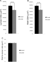
Tempol treatment increased the lumen (A) and wall cross-sectional area (B) of the carotid artery in SHRSP (n=7 for both groups). Surprisingly, the wall-to-lumen ratio was not altered by tempol treatment (C). Morphometry was performed with images captured at 4× objective and analyzed using AxioVision Rel 4.6 (Carl Zeiss Inc).
Fig 8
Fig 8
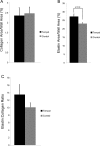
Tempol treatment did not change the area of total collagen in the vessel wall, as measured by percentage of wall area occupied by collagen (A) after Picrosirius red staining and analysis by polarized microscopy. Percent area occupied by elastin in the vessel wall was increased by tempol treatment in SHRSP (B). Elastin fibers were identified using Van Gieson's method for elastic fibers staining. Elastin:collagen ratio, an indicator of vascular compliance, was not altered by tempol treatment (C) (n=7 for both groups).

Similar articles

Cited by

References

    1. Alberts MJ, Ovbiagele B. Current strategies for ischemic stroke prevention: role of multimodal combination therapies. J Neurol. 2007;254(10):1414–26. - PubMed
    1. Baumbach GL, Ghoneim S. Vascular remodeling in hypertension. Scanning Microsc. 1993;7(1):137–42. discussion 143. - PubMed
    1. Baumbach GL, Hajdu MA. Mechanics and composition of cerebral arterioles in renal and spontaneously hypertensive rats. Hypertension. 1993;21(6 Pt 1):816–26. - PubMed
    1. Bonacasa B, Sanchez ML, Rodriguez F, Lopez B, Quesada T, Fenoy FJ, Hernandez I. 2-Methoxyestradiol attenuates hypertension and coronary vascular remodeling in spontaneously hypertensive rats. Maturitas. 2008;61(4):310–6. - PubMed
    1. Briones AM, Rodriguez-Criado N, Hernanz R, Garcia-Redondo AB, Rodrigues-Diez RR, Alonso MJ, Egido J, Ruiz-Ortega M, Salaices M. Atorvastatin prevents angiotensin II-induced vascular remodeling and oxidative stress. Hypertension. 2009;54(1):142–9. - PubMed

Publication types

MeSH terms