

Differential localization of vesicular glutamate transporters and peptides in corneal afferents to trigeminal nucleus caudalis
- PMID: 20593358
- PMCID: PMC2933108
- DOI: 10.1002/cne.22414
Differential localization of vesicular glutamate transporters and peptides in corneal afferents to trigeminal nucleus caudalis
Abstract
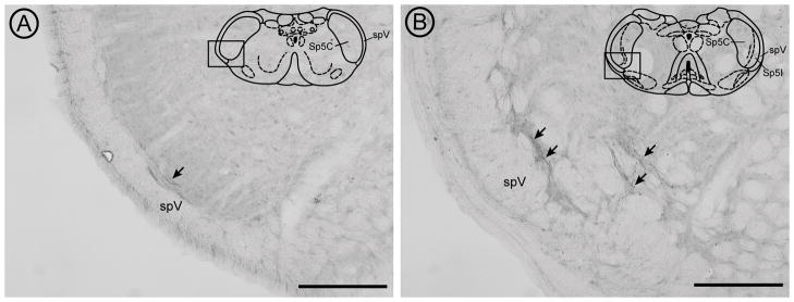
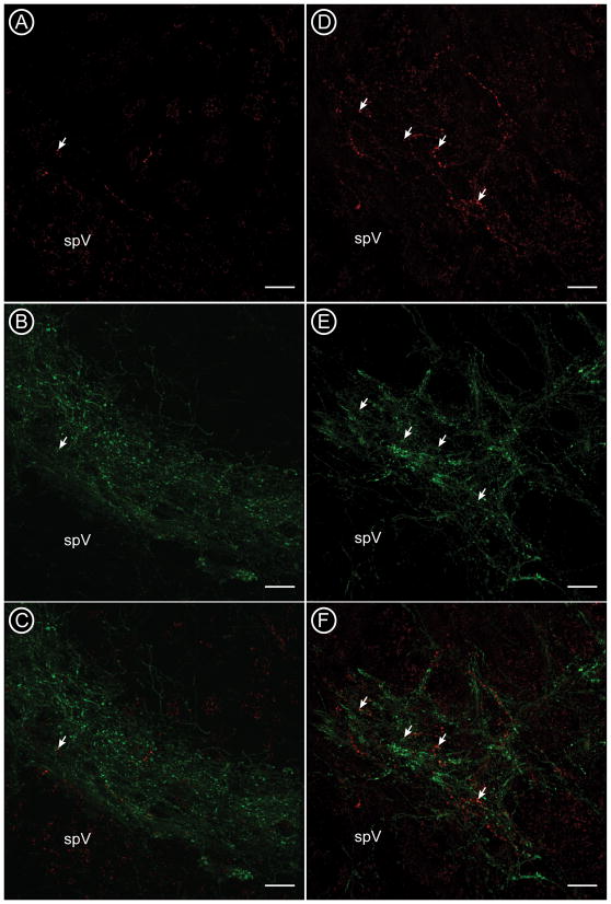
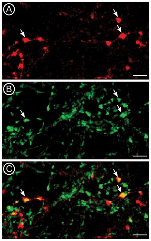
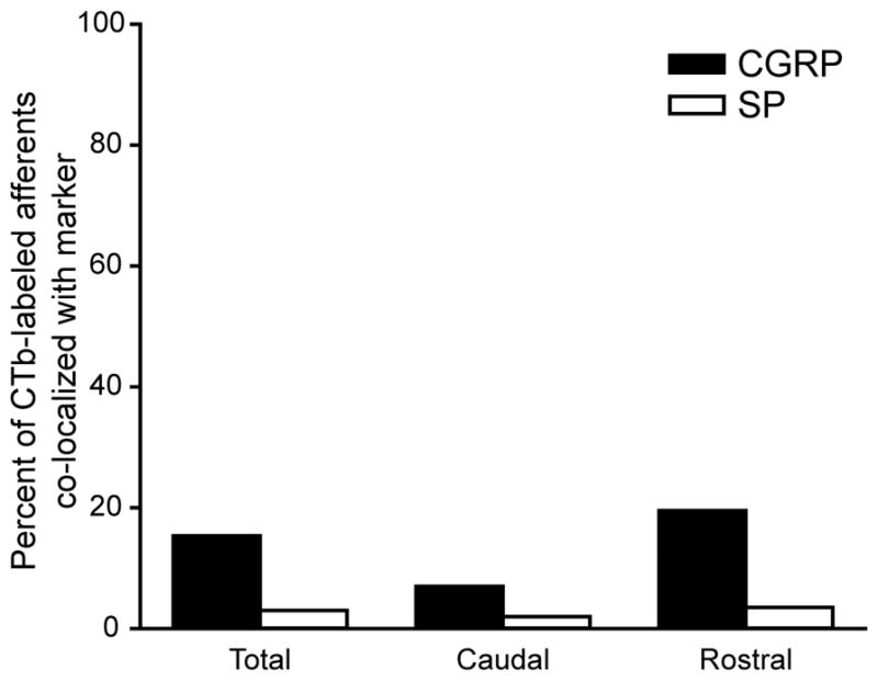
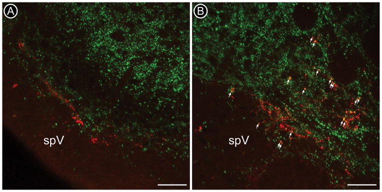
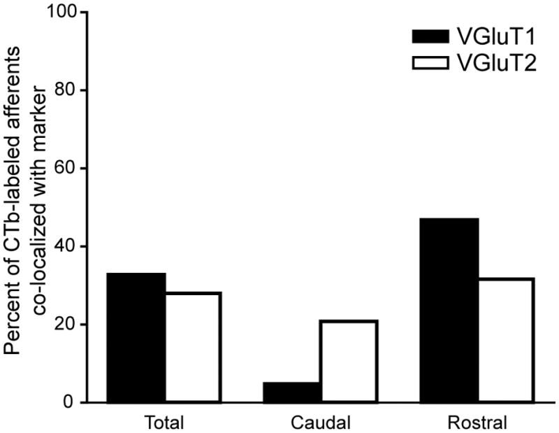
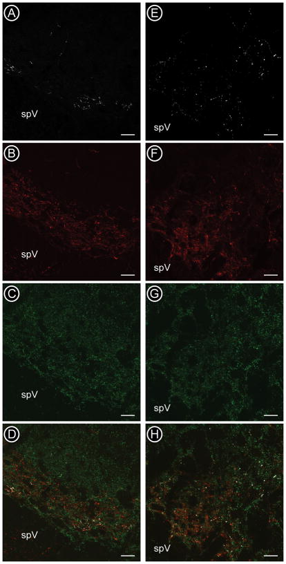
Trigeminal afferents convey nociceptive information from the corneal surface of the eye to the trigeminal subnucleus caudalis (Vc). Trigeminal afferents, like other nociceptors, are thought to use glutamate and neuropeptides as neurotransmitters. The current studies examined whether corneal afferents contain both neuropeptides and vesicular glutamate transporters. Corneal afferents to the Vc were identified by using cholera toxin B (CTb). Corneal afferents project in two clusters to the rostral and caudal borders of the Vc, regions that contain functionally distinct nociceptive neurons. Thus, corneal afferents projecting to these two regions were examined separately. Dual immunocytochemical studies combined CTb with either calcitonin gene-related peptide (CGRP), substance P (SP), vesicular glutamate transporter 1 (VGluT1), or VGluT2. Corneal afferents were more likely to contain CGRP than SP, and corneal afferents projecting to the rostral region were more likely to contain CGRP than afferents projecting caudally. Overall, corneal afferents were equally likely to contain VGluT1 or VGluT2. Together, 61% of corneal afferents contained either VGluT1 or VGluT2, suggesting that some afferents lack a VGluT. Caudal corneal afferents were more likely to contain VGluT2 than VGluT1, whereas rostral corneal afferents were more likely to contain VGluT1 than VGluT2. Triple-labeling studies combining CTb, CGRP, and VGluT2 showed that very few corneal afferents contain both CGRP and VGluT2, caudally (1%) and rostrally (2%). These results suggest that most corneal afferents contain a peptide or a VGluT, but rarely both. Our results are consistent with a growing literature suggesting that glutamatergic and peptidergic sensory afferents may be distinct populations.
Figures
Similar articles
-
Differential content of vesicular glutamate transporters in subsets of vagal afferents projecting to the nucleus tractus solitarii in the rat.J Comp Neurol. 2014 Feb 15;522(3):642-53. doi: 10.1002/cne.23438. J Comp Neurol. 2014. PMID: 23897509 Free PMC article.
-
VGLUT1 or VGLUT2 mRNA-positive neurons in spinal trigeminal nucleus provide collateral projections to both the thalamus and the parabrachial nucleus in rats.Mol Brain. 2018 Apr 12;11(1):22. doi: 10.1186/s13041-018-0362-y. Mol Brain. 2018. PMID: 29650024 Free PMC article.
-
The expression of vesicular glutamate transporters VGLUT1 and VGLUT2 in neurochemically defined axonal populations in the rat spinal cord with emphasis on the dorsal horn.Eur J Neurosci. 2003 Jan;17(1):13-27. doi: 10.1046/j.1460-9568.2003.02406.x. Eur J Neurosci. 2003. PMID: 12534965
-
Vesicular glutamate transporter isoforms: The essential players in the somatosensory systems.Prog Neurobiol. 2018 Dec;171:72-89. doi: 10.1016/j.pneurobio.2018.09.006. Epub 2018 Sep 28. Prog Neurobiol. 2018. PMID: 30273635 Review.
-
The role of corneal afferent neurons in regulating tears under normal and dry eye conditions.Exp Eye Res. 2013 Dec;117:79-87. doi: 10.1016/j.exer.2013.08.011. Epub 2013 Aug 28. Exp Eye Res. 2013. PMID: 23994439 Free PMC article. Review.
Cited by
-
VGLUTs in Peripheral Neurons and the Spinal Cord: Time for a Review.ISRN Neurol. 2013 Nov 20;2013:829753. doi: 10.1155/2013/829753. ISRN Neurol. 2013. PMID: 24349795 Free PMC article. Review.
-
Corneal pain activates a trigemino-parabrachial pathway in rats.Brain Res. 2014 Mar 6;1550:18-26. doi: 10.1016/j.brainres.2014.01.002. Epub 2014 Jan 10. Brain Res. 2014. PMID: 24418463 Free PMC article.
-
Diurnal changes in perineuronal nets and parvalbumin neurons in the rat medial prefrontal cortex.Brain Struct Funct. 2021 May;226(4):1135-1153. doi: 10.1007/s00429-021-02229-4. Epub 2021 Feb 14. Brain Struct Funct. 2021. PMID: 33585984 Free PMC article.
-
Non-peptidergic small diameter primary afferents expressing VGluT2 project to lamina I of mouse spinal dorsal horn.Mol Pain. 2011 Dec 8;7:95. doi: 10.1186/1744-8069-7-95. Mol Pain. 2011. PMID: 22152428 Free PMC article.
-
Differential content of vesicular glutamate transporters in subsets of vagal afferents projecting to the nucleus tractus solitarii in the rat.J Comp Neurol. 2014 Feb 15;522(3):642-53. doi: 10.1002/cne.23438. J Comp Neurol. 2014. PMID: 23897509 Free PMC article.
References
-
- Aicher SA, Mitchell JL, Swanson KC, Zadina JE. Endomorphin-2 axon terminals contact mu-opioid receptor-containing dendrites in trigeminal dorsal horn. Brain Res. 2003;977(2):190–8. - PubMed
-
- Belmonte C, Acosta MC, Gallar J. Neural basis of sensation in intact and injured corneas. Exp Eye Res. 2004;78(3):513–25. - PubMed
-
- Bereiter DA, Bereiter DF. N-methyl-D-aspartate and non-N-methyl-D-aspartate receptor antagonism reduces Fos-like immunoreactivity in central trigeminal neurons after corneal stimulation in the rat. Neuroscience. 1996;73(1):249–58. - PubMed
-
- Bereiter DA, Bereiter DF, Tonnessen BH, Maclean DB. Selective blockade of substance P or neurokinin A receptors reduces the expression of c-fos in trigeminal subnucleus caudalis after corneal stimulation in the rat. Neuroscience. 1998;83(2):525–34. - PubMed
-
- Bereiter DA, Hirata H, Hu JW. Trigeminal subnucleus caudalis: beyond homologies with the spinal dorsal horn. Pain. 2000;88(3):221–4. - PubMed
Publication types
MeSH terms
Substances
Grants and funding
LinkOut - more resources
Full Text Sources
Research Materials
Miscellaneous
