. 2010 Sep 3;285(36):28298-308.
doi: 10.1074/jbc.M110.101717. Epub 2010 Jun 28.

A small ubiquitin-related modifier-interacting motif functions as the transcriptional activation domain of Krüppel-like factor 4

Affiliations

A small ubiquitin-related modifier-interacting motif functions as the transcriptional activation domain of Krüppel-like factor 4

James X Du et al. J Biol Chem. .

Abstract

The zinc finger transcription factor, Krüppel-like factor 4 (KLF4), regulates numerous biological processes, including proliferation, differentiation, and embryonic stem cell self-renewal. Although the DNA sequence to which KLF4 binds is established, the mechanism by which KLF4 controls transcription is not well defined. Small ubiquitin-related modifier (SUMO) is an important regulator of transcription. Here we show that KLF4 is both SUMOylated at a single lysine residue and physically interacts with SUMO-1 in a region that matches an acidic and hydrophobic residue-rich SUMO-interacting motif (SIM) consensus. The SIM in KLF4 is required for transactivation of target promoters in a SUMO-1-dependent manner. Mutation of either the acidic or hydrophobic residues in the SIM significantly impairs the ability of KLF4 to interact with SUMO-1, activate transcription, and inhibit cell proliferation. Our study provides direct evidence that SIM in KLF4 functions as a transcriptional activation domain. A survey of transcription factor sequences reveals that established transactivation domains of many transcription factors contain sequences highly related to SIM. These results, therefore, illustrate a novel mechanism by which SUMO interaction modulates the activity of transcription factors.


Figures

FIGURE 1.
FIGURE 1.
The mouse KLF4 is SUMOylated at lysine residue 275. A, conservation across species of the SM (underlines) in KLF4 is shown. The SM is located between amino acid residues 274 and 277 of the mouse KLF4. The conserved SUMO target lysine residues in the SM are italicized. B, SUMOylation of KLF4 by GFP-tagged or His-tagged SUMO-1 is shown. pCMV-Myc-KLF4 was co-transfected into COS-1 cells with GFP-SUMO-1 (lanes 1, 4, and 7), His-SUMO-1 (lanes 2, 5, and 8), or vector alone (lanes 3, 6, and 9). Lysates were immunoprecipitated with a rabbit Myc antibody followed by Western blotting with mouse Myc (lanes 1–3), GFP (lanes 4–6), or His (lanes 7–9) antibody. Asterisks indicate GFP-SUMO-1-conjugated Myc-KLF4, and arrowheads indicate His-SUMO-1-conjugated Myc-KLF4. The positions of the molecular mass markers in kDa are shown to the left of the blots. IB, immunoblot. C, identification of the SUMOylation site within KLF4 is shown. COS-1 cells were co-transfected with GFP-SUMO-1 and one of the following constructs: pCMV-Myc-KLF4 (WT), pCMV-Myc-KLF4-K273R (K273R), pCMV-Myc-KLF4-K275R (K275R), or pCMV-Myc-KLF4-E277G (E277G). Lysates were immunoprecipitated with a rabbit Myc antibody followed by Western blotting with a mouse Myc (lanes 1–4) or GFP (lanes 5–8) antibody. Arrow, GFP-SUMO-1-conjugated Myc-KLF4 or Myc-KLF4-K273R. D, SUMOylation of untagged KLF4. COS-1 cells, which express negligible amount of endogenous KLF4, were co-transfected with GFP-SUMO-1 and either pMT3-KLF4 (WT) or pMT3-KLF4-K275R (K275R). Lysates were immunoprecipitated with a rabbit KLF4 antibody and blotted with a mouse GFP antibody. SUMOylated KLF4 is indicated by the asterisk on the top panel. The bottom panel shows levels of KLF4 or KLF4-K275R in the immune complexes as probed by a goat KLF4 antibody.
FIGURE 2.
FIGURE 2.
SUMOylation of KLF4 does not reduce its protein levels. pCMV-Script (Vector), pCMV-Myc-KLF4 (WT), pCMV-Myc-KLF4-K273R (K273R), pCMV-Myc-KLF4-K275R (K275R), or pCMV-Myc-KLF4-E277G (E277G) were co-transfected into COS-1 cells with either vector alone (A), His-SUMO-1 (B), or PIAS1 (C). Lysates were immunoblotted with mouse Myc (upper panel) or β-actin (lower panel) antibody. D and E, pCMV-Myc-KLF4 (WT) or pCMV-Myc-KLF4-K275R (K275R) was transfected into HEK293T cells and subjected to cycloheximide chase assay for up to 3 h. D, shown is an immunoblot with Myc (upper panel) and β-actin (lower panel) antibody. E, corresponding quantitative chemiluminescence measurements after normalization with actin control are shown. The relative protein levels at 0 h were set as 1, and the average protein levels and S.D. (n = 3) at the other time points relative to those at 0 h are shown.
FIGURE 3.
FIGURE 3.
KLF4 physically interacts with SUMO-1 through a SIM. A, conservation across species of the putative SIM in KLF4 is shown. The acidic stretch is shown in bold and italics, and the hydrophobic core is shown in bold and underlines. B, HEK293T cells were transfected with vector only, pCMV-Myc-KLF4, or pCMV-Myc-KLF4(1–100), a truncated mutant containing only the amino-terminal 100 residues. The corresponding lysates were either incubated with purified recombinant GST-SUMO-1 (lanes 3–5) or not (lanes 1 and 2). The GST pulldown assay was conducted followed by Western blotting with mouse Myc (upper and middle panels) and SUMO-1 (lower panel) antibodies. C, lysates from HEK293T cells transfected with pCMV-Myc-KLF4 (WT) (lane 2) or pCMV-Myc-KLF4-L101A/I106A (LI) (lane 3) were incubated with purified GST-SUMO-1. An equal portion of WT lysate not incubated with GST-SUMO-1 (lane 1) served as a control. GST pulldown assay was followed by Western blotting with mouse anti-Myc and SUMO-1. D, lysates from Klf4-null (Klf4−/−) mouse embryonic fibroblasts (39) co-transfected with Myc-SUMO-1ΔGG and pMT3 (-), pMT3-KLF4 (WT), pMT3-KLF4-E93V/E95V/E96V (EEE), or pMT3-KLF4-D99V/D102V/D104V (DDD) were immunoprecipitated (IP) with a rabbit KLF4 antibody followed by Western blotting with mouse Myc or rabbit KLF4 antibody. E, GFP or GFP linked to full-length KLF4 SIM (GFP-SIM) or the carboxyl terminus of SIM (GFP-SIMΔ) (upper panel) were transfected into HEK293T cells, and lysates were incubated with purified GST-SUMO-1 followed by a GST pulldown assay and Western blotting with mouse GFP or SUMO-1 antibody.
FIGURE 4.
FIGURE 4.
The KLF4 SIM is a transcriptional activation domain. A, localization of the KLF4 transcriptional activation domain is shown. Left, shown is a schematic of GAL4-DBD fused to full-length KLF4 (GAL4-DBD-KLF4), the KLF4 carboxyl terminus (residues 350–483) that contains the zinc finger DNA binding domain (GAL4-DBD-ZF), KLF4 amino-terminal 349 residues (GAL4-DBD-1–349), the KLF4 first 158 residues (GAL4-DBD-1–158), and KLF4 SIM (residues 92–110) (GAL4-DBD-SIM). Right, the GAL4-DBD constructs indicated on the left were transformed into AH109 yeast strain, and their ability to transactivate both Ade and His reporters was detected by growth in synthetic dropout medium lacking adenine and histidine (ADE HIS). Vec, vector (GAL4-DBD) alone. ZF, zinc finger. B, shown is localization of the transactivation domain of KLF4 to it SIM. Left, a schematic of wild type or mutated SIMs fused to GAL4-DBD is shown. The alanine residues used to substitute for the acidic or hydrophobic residues are underlined. Right, the GAL4-DBD-SIM constructs indicated on the left were transformed into AH109 yeast strain, and their ability to transactivate the single reporter Ade or both Ade and His reporters was detected by growth in synthetic dropout medium lacking either only adenine (ADE) or both adenine and histidine (ADE HIS). C, shown is quantification of the transcriptional activation activity of KLF4 SIM or SIM mutants fused to the GAL4-DBD by α-galactosidase assay using MEL1 as a reporter. Left, a representative experiment in triplicate is shown. The intensity of yellow color reflects the strength of transactivation. The residual yellowish color from vector alone was due to endogenous α-galactosidase in yeast host and served as a blank control. Right, quantitative results from the α-galactosidase assay are shown on the left. All GAL4-DBD-SIM fusion proteins were expressed at similar levels, as judged by immunoblotting against the pre-existing Myc tag (bottom right). ***, p < 0.001 compared with wild type SIM by two-tailed t test.
FIGURE 5.
FIGURE 5.
The KLF4 SIM is crucial for transcriptional activation in mammalian cells. Dual luciferase reporter assays were performed with C/EBPβ or Lefty1-luciferase reporter plasmids and expression constructs of KLF4 or its mutants in HEK293T cells as described under “Experimental Procedures”. A and B, shown is co-transfection of vector (Vec) alone, pCMV-Myc-KLF4 (WT), or pCMV-Myc-L101A/I106A (LI) with C/EBPβ-luciferase (A) or Lefty1-luciferase (B). The expression levels of Myc-KLF4 (WT) and Myc-KLF4-L101A/I106A (LI) were shown by Western blotting against Myc and β-actin. C and D, shown is co-transfection of vector (Vec) alone, pMT3-KLF4 (WT), pMT3-KLF4-E93V/E95V/E96V (EEE), or pMT3-D99V/D102V/D104V (DDD) with C/EBPβ-luciferase (C) and Lefty1-luciferase (D). The expression levels of both the EEE and DDD mutants have previously been documented (34). Shown are the means and S.D. of four independent experiments. *, p < 0.05; **, p < 0.01; ***, p < 0.001; by two-tailed t test. Asterisks not associated with brackets are comparisons to vector alone. RLU is relative luciferase unit.
FIGURE 6.
FIGURE 6.
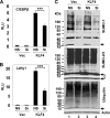
Reduction of SUMO-1 inhibits KLF4 transcriptional activity. HEK293T cells were transfected with the C/EBPβ-luciferase (A) or Lefty1-luciferase (B) reporter, vector (Vec) or pCMV-Myc-KLF4 (KLF4), nonspecific siRNA (NS) or the dual siRNA mixture against SUMO-1 (Si), and the control renilla luciferase plasmid. Dual luciferase assays were performed, and the normalized luciferase activity presented as relative luciferase units (RLU). Shown are the means and S.D. of three independent experiments. ***, p < 0.001 by two-tailed t test. C, a fraction of lysates from the corresponding cells was subjected to Western blotting with mouse antibodies against SUMO-1 (upper panel), SUMO-2/3 (middle panel), and ubiquitin (lower panel). β-actin was used as a loading control. The arrows indicate free SUMO-1 (upper panel), SUMO-2/3 (middle panel), and ubiquitin (lower panel). A short exposure of free SUMO-2/3 in the middle panel is also provided.
FIGURE 7.
FIGURE 7.
KLF4 SIM is crucial for its anti-proliferative activity. A, COS-1 cells were transfected with vector alone (Vec), pCMV-Myc-KLF4 (WT), or pCMV-Myc-KLF4-L101A/I106A (LI), labeled with BrdU, and stained with mouse BrdU and rabbit Myc antibodies. Hoechst dye was used to reveal the nuclei. B, cells were transfected with pMT3 (Vec), pMT3-KLF4 (WT), pMT3-KLF4-E93V/E95V/E96V (EEE), or pMT3-KLF4-D99V/D102V/D104V (DDD), labeled with BrdU, and stained with mouse BrdU and rabbit KLF4 antibodies. Shown are several representative cells in each panel. Between 100 and 300 cells were observed for each construct, and the percentages of KLF4-positive cells (green) that were also positive for BrdU (yellow) are shown in the charts. *, p < 0.05 and ***, p < 0.001; by two-tailed t test. Asterisks not associated with brackets are comparisons to vector alone.

Similar articles

Cited by

References

    1. Triezenberg S. J. (1995) Curr. Opin. Genet. Dev. 5, 190–196 - PubMed
    1. Xiao H., Friesen J. D., Lis J. T. (1994) Mol. Cell. Biol. 14, 7507–7516 - PMC - PubMed
    1. Chang J., Kim D. H., Lee S. W., Choi K. Y., Sung Y. C. (1995) J. Biol. Chem. 270, 25014–25019 - PubMed
    1. Seipel K., Georgiev O., Schaffner W. (1994) Biol. Chem. Hoppe Seyler 375, 463–470 - PubMed
    1. Sekiyama N., Ikegami T., Yamane T., Ikeguchi M., Uchimura Y., Baba D., Ariyoshi M., Tochio H., Saitoh H., Shirakawa M. (2008) J. Biol. Chem. 283, 35966–35975 - PubMed

Publication types

MeSH terms

LinkOut - more resources