. 2010 Sep;11(9):1180-90.
doi: 10.1111/j.1600-0854.2010.01092.x. Epub 2010 Jun 21.

Spatial regulation of Golgi phosphatidylinositol-4-phosphate is required for enzyme localization and glycosylation fidelity

Affiliations

Spatial regulation of Golgi phosphatidylinositol-4-phosphate is required for enzyme localization and glycosylation fidelity

Fei Ying Cheong et al. Traffic. 2010 Sep.

Abstract

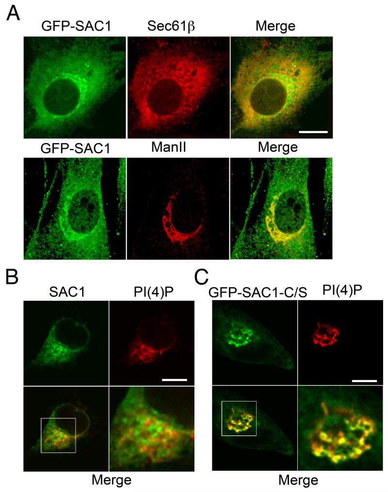
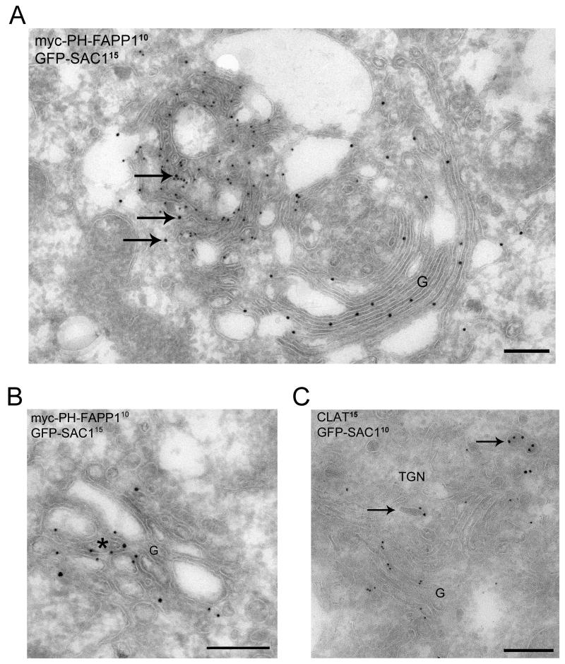
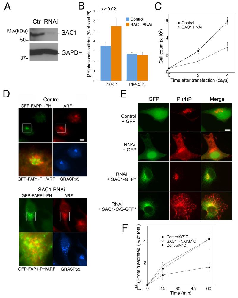
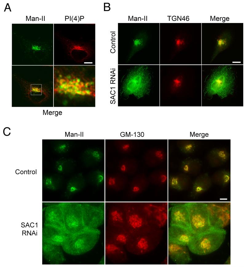
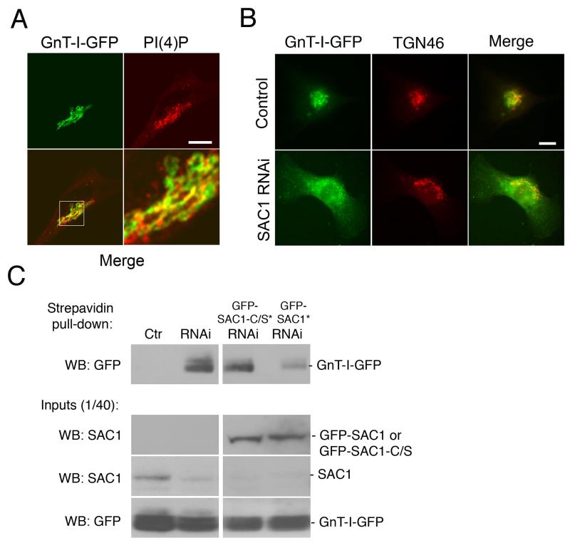
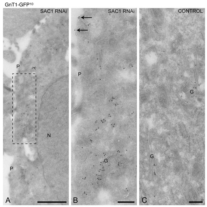
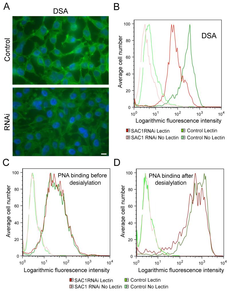
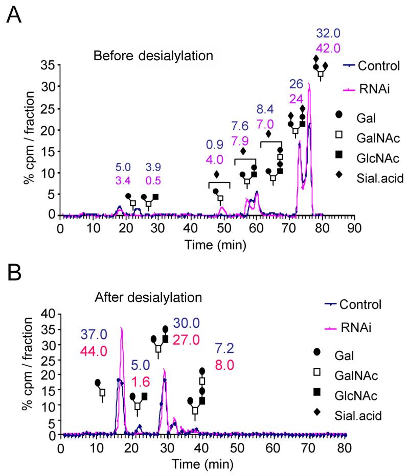
The enrichment of phosphatidylinositol-4-phosphate (PI(4)P) at the trans Golgi network (TGN) is instrumental for proper protein and lipid sorting, yet how the restricted distribution of PI(4)P is achieved remains unknown. Here, we show that lipid phosphatase Suppressor of actin mutations 1 (SAC1) is crucial for the spatial regulation of Golgi PI(4)P. Ultrastructural analysis revealed that SAC1 is predominantly located at cisternal Golgi membranes but is absent from the TGN, thus confining PI(4)P to the TGN. RNAi-mediated knockdown of SAC1 caused changes in Golgi morphology and mislocalization of Golgi enzymes. Enzymes involved in glycan processing such as mannosidase-II (Man-II) and N-acetylglucosamine transferase-I (GnT-I) redistributed to aberrant intracellular structures and to the cell surface in SAC1 knockdown cells. SAC1 depletion also induced a unique pattern of Golgi-specific defects in N-and O-linked glycosylation. These results indicate that SAC1 organizes PI(4)P distribution between the Golgi complex and the TGN, which is instrumental for resident enzyme partitioning and Golgi morphology.


Figures

Figure 1
Figure 1. Localization of SAC1 in proliferating cells
(A) NIH3T3 cells expressing GFP-SAC1 (green) were costained with antibodies against Sec61β (red) or Man-II (red) and examined by confocal immunofluorescence microscopy. (B,C) NIH3T3 were stained with antibodies against SAC1 (green) (B), or transfected with a construct for expressing GFP-SAC1-C/S (green) (C), costained with antibodies against PI(4)P (red) and examined by confocal immunofluorescence microscopy. The boxed area is enlarged in the adjacent panel. Scale bars, 15 μm.
Figure 2
Figure 2. SAC1 localizes to the Golgi complex but is absent from TGN membranes
(A) Hela cells expressing GFP-SAC1 and the PI(4)P probe myc-PH-FAPP1 were double labeled for the myc-epitope (10 nm gold particles) and GFP (15 nm gold particles). The GFP-SAC1 in myc-PH-FAPP1-positive regions is marked with arrows. (B) Limited overlap of myc-PH-FAPP1 (10 nm protein A-gold) and GFP-SAC1 (15 nm protein A-gold) at a continuous membrane segment, indicated by an asterisk. (C) Hela cells expressing GFP-SAC1 were double-labeled for clathrin (15 nm gold particles) and GFP (10 nm gold particles). Labeling patterns were studied in moderately expressing HeLa cells. GFP-SAC1 label was readily detected throughout the Golgi complex, but virtually absent from the TGN membranes. The position of the TGN relative to the Golgi complex is marked by the presence of clathrin (arrows). G = Golgi complex, Scale bars, 200 nm
Figure 3
Figure 3. SAC1 depletion causes changes in PI(4)P distribution
(A, B) Hela cells were transfected with siRNAs directed against SAC1 or control RNAs. 72 h after transfection, cells were harvested and analyzed by immunoblotting using antibodies against SAC1 (A), or labeled with [3H]myo-inositol for 48 h and analyzed for phosphoinositide contents (B). The depicted data are means +/− SD, n=4 (SAC1 knockdown efficiencies in the RNAi samples > 90%). (C) Hela cells were transfected with siRNAs directed against SAC1 or control RNAs. Equal numbers of cells were seeded on plates and cell proliferation was examined at the times indicated. The depicted data are means +/− SD from triplicates derived in three independent sets of experiments. (D) Hela cells were transfected with siRNAs directed against SAC1 or control RNAs. Cells were cotransfected with a vector for expressing GFP-PH-FAPP1 (green). 72 h after transfection cells were stained with antibodies against ARF (red) and GRASP65 (blue) and analyzed by immunofluorescence microscopy. The boxed areas are enlarged in the adjacent panels. Scale bars, 10 μm. (E) Hela cells were transfected with siRNAs directed against SAC1 or control RNAs. Where indicated, cells were either cotransfected with a vector for expressing GFP (green) or constructs for expressing siRNA-resistant versions of GFP-SAC1 (green) or GFP-SAC1-C/S (green). 72 h after transfection cells were stained with antibodies against PI(4)P (red) and analyzed by immunofluorescence microscopy. Transfected cells were identified by GFP fluorescence. Scale bars, 10 μm. (F) Hela cells were transfected with siRNAs directed against SAC1 or control RNAs. 72 h after transfection, cells were pulse-labeled with [35S]Met/Cys, incubated at 18°C for 3 h and then shifted to 37°C. At the indicated times, proteins in the culture supernatants and the cell lysates were precipitated, collected on filters and quantified by scintillation counting. As an additional control, the secretion assay was performed at 4°C. The depicted data are means +/− SD from triplicates derived in three independent sets of experiments.
Figure 4
Figure 4. Mislocalization of Man-II in SAC1 knockdown cells
(A) HeLa cells were stained with antibodies against Man-II (green), costained with antibodies against PI(4)P (red) and examined by confocal immunofluorescence microscopy. The boxed area is enlarged in the adjacent panel. (B) Hela cells were transfected with siRNAs against SAC1 or control RNAs. 72 h after transfection, cells were stained with antibodies against Man-II (green), costained with antibodies against TGN46 (red) and analyzed by immunofluorescence microscopy. (C) Hela cells were transfected with siRNAs directed against SAC1 or control RNAs containing three point mutations. 72 h after transfection, cells were costained with antibodies against Man-II (green) and GM130 (red) and analyzed by immunofluorescence microscopy. Scale bars, 10 μm.
Figure 5
Figure 5. SAC1 knockdown triggers mislocalization of medial Golgi marker GnT-I-GFP
(A) HeLa cells were transfected with a plasmid for expressing GnT-I-GFP (green), costained with antibodies against PI(4)P (red) and examined by confocal immunofluorescence microscopy. The boxed area is enlarged in the adjacent panel. Scale bars, 10 μm. (B) Hela cells were cotransfected with a plasmid for expressing GnT-I-GFP (green) and siRNAs against SAC1 or control RNAs. 72 h after transfection, cells were costained with antibodies against TGN46 (red) and analyzed by immunofluorescence microscopy. Scale bars, 10 μm. (C) Biotinylation of cell surface proteins. Hela cells were transfected with plasmids for expressing GnT-I-GFP and siRNAs against SAC1 or control RNAs. Where indicated cells were cotransfected with plasmids for expressing siRNA-resistant versions of GFP-SAC1 (green) or GFP-SAC1-C/S (green). 72 h after transfection, cell surface proteins were biotinylated, isolated from detergent extracts by streptavidin agarose and separated by SDS-PAGE. Biotinylated GnT-I-GFP was identified by immunoblotting using specific polyclonal antibodies against GFP.
Figure 6
Figure 6. SAC1 depletion causes dispersion of GnT-I-GFP
Hela cells expressing GnT-I-GFP were transduced with siRNAs against SAC1 (A,B) or control RNAs (C) after which ultrathin cryosections were prepared that were immunogold-labeled for GFP to localize GnT-I-GFP (10 nm gold particles). (A,B) The boxed area in A is enlarged in B. SAC1 depletion caused a redistribution of GnT-I-GFP to numerous small-sized vesicles that were dispersed throughout the cytoplasm. In addition, some labeling was found at the plasma membrane (arrows in B). (C) In control cells GnT-I-GFP shows a restricted localization to the Golgi complex. G = Golgi complex, N = nucleus, P = plasma membrane Bars (A), 1 m; (B,C), 200 nm
Figure 7
Figure 7. SAC1 knockdown cells display altered N-linked glycan processing
(A) Control and SAC1 knockdown cells were fixed and incubated with biotinylated DSA, stained with FITC-Avidin and examined for cell surface staining. Scale bar, 15 μm. (B-D). FACS analysis of cell surface binding of lectins. Cells were incubated with or without the biotinylated lectins followed by incubation with FITC conjugated avidin. Green profiles indicate control cells, (light green, without lectin; dark green, with lectin). Red profiles indicate SAC1 knockdown cells, (light red, without lectin; dark red, with lectin). Depicted are the results for DSA-binding (B) and PNA-binding before (C) or after desialylation. Lectin specificities: DSA, linear, unbranched poly-N-acetyl-lactosamine [Galβ1-4GlcNAcβ1-3]n repeats; PNA, Core 1 O-glycan,Galβ1-3GalNAcα.
Figure 8
Figure 8. SAC1 knockdown cells show changes in O-linked glycans
(A, B) Control and SAC1 knockdown cells were labeled with 3H-galactose in the presence of GAP. 3H-GAP products were then purified from the medium and analyzed using HPLC before (A) or after desialylation (B). The numbers indicate the percentage of total labeled glycans.

Similar articles

Cited by

References

    1. Wang YJ, Wang J, Sun HQ, Martinez M, Sun YX, Macia E, Kirchhausen T, Albanesi JP, Roth MG, Yin HL. Phosphatidylinositol 4 phosphate regulates targeting of clathrin adaptor AP-1 complexes to the Golgi. Cell. 2003;114:299–310. - PubMed
    1. Hanada K, Kumagai K, Yasuda S, Miura Y, Kawano M, Fukasawa M, Nishijima M. Molecular machinery for non-vesicular trafficking of ceramide. Nature. 2003;426:803–809. - PubMed
    1. D’Angelo G, Polishchuk E, Di Tullio G, Santoro M, Di Campli A, Godi A, West G, Bielawski J, Chuang CC, van der Spoel AC, Platt FM, Hannun YA, Polishchuk R, Mattjus P, De Matteis MA. Glycosphingolipid synthesis requires FAPP2 transfer of glucosylceramide. Nature. 2007;449:62–67. - PubMed
    1. Halter D, Neumann S, van Dijk SM, Wolthoorn J, de Maziere AM, Vieira OV, Mattjus P, Klumperman J, van Meer G, Sprong H. Pre- and post-Golgi translocation of glucosylceramide in glycosphingolipid synthesis. J Cell Biol. 2007;179:101–115. - PMC - PubMed
    1. Wang J, Sun HQ, Macia E, Kirchhausen T, Watson H, Bonifacino JS, Yin HL. PI4P promotes the recruitment of the GGA adaptor proteins to the trans-Golgi network and regulates their recognition of the ubiquitin sorting signal. Mol Biol Cell. 2007;18:2646–2655. - PMC - PubMed

Substances