. 2010 Sep;45(1):75-83.
doi: 10.1016/j.mcn.2010.06.001. Epub 2010 Jun 13.

SIDL interacts with the dendritic targeting motif of Shal (K(v)4) K+ channels in Drosophila

Affiliations

SIDL interacts with the dendritic targeting motif of Shal (K(v)4) K+ channels in Drosophila

Fengqiu Diao et al. Mol Cell Neurosci. 2010 Sep.

Abstract

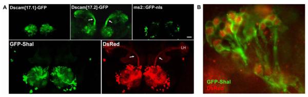
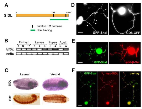
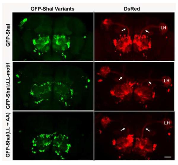
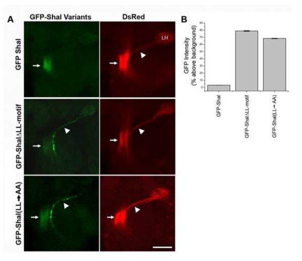
Shal K(+) (K(v)4) channels in mammalian neurons have been shown to be localized exclusively to somato-dendritic regions of neurons, where they function as key determinants of dendritic excitability. To gain insight into the mechanisms underlying dendritic localization of K(v)4 channels, we use Drosophila melanogaster as our model system. We show that Shal K(+) channels display a conserved somato-dendritic localization in vivo in Drosophila. From a yeast-2-hybrid screen, we identify the novel interactor, SIDL (for Shal Interactor of Di-Leucine Motif), as the first target protein reported to bind the highly conserved di-leucine motif (LL-motif) implicated in dendritic targeting. We show that SIDL is expressed primarily in the nervous system, co-localizes with GFP-Shal channels in neurons, and interacts specifically with the LL-motif of Drosophila and mouse Shal channels. We disrupt the Shal-SIDL interaction by mutating the LL-motif on Shal channels, and show that Shal K(+) channels are then mislocalized to some, but not all, axons in vivo. These results suggest that there are multiple mechanisms underlying Shal K(+) channel targeting, one of which depends on the LL-motif. The identification of SIDL may provide the first step for future investigation into the molecular machinery regulating the LL-motif-dependent targeting of K(+) channels.


Figures

Figure 1
Figure 1. Somato-Dendritic Localization of GFP-Shal is Conserved in Drosophila and Dependent on the Conserved LL-motif
(A) Top panels, GAL4-GH146 was used to drive expression of three UAS-transgenes shown previously to localize to distinct subcellular domains. Adult brains were dissected, fixed, and subjected to confocal microscopy to examine GFP localization in PNs. As expected, Dscam[17.1]-GFP (top left) is restricted to the dendrite rich interior of the antennal lobe, while Dscam[17.2]-GFP (top middle) is strongly expressed in axons present in the inner antennocerebral tract (iACT, see arrows) and dendrites. GFP fused to the nuclear localization signal (ms2::GFP-nls, top right) indicates the location of PN cell body nuclei present in clusters around the periphery of the antennal lobe. Bottom panels, GAL4-GH146 was used to drive expression of UAS-GFP-Shal (left) and UAS-DsRed (right). GFP-Shal signal was detected in PN cell bodies and dendrites, but not in axons. DsRed expressed in the same brain is shown (right) with arrows indicating the axonal iACT projecting to the lateral horn (LH). Shown are representative images, composed of collapsed confocal projections in order to display labeling in all cellular compartments. Note that collapsed images reveal more background, especially evident in the mushroom bodies. Scale bar represent 50 μm. (B) Under higher magnification (630X) it is evident that GFP-Shal channels are present in cell bodies as well as in dendritic tracts of the AL. These results suggest that somato-dendritic localization of GFP-Shal is conserved in Drosophila.
Figure 2
Figure 2. SIDL Interacts Specifically with the Highly Conserved Di-Leucine Motif of Shal Channels
(A) Shown are direct Y2H assay results testing interaction of the C-terminal 382 residues of SIDL (SIDLC) with full-length and deletion constructs of the C-terminal tails of Shal, Kv4.2, Shaker, Shaw, and Shab channel subunits (amino acid numbers indicated). Interaction strength, determined by reporter gene expression, is indicated by number of “+” symbols; “−” indicates no reporter gene expression. Note that the 16-residue dileucine motif (red) of Shal channels is required and sufficient for binding SIDLC. (B-C) Shown are immunoblots from GST-pull down assays using GST-fusion proteins indicated above each lane, incubated with purified myc-SIDLC protein (B), or embryonic membrane extracts (C). (B) Significant myc-SIDLC protein was pulled-down by GST-Shal1C and GST-Shal2C, but not by GST alone or GST-Shal2CΔ9. Immunoblots were probed with anti-GST (B) or Ponceau S (C) to show the presence of GST proteins in each assay. (C) Note that GST-SIDLC pulled-down endogenous Shal protein (Shal(bound)) from embryonic membrane extracts, even with far less GST-SIDLC protein present than GST alone. Immunoblot of unbound fractions is also shown using anti-syntaxin (syx(unbound)) as a control for the presence of membrane extract proteins
Figure 3
Figure 3. SIDL is Primarily Expressed in the Nervous System and Co-Localizes with GFP-Shal in Dendrites
(A) SIDL protein illustration showing Shal binding region identified in Y2H (765-1146 amino acids; green) and the two putative transmembrane domains predicted at residues 787-807 and 1079-1099 (see text). (B) RT-PCR for SIDL and actin in RNA isolated from Drosophila at different developmental stages: embryo (hours AEL at 25°C indicated), larvae (instar stage indicated), pupae (days aged indicated), and adult (heads (H) or bodies (B)). (C) RNA in situ hybridization for SIDL in representative 12-hour embryos; immunostaining for neuron-specific elav protein is shown for reference. SIDL appears to be expressed primarily in the nervous system. (D) Small clusters and single neurons with processes shown for primary cultures grown from transgenic lines expressing GFP-Shal (left) or CD8-GFP (right). Note that GFP-Shal is present in puncta along neuronal processes (see arrowheads for examples), while CD8-GFP is expressed throughout processes. (E) Shown is a single representative neuron from primary cultures made from a transgenic line expressing GFP-Shal (green) and nod-β-Gal (red). GFP-Shal positive processes all show immunostaining for the dendritic marker, nod-β-Gal. (F) A single representative neuron in primary culture from a transgenic strain expressing GFP-Shal and myc-SIDL in neurons. Immunostaining shows that GFP-Shal (left-green) and myc-SIDL (middle-red) co-localize (right-yellow) in cell bodies and in puncta along dendritic processes. Scale bars represent 5 μm for (D,E) and 10 μm for (F).
Figure 4
Figure 4. GFP-ShalΔLL-motif and GFP-Shal(LL→AA) Channels are Not Present in iACT Axons
GAL4-GH146 was used to drive expression of UAS-DsRed and either UAS-GFP-Shal, UAS-GFP-ShalΔLL-motif, or UAS-GFP-Shal(LL→AA) in vivo. DsRed expression (right column) shows diffuse localization in PN cell bodies in clusters around the antennal lobe, in dendrites in the antennal lobe, and axons projecting in the iACT (arrows) to the lateral horn (LH). All GFP-Shal variants (left column) appear restricted to cell bodies and dendrites, and absent from the iACT. Images are representative of a total of 29 brains for GFP-Shal, 11 brains for GFP-ShalΔLL-motif, and 8 brains for GFP-Shal(LL→AA). Images shown are collapsed Z-series confocal projections to display PN cell bodies, dendrites, and iACT axons together. Note the more punctate signal evident for GFP-ShalΔLL-motif and GFP-Shal(LL→AA) channels, compared with GFP-Shal. Scale bars represent 50um.
Figure 5
Figure 5. GFP-ShalΔLL-motif and GFP-Shal(LL→AA) Channels are Mis-Targeted to mACT Axons
(A) GAL4-GH146 was used to drive expression of UAS-DsRed (right column) and either UAS-GFP-Shal, UAS-GFP-ShalΔLL-motif, or UAS-GFP-Shal(LL→AA) (left column) in vivo. Shown are single plane confocal images focused on mACT axons (arrowheads) which project to the lateral horn (LH). The proximal segment of axons in the iACT can also be seen in this focal plane (see arrows). GFP-Shal is observed in the proximal segment of the iACT, but not in the mACT. GFP-ShalΔLL-motif and GFP-Shal(LL→AA) are found in both the proximal segment of iACT axons (arrows) and the axons of the mACT (arrowheads); images are representative of 11 and 8 brains examined for GFP-ShalΔLL-motif and GFP-Shal(LL→AA), respectively. (B) Quantification of GFP signal intensity along the mACT, expressed as the percentage over background fluorescence. There is significantly more GFP-ShalΔLL-motif (78.93% ± 0.54, N=20 antennal lobes) and GFP-Shal(LL→AA) (68.44% ± 0.35, N=15 antennal lobes) localized to the mACT when compared to GFP-Shal (3.16 ± 0.05, N= 10 antennal lobes).
Figure 6
Figure 6. GFP-ShalΔLL-motif Channels are Mis-Targeted to Axons in Class IV da Neurons
ppk-GAL4 was used to drive expression of either UAS-mCD8-GFP (CD8-GFP; left column), UAS-GFP-Shal (GFP-Shal; middle column), or UAS-GFP-ShalΔLL-motif (GFP-ShalΔLL-motif; right column) in vivo. Shown are conventional epi-fluorescent images from 2nd instar larvae taken at focal planes to examine either the v’ada (top row) da neuron in the ventral’ cluster, or the VNC (bottom row). Arrows indicate the axon in the CD8-GFP expressing v’ada neuron (top-left), and the punctate GFP-ShalΔLL-motif signal seen in v’ada axons extending from the cell body (top-right). GFP signal was consistently observed in the VNC of larvae expressing CD8-GFP or GFP-ShalΔLL-motif, but not in GFP-Shal transformants. Scale bars represent 5μm for top row, and 10μm for bottom row.

Similar articles

Cited by

References

    1. Andersen R, Li Y, Resseguie M, Brenman JE. Calcium/calmodulin-dependent protein kinase II alters structural plasticity and cytoskeletal dynamics in Drosophila. J Neurosci. 2005;25:8878–88. - PMC - PubMed
    1. Ashraf SI, McLoon AL, Sclarsic SM, Kunes S. Synaptic protein synthesis associated with memory is regulated by the RISC pathway in Drosophila. Cell. 2006;124:191–205. - PubMed
    1. Baro DJ, Ayali A, French L, Scholz NL, Labenia J, Lanning CC, Graubard K, Harris-Warrick RM. Molecular underpinnings of motor pattern generation: differential targeting of shal and shaker in the pyloric motor system. J Neurosci. 2000;20:6619–30. - PMC - PubMed
    1. Birnbaum SG, Varga AW, Yuan LL, Anderson AE, Sweatt JD, Schrader LA. Structure and function of Kv4-family transient potassium channels. Physiol Rev. 2004;84:803–33. - PubMed
    1. Boiko T, Vakulenko M, Ewers H, Yap CC, Norden C, Winckler B. Ankyrin-dependent and -independent mechanisms orchestrate axonal compartmentalization of L1 family members neurofascin and L1/neuron-glia cell adhesion molecule. J Neurosci. 2007;27:590–603. - PMC - PubMed

Publication types

MeSH terms

Substances