. 2010 Sep;84(17):8422-32.
doi: 10.1128/JVI.00599-10. Epub 2010 Jun 10.

Vaccinia virus A25 and A26 proteins are fusion suppressors for mature virions and determine strain-specific virus entry pathways into HeLa, CHO-K1, and L cells

Affiliations

Vaccinia virus A25 and A26 proteins are fusion suppressors for mature virions and determine strain-specific virus entry pathways into HeLa, CHO-K1, and L cells

Shu-Jung Chang et al. J Virol. 2010 Sep.

Abstract

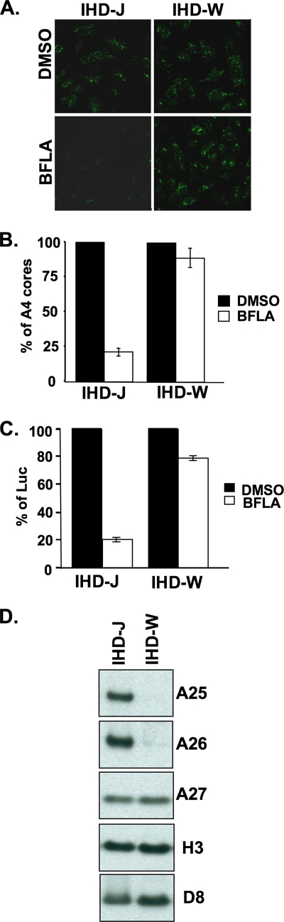
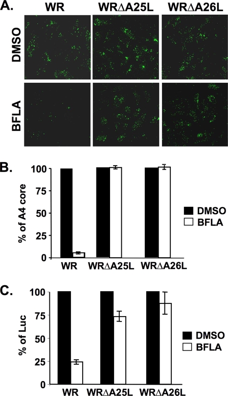
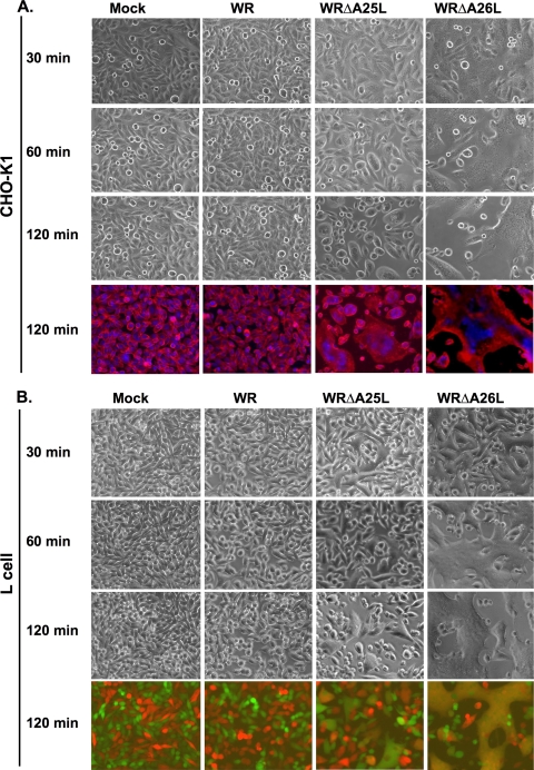
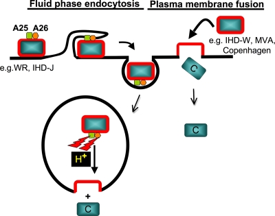
Mature vaccinia virus enters cells through either fluid-phase endocytosis/macropinocytosis or plasma membrane fusion. This may explain the wide range of host cell susceptibilities to vaccinia virus entry; however, it is not known how vaccinia virus chooses between these two pathways and which viral envelope proteins determine such processes. By screening several recombinant viruses and different strains, we found that mature virions containing the vaccinia virus A25 and A26 proteins entered HeLa cells preferentially through a bafilomycin-sensitive entry pathway, whereas virions lacking these two proteins entered through a bafilomycin-resistant pathway. To investigate whether the A25 and A26 proteins contribute to entry pathway specificity, two mutant vaccinia viruses, WRDeltaA25L and WRDeltaA26L, were subsequently generated from the wild-type WR strain. In contrast to the WR strain, both the WRDeltaA25L and WRDeltaA26L viruses became resistant to bafilomycin, suggesting that the removal of the A25 and A26 proteins bypassed the low-pH endosomal requirement for mature virion entry. Indeed, WRDeltaA25L and WRDeltaA26L virus infections of HeLa, CHO-K1, and L cells immediately triggered cell-to-cell fusion at a neutral pH at 1 to 2 h postinfection (p.i.), providing direct evidence that viral fusion machinery is readily activated after the removal of the A25 and A26 proteins to allow virus entry through the plasma membrane. In summary, our data support a model that on vaccinia mature virions, the viral A25 and A26 proteins are low-pH-sensitive fusion suppressors whose inactivation during the endocytic route results in viral and cell membrane fusion. Our results also suggest that during virion morphogenesis, the incorporation of the A25 and A26 proteins into mature virions may help restrain viral fusion activity until the time of infections.


Figures

FIG. 1.
FIG. 1.
BFLA sensitivity of IA27L and IA27L-A26WR viruses. (A) Immunofluorescence microscopy of virus-uncoating assays with HeLa cells pretreated with DMSO or 25 nM BFLA and infected with the IA27L or IA27L-A26WR virus. Anti-A4 Ab was used to stain uncoated viral cores. (B) Quantitative results of virus-uncoating assays shown in A. Viral core numbers in 30 cells were counted in each panel in A to obtain an average number of viral cores per cell. The viral core number obtained from cells treated with DMSO (−) was used as the 100% control. The percentage of A4 cores was calculated as follows: 100 × (viral core number per cell in BFLA-treated cells/viral core number per cell in DMSO-treated cells). The experiments were repeated three times, and the standard deviations are shown. (C) Acid bypass experiments with HeLa cells infected with the IA27L or IA27L-A26WR virus. HeLa cells were pretreated with DMSO (−) or with 25 nM BFLA (+) and subsequently infected with the IA27L or IA27L-A26WR virus at 4°C for 60 min. Cells were then treated with neutral (pH 7.4) or acidic (pH 4.7) buffer for 3 min, washed with PBS, and harvested for luciferase (Luc) assays at 2 h p.i. Luciferase activity obtained from cells treated with DMSO (−) was used as the 100% control. (D) Silver staining of SDS-PAGE gels containing 1 μg of purified IA27L and IA27L-A26WR MV particles. Arrows marked the A25 and A26 protein bands identified by mass spectrometry. (E) Immunoblot analyses of purified IA27L and IA27L-A26WR MV particles with anti-A25 (1:5,000), anti-A26 (1:1,000), anti-A27 (1:500), anti-H3 (1:1,000), and anti-D8 (1:1,000) Abs.
FIG. 2.
FIG. 2.
BFLA sensitivity of two vaccinia virus strains, IHD-J and IHD-W. (A) Immunofluorescence microscopy of virus-uncoating assays in HeLa cells pretreated with DMSO or 25 nM BFLA and infected with IHD-J or IHD-W. Anti-A4 Ab was used to stain uncoated viral cores. (B) Quantitative results of virus-uncoating assays shown in A. (C) Viral early luciferase assays of HeLa cells pretreated with DMSO or 25 nM BFLA, subsequently infected with IHD-J and IHD-W, and harvested at 2 h p.i. for luciferase assays. The viral luciferase activity of infected cells in the absence of BFLA (DMSO) was set to 100%. (D) Immunoblot analyses of purified IHD-J and IHD-W MV particles (1 μg) with anti-A25 (1:5,000), anti-A26 (1:1,000), anti-A27 (1:500), anti-H3 (1:1,000), and anti-D8 (1:1,000) Abs.
FIG. 3.
FIG. 3.
(A) Anti-A25 Ab blocked vaccinia virus plaque formation. BSC40 cells were infected with vaccinia virus wt strain WR in the presence of preimmune (1:100), anti-vaccinia virus MV (anti-VV) (1:100), or anti-A25 (1:100) Ab; washed; and overlaid with 1% agar. The plaques at 2 days p.i. were fixed and stained with 1% crystal violet before counting. (B) Schematic representation of vaccinia virus genomes containing ORFs A24R to A28L. Wild-type strain WR is shown on the top, with arrows pointing toward the direction of transcription. In WRΔA25L and WRΔA26L viruses, the viral A25L or A26L ORF was substituted with a dual-expression cassette, Luc-Gpt, containing a luciferase (Luc) gene driven by a viral early promoter and the Eco-Gpt (Gpt) gene driven by the viral p7.5 promoter. (C) Immunoblot analyses of A25 and A26 protein expressions in cells and on MV particles. HeLa cell lysates (Lysates) or purified MV particles (MV) were separated by SDS-PAGE and probed with anti-A25, anti-A26, anti-A27, and anti-D8 Abs. (D) One-step growth curve analyses of wild-type (WR) vaccinia virus and WRΔA25L and WRΔA26L mutant viruses. HeLa cells were infected at an MOI of 5 PFU per cell and harvested at 0, 2, 4, 8, 16, 24, and 48 h p.i. for plaque determination assays. (E) Extracellular virus titers were determined from culture medium collected from the infected cells shown in D. EEV titers were determined in the presence of anti-L1 MAb 2D5 (1:500) to block contaminating MV from forming plaques. The experiments were performed twice.
FIG. 4.
FIG. 4.
BFLA sensitivity of wild-type vaccinia virus strain WR and the WRΔA25L and WRΔA26L mutant viruses. (A) Immunofluorescence microscopy of virus-uncoating assays with HeLa cells pretreated with DMSO or 25 nM BFLA and infected with wild-type vaccinia virus WR and the WRΔA25L and WRΔA26L mutant viruses. Anti-A4 Ab was used to stain uncoated viral cores. (B) Quantitative results of virus-uncoating assays shown in A. (C) Viral early luciferase assays of HeLa cells pretreated with DMSO or 25 nM BFLA, subsequently infected with wild-type WR, WRΔA25L, and WRΔA26L viruses, and harvested at 2 h p.i. for luciferase assays. The viral luciferase activity of infected cells in the absence of BFLA (DMSO) was set to 100%.
FIG. 5.
FIG. 5.
Cell fusion from without at neutral pH on HeLa cells triggered by MV of WRΔA25L and WRΔA26L viruses but not by wt WR. (A) RFP- and GFP-expressing HeLa cells were mixed at a ratio of 1:1 during cell seeding. These cells were infected with purified wt WR, WRΔA25L, and WRΔA26L viruses at an MOI of 100 PFU per cell at 37°C for 60 min at a neutral pH as described in Materials and Methods, fixed at 2 h p.i., and photographed. Merge, red, and green immunofluorescence images were overlays with cell morphology. (B) Quantification of cell fusion from without (A), as described in Materials and Methods. Percentages of cell fusion were quantified into four different categories based on the numbers of cell nuclei per cell (1, 2 to 10, 11 to 20, or >20 nuclei/cell). (C) Wild-type WR MV triggered cell fusion from without only after low-pH (pH 4.7) treatment. GFP- and RFP-expressing HeLa cells were infected with wild-type WR as described above (A) and treated briefly with low-pH buffer (pH 4.7) for 3 min, and cell fusion was developed and photographed at 2 h p.i.
FIG. 6.
FIG. 6.
Kinetic analyses of cell fusion from without at neutral pH on CHO-K1 and L cells triggered by MV of WRΔA25L and WRΔA26L viruses but not by wt WR. (A) CHO-K1 cells were infected with WR, WRΔA25L, and WRΔA26L viruses at an MOI of 100 PFU per cell at 37°C for 60 min at a neutral pH as described in the legend of Fig. 5A. Cells were photographed at 30, 60, and 120 min p.i. to monitor the occurrence of flat fused cells. At the end of the experiment (120 min p.i.), cells were fixed, and nuclei were stained with DAPI (0.5 μg/ml). The plasma membrane was stained with a fluorescent dye, PKH26, to visualize cell shapes. (B) RFP- and GFP-expressing L cells were cocultured and infected with wt WR, WRΔA25L, and WRΔA26L viruses at a neutral pH as described above. Cells were photographed at 30, 60, and 120 min p.i. to monitor the occurrence of flat fused cells. At the end of the experiment (120 min p.i.), cells were fixed for photography, and merged images of red and green immunofluorescence are shown. Yellow images represent areas of fused cells.
FIG. 7.
FIG. 7.
Peptide-blocking cell fusion assays. RFP- and GFP-expressing L cells were mock infected or infected with WRΔA26L virus with or without peptides at different concentrations (0.1, 0.5, 1.0, or 2.0 mg/ml), as described in Materials and Methods, and photographed at 2 h p.i. The A26 peptide corresponds to the C-terminal coiled-coil region (aa 441 to 472) of the A26 protein. Another peptide with no homology with A26 amino acid sequences was included as the control. Merged images of red and green immunofluorescence are shown. Yellow images represent areas of fused cells.
FIG. 8.
FIG. 8.
Vaccinia virus strains MVA and Copenhagen lacking intact A25 and A26 proteins triggered cell fusion at neutral pH. (A) Immunoblots of purified MV from strains WR, VV-hr (Copenhagen strain), and MVA with Abs recognizing viral A25, A26, A27, and H3 proteins. (B) Cell fusion assays at 2 h p.i. after infections with strains IHD-J, IHD-W, VV-hr (Copenhagen), and MVA. Merged images of red and green immunofluorescence are shown. Yellow images representing areas of fused cells were observed for all strains except IHD-J.
FIG. 9.
FIG. 9.
Working model of vaccinia virus entry pathways controlled by viral A25 and A26 proteins. We propose that viral A25 and A26 proteins function as cell fusion suppressors for vaccinia virus MV particles. Vaccinia virus strains WR and IHD-J contain the A25 and A26 proteins to suppress viral fusion with the plasma membrane of cells. These MV are internalized into intracellular vesicles, of which the acidic environments (H+) nullify the fusion suppressor activity of the A25 and A26 proteins, leading to viral membrane fusion with endosomal membranes to release viral cores into the cytoplasm. In contrast, MV of vaccinia virus strains lacking the A25 and A26 proteins, such as strains IHD-W, MVA, and Copenhagen, enter HeLa cells through fusion with the plasma membrane in neutral-pH environments. C, viral core.

Similar articles

Cited by

References

    1. Armstrong, J. A., D. H. Metz, and M. R. Young. 1973. The mode of entry of vaccinia virus into L cells. J. Gen. Virol. 21:533-537. - PubMed
    1. Bengali, Z., A. C. Townsley, and B. Moss. 2009. Vaccinia virus strain differences in cell attachment and entry. Virology 389:132-140. - PMC - PubMed
    1. Carter, G. C., M. Law, M. Hollinshead, and G. L. Smith. 2005. Entry of the vaccinia virus intracellular mature virion and its interactions with glycosaminoglycans. J. Gen. Virol. 86:1279-1290. - PubMed
    1. Chang, A., and D. H. Metz. 1976. Further investigations on the mode of entry of vaccinia virus into cells. J. Gen. Virol. 32:275-282. - PubMed
    1. Ching, Y. C., C. S. Chung, C. Y. Huang, Y. Hsia, Y. L. Tang, and W. Chang. 2009. Disulfide bond formation at the C termini of vaccinia virus A26 and A27 proteins does not require viral redox enzymes and suppresses glycosaminoglycan-mediated cell fusion. J. Virol. 83:6464-6476. - PMC - PubMed

Publication types

LinkOut - more resources