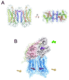
Oligomeric forms of G protein-coupled receptors (GPCRs)
- PMID: 20538466
- PMCID: PMC2937196
- DOI: 10.1016/j.tibs.2010.05.002
Oligomeric forms of G protein-coupled receptors (GPCRs)
Abstract
Oligomerization is a general characteristic of cell membrane receptors that is shared by G protein-coupled receptors (GPCRs) together with their G protein partners. Recent studies of these complexes, both in vivo and in purified reconstituted forms, unequivocally support this contention for GPCRs, perhaps with only rare exceptions. As evidence has evolved from experimental cell lines to more relevant in vivo studies and from indirect biophysical approaches to well defined isolated complexes of dimeric receptors alone and complexed with G proteins, there is an expectation that the structural basis of oligomerization and the functional consequences for membrane signaling will be elucidated. Oligomerization of cell membrane receptors is fully supported by both thermodynamic calculations and the selectivity and duration of signaling required to reach targets located in various cellular compartments.
Copyright © 2010 Elsevier Ltd. All rights reserved.
Figures


Similar articles
-
GPCRs and Signal Transducers: Interaction Stoichiometry.Trends Pharmacol Sci. 2018 Jul;39(7):672-684. doi: 10.1016/j.tips.2018.04.002. Epub 2018 May 5. Trends Pharmacol Sci. 2018. PMID: 29739625 Free PMC article. Review.
-
Functional crosstalk between GPCRs: with or without oligomerization.Curr Opin Pharmacol. 2010 Feb;10(1):6-13. doi: 10.1016/j.coph.2009.10.009. Epub 2009 Dec 3. Curr Opin Pharmacol. 2010. PMID: 19962942 Review.
-
The Dynamics of GPCR Oligomerization and Their Functional Consequences.Int Rev Cell Mol Biol. 2018;338:141-171. doi: 10.1016/bs.ircmb.2018.02.005. Epub 2018 Apr 7. Int Rev Cell Mol Biol. 2018. PMID: 29699691 Review.
-
Structural features of the G-protein/GPCR interactions.Biochim Biophys Acta. 2014 Jan;1840(1):16-33. doi: 10.1016/j.bbagen.2013.08.027. Epub 2013 Sep 7. Biochim Biophys Acta. 2014. PMID: 24016604 Review.
-
Oligomerization of GPCRs involved in endocrine regulation.J Mol Endocrinol. 2016 Jul;57(1):R59-80. doi: 10.1530/JME-16-0049. Epub 2016 May 5. J Mol Endocrinol. 2016. PMID: 27151573 Review.
Cited by
-
Crystal structure of oligomeric β1-adrenergic G protein-coupled receptors in ligand-free basal state.Nat Struct Mol Biol. 2013 Apr;20(4):419-25. doi: 10.1038/nsmb.2504. Epub 2013 Feb 24. Nat Struct Mol Biol. 2013. PMID: 23435379 Free PMC article.
-
Role of G-Proteins and GPCRs in Cardiovascular Pathologies.Bioengineering (Basel). 2023 Jan 6;10(1):76. doi: 10.3390/bioengineering10010076. Bioengineering (Basel). 2023. PMID: 36671648 Free PMC article. Review.
-
Novel structural and functional insights into M3 muscarinic receptor dimer/oligomer formation.J Biol Chem. 2013 Nov 29;288(48):34777-90. doi: 10.1074/jbc.M113.503714. Epub 2013 Oct 16. J Biol Chem. 2013. PMID: 24133207 Free PMC article.
-
Clustering of glycoprotein VI (GPVI) dimers upon adhesion to collagen as a mechanism to regulate GPVI signaling in platelets.J Thromb Haemost. 2017 Mar;15(3):549-564. doi: 10.1111/jth.13613. Epub 2017 Feb 16. J Thromb Haemost. 2017. PMID: 28058806 Free PMC article.
-
Structural determinants of the supramolecular organization of G protein-coupled receptors in bilayers.J Am Chem Soc. 2012 Jul 4;134(26):10959-65. doi: 10.1021/ja303286e. Epub 2012 Jun 25. J Am Chem Soc. 2012. PMID: 22679925 Free PMC article.
References
-
- Gilman AG. G proteins: transducers of receptor-generated signals. Annual review of biochemistry. 1987;56:615–649. - PubMed
-
- Rodbell M. The role of hormone receptors and GTP-regulatory proteins in membrane transduction. Nature. 1980;284:17–22. - PubMed
-
- Romano C, et al. Metabotropic glutamate receptor 5 is a disulfide-linked dimer. J Biol Chem. 1996;271:28612–28616. - PubMed
-
- Farrens DL, et al. Requirement of rigid-body motion of transmembrane helices for light activation of rhodopsin. Science. 1996;274:768–770. - PubMed
Publication types
MeSH terms
Substances
Grants and funding
LinkOut - more resources
Full Text Sources
Research Materials

