Review
. 2010 Jun;5(6):867-81.
doi: 10.2217/fmb.10.40.

Expanding knowledge of P3 proteins in the poliovirus lifecycle

Affiliations
Review

Expanding knowledge of P3 proteins in the poliovirus lifecycle

Craig E Cameron et al. Future Microbiol. 2010 Jun.

Abstract

Poliovirus is the most extensively studied member of the order Picornavirales, which contains numerous medical, veterinary and agricultural pathogens. The picornavirus genome encodes a single polyprotein that is divided into three regions: P1, P2 and P3. P3 proteins are known to participate more directly in genome replication, for example by containing the viral RNA-dependent RNA polymerase (RdRp or 3Dpol), among several other proteins and enzymes. We will review recent data that provide new insight into the structure, function and mechanism of P3 proteins and their complexes, which are required for initiation of genome replication. Replication of poliovirus genomes occurs within macromolecular complexes, containing viral RNA, viral proteins and host-cell membranes, collectively referred to as replication complexes. P2 proteins clearly contribute to interactions with the host cell that are required for virus multiplication, including formation of replication complexes. We will discuss recent data that suggest a role for P3 proteins in formation of replication complexes. Among the least understood steps of the poliovirus lifecycle is encapsidation of genomic RNA. We will also describe data that suggest a role for P3 proteins in this step.


Figures

Figure 1
Figure 1. Poliovirus genome. The 5′-end of the genome is covalently linked to a peptide (VPg) encoded by the 3B region of the genome. The 3′-end contains a poly(rA) tail
Three cis-acting replication elements are known. oriL is located in the 5′-NTR. oriR is located in the 3′-NTR and is not absolutely required for poliovirus genome replication [101,102]. oriI is located in the 2C-coding sequence for poliovirus and is also referred to as 2C-cis-acting genome replication element; the position of this element is virus dependent. oriI is required for initiation of minus-strand RNA synthesis. Translation initiation employs an IRES. The single open reading frame encodes a polyprotein. P1 encodes virion structural proteins as indicated. P2 encodes proteins thought to participate in virus–host interactions required for genome replication. The 2A protein is a protease that releases the structural protein precursor from the polyprotein. This protein also cleaves cellular proteins to enhance virus multiplication. Protein 2B, 2C and 2BC interact with membranes and contribute to the formation of replication complexes. The 2C protein is ATPase; this activity is required for 2C to contribute to replication complex formation. P3 encodes proteins thought to participate directly in genome replication. Polyprotein processing is mediated by protease activity residing in 2A, 3C and/or 3CD proteins. IRES: Internal ribosome entry site; NTR: Nontranslated region; VPg: Virion protein genome linked.
Figure 2
Figure 2. P3 precursor processing pathways and functions of proteins encoded in the P3 region
Processing of the P3 precursor occurs by two independent pathways, major (A) and minor (B). In the major pathway (A), processing between 3B and 3C yields 3AB and 3CD. In the minor pathway (B), processing between 3A and 3B yields 3A and 3BCD. 3BCD processing yields 3BC and 3D; 3BC processing yields 3B and 3C. Known functions for each of the P3 proteins are listed; those colored red are described in the text. RC: Replication complex; RdRp: RNA-dependent RNA polymerase. Adapted from [10,11].
Figure 3
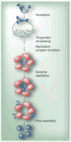
Figure 3. Poliovirus multiplication cycle
Genomic RNA is first translated to produce the poliovirus polyprotein. The polyprotein is co- and post-translationally processed to produce the various precursors and processed proteins that are needed for poliovirus multiplication. Replication complexes are formed, followed by genome replication. Replicated RNA enters either the translation–replication cycle or viral particle assembly step. For virus assembly, RNA encapsidation and virus particle maturation must occur. Adapted from [78].
Figure 4
Figure 4. Structures of the poliovirus proteins
Poliovirus proteins (A) 3A (PDB 1NG7 [40]), (B) 3B (VPg)(2BBP [39]), (C) 3Cpro (1L1N [38]), (D) 3Dpol (1RA6 [41]) and (E) 3CD (2IJD [21]) are shown. The 3A (soluble domain, residues 1–59) and 3B structures were determined by nuclear magnetic resonance; the other three are crystallographic structures. VPg: Virion protein genome linked.
Figure 5
Figure 5. Model for 3AB and 3A topologies on membranes
The hydrophobic domain of 3A interacts with membranes in the context of 3AB. Once 3B is cleaved and released, 3A equilibrates to both trans- and nontrans-membrane forms. Adapted from [18].
Figure 6
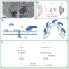
Figure 6. Proposed mechanisms for poliovirus replication complex formation
(A) Poliovirus (PV)-induced vesicles. Electron micrograph of uninfected (left panel) and PV-infected (right panel) HeLa cells. Uninfected HeLa cells show normal intracellular membrane structures. PV-infected HeLa cells show PV-induced vesicles that cluster perinuclearly. HeLa cells were infected with PV at MOI of 10, fixed 7 h postinfection and visualized by electron microscopy. Scale bar: 2 μm. (B) Model for COP-coated vesicular trafficking from ER to Golgi. COPII vesicles originate from the ER and transport and fuse to ERGIC. COPI vesicles originate from ERGIC and transport and fuse to the Golgi. (C) Proposed mechanism for Arf-mediated vesicle formation. The GTPase-inactive form of Arf, Arf-GDP, prefers to be soluble in the cytosol. Once Arf-GDP binds to the membrane, its membrane association is stabilized by the Arf–p24 protein–protein interaction. Arf-GDP is then converted to the active form of Arf, Arf-GTP, by a GEF. Arf-GTP recruits cargo and coatomer proteins, such as COPI, and curvature is induced to form a vesicle. (D) Proposed mechanism for 3A and 3CD regulation of Arf-mediated vesicle formation. 3A and BFA inhibit the normal, cellular pathway required for Arf activation by preventing GEF function (left). In this case the GEF is GBF1. Poliovirus may have evolved a virus-specific mechanism for Arf activation. For example, 3A and/or 3CD may bind to Arf-GDP–GEF complex, causing activation of Arf and COPI recruitment by a mechanism that may no longer be sensitive to GAPs. It is also possible that COPI is not recruited at all. ER: Endoplasmic reticulum; ERGIC: ER–Golgi intermediate compartment; GAP: GTPase-activating protein; GEF: Guanine nucleotide exchange factor.
Figure 7
Figure 7. Two binding sites for VPg(3B) in 3Dpol/VPg(3B) complexes
The complex structure of 3Dpol/VPg(3B) from FMDV is from PDB 2F8E [42] (A) and that of CVB3 is from PDB 3CDW [43] (B). VPg(3B) is shown in black, and 3Dpol is shown in white. Two different views are shown to emphasize the different binding sites for VPg(3B) in the two complexes. The views on the right can be obtained by rotating the left view 90° counterclockwise. In FMDV, the VPg(3B) peptide occupied the active site of the polymerase with the tyrosyl residue (Tyr3) positioned at the catalytic center. In the CVB3 complex, the VPg(3B) peptide binds to the back of the thumb of the polymerase. CVB3: Coxsackievirus B3; FMDV: Foot-and-mouth disease virus; VPg: Virion protein genome linked.
Figure 8
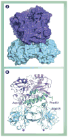
Figure 8. Model of 3C2–3D complex
(A) Surfaces of 3C dimer and 3Dpol are shown in cyan and purple, respectively. (B) Ribbon diagram of the model shown in (A). The polymerase subdomains are depicted in different colors: fingers (purple), palm (magenta) and thumb (green). 3Dpol residues Asp406, Arg455 and Phe461, known to be required for uridylylation, are highlighted. Adapted from [20].
Figure 9
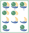
Figure 9. The same heteromeric complex from different viruses may form different subassemblies
A cartoon is shown of three heterotrimeric complexes. These complexes are identical in structure. However, the interactions used to stabilize the complex (lines between subunits of the complex) are distributed differently. As a result, the stable subassemblies will differ as indicated.
Figure 10
Figure 10. Assembly and organization of the picornavirus 3B (VPg) uridylylation complex
(A) Model for organization of 3C2–oriI complex. Protein 3C exists in a monomer–dimer equilibrium. OriI contains two 3C-binding sites. Binding of oriI to 3C shifts the equilibrium in favor of a 3C dimer (3C2). A consequence of 3C binding to oriI is destabilization of the secondary structure associated with unbound oriI, thus extending the loop. (B) Model for initiation of negative-strand RNA synthesis. 3(B)CD proteins bound to oriL and oriR interact with 3D and/or dimerized 3CDs bound to oriI. Multiple interactions among 3CDs on oriL, oriI and oriR facilitate circularization of the poliovirus genome and recruit both the 5′- and 3′-ends to oriI. Adapted with modifications from [103,104].

Similar articles

Cited by

References

    1. Racaniello VR. Picornaviridae: the viruses and their replication. In: Knipe DM, Howley PM, editors. Fields Virology. 5. Lippincott Williams & Wilkins; Philadelphia, PA, USA: 2007. pp. 765–838.
    1. Le Gall O, Christian P, Fauquet CM, et al. Picornavirales, a proposed order of positive-sense single-stranded RNA viruses with a pseudo-T = 3 virion architecture. Arch Virol. 2008;153(4):715–727. - PubMed
    1. Paul AV, Belov GA, Ehrenfeld E, Wimmer E. Model of picornavirus RNA replication. In: Cameron CE, Götte M, Raney KD, editors. Viral Genome Replication. 1. Vol. 3. Springer; Philadelphia, PA: 2009. p. 23.
    1. Steil BP, Barton DJ. Cis-active RNA elements (CREs) and picornavirus RNA replication. Virus Res. 2009;139(2):240–252. - PMC - PubMed
    1. Arnold E, Luo M, Vriend G, et al. Implications of the picornavirus capsid structure for polyprotein processing. Proc Natl Acad Sci USA. 1987;84(1):21–25. - PMC - PubMed

Publication types

MeSH terms