. 2010 Aug;84(16):8111-23.
doi: 10.1128/JVI.00459-10. Epub 2010 Jun 2.

Role of noncovalent SUMO binding by the human cytomegalovirus IE2 transactivator in lytic growth

Affiliations

Role of noncovalent SUMO binding by the human cytomegalovirus IE2 transactivator in lytic growth

Eui Tae Kim et al. J Virol. 2010 Aug.

Abstract

The 86-kDa immediate-early 2 (IE2) protein of human cytomegalovirus (HCMV) is a promiscuous transactivator essential for viral gene expression. IE2 is covalently modified by SUMO at two lysine residues (K175 and K180) and also interacts noncovalently with SUMO. Although SUMOylation of IE2 has been shown to enhance its transactivation activity, the role of SUMO binding is not clear. Here we showed that SUMO binding by IE2 is necessary for its efficient transactivation function and for viral growth. IE2 bound physically to SUMO-1 through a SUMO-interacting motif (SIM). Mutations in SIM (mSIM) or in both SUMOylation sites and SIM (KR/mSIM), significantly reduced IE2 transactivation effects on viral early promoters. The replication of IE2 SIM mutant viruses (mSIM or KR/mSIM) was severely depressed in normal human fibroblasts. Analysis of viral growth curves revealed that the replication defect of the mSIM virus correlated with low-level accumulation of SUMO-modified IE2 and of viral early and late proteins. Importantly, both the formation of viral transcription domains and the association of IE2 with viral promoters in infected cells were significantly reduced in IE2 SIM mutant virus infection. Furthermore, IE2 was found to interact with the SUMO-modified form of TATA-binding protein (TBP)-associated factor 12 (TAF12), a component of the TFIID complex, in a SIM-dependent manner, and this interaction enhanced the transactivation activity of IE2. Our data demonstrate that the interaction of IE2 with SUMO-modified proteins plays an important role for the progression of the HCMV lytic cycle, and they suggest a novel viral mechanism utilizing the cellular SUMO system.


Figures

FIG. 1.
FIG. 1.
Interaction of IE2 with SUMO through a SIM. (A) SUMO-related domain structures of IE2. Two SUMO conjugation sites (K175 and K180), a SIM (between residues 200 and 208), and the protein inhibitor of the activated STAT1 (PIAS1)-binding region (PIAS1) are indicated. Two transactivation domains identified using the GAL4 swap assays (48) are represented as black boxes. The regions of IE2 required for transactivation (residues 1 to 98 and 195 to 579) are also shown. The numbers are amino acid positions of the protein. (B) In vitro binding of IE2 with SUMO-1 through a SIM. The proteins were purified from bacteria and were used in GST pulldown assays. Five micrograms of GST-IE2(135-289) or its SIM mutant version, IE2(135-289/mSIM), was immobilized on glutathione-Sepharose beads and was incubated with 0.5 μg of His-SUMO-1. The bound proteins were fractionated by SDS-PAGE and were detected by immunoblotting with an anti-His Ab (top), and 17% of the GST or GST fusion protein used in the reaction was also fractionated by SDS-PAGE and stained by Coomassie blue (bottom). Five percent of the His-SUMO-1 used in the binding assays is shown as an input control. (C) Competition assays. The in vitro binding assay for which results are shown in panel B was conducted in the presence of increasing amounts of untagged SUMO-1 or BSA (1:1, 1:3, and 1:5 molar ratio with His-SUMO-1). The bead-bound proteins were detected by immunoblotting (IB) with an anti-His Ab (left, top) or with anti-SUMO-1 Ab (left, bottom). Two micrograms of the SUMO-1 and BSA proteins used is shown by Coomassie blue staining (right, top). The relative amounts of His-SUMO-1 bound to IE2 in the presence of competitors were quantified and are shown in the graph (right, bottom).
FIG. 2.
FIG. 2.
Effect of the SIM mutation on the transactivation function of IE2. (A) (Top) HF (2 × 10⁵) were electroporated with 0.5 μg of a plasmid containing a luciferase reporter gene driven by the HCMV UL112-113 promoter (pUL112-113-Luc) or polymerase promoter (pPol-Luc) and 1.5 μg of a plasmid expressing wild-type or mutant (KR, mSIM, or mSIM/KR) IE2. At 48 h after transfection, whole-cell lysates were prepared and assayed for luciferase activity. Luciferase activities are indicated as fold activation over the basal level of a parent vector. The results shown are the mean values with standard errors for three independent experiments. (Bottom) The expression levels of HA-IE2 proteins and β-actin in cell lysates of a representative assay are shown in immunoblots. (B) (Top) ChIP assays. HF in 150-mm-diameter dishes were electroporated with 2 μg of a plasmid containing the Pol-Luc reporter gene and 6 μg of a plasmid expressing wild-type or mSIM IE2. At 48 h after transfection, the ChIP assays (see Materials and Methods) were performed with an anti-RNA polymerase II (anti-Pol II) Ab and IgG (as a control). (Bottom) The expression levels of wild-type or mutant HA-IE2 proteins and β-actin in cell lysates are shown in immunoblots.
FIG. 3.
FIG. 3.
Generation of the IE2 SIM mutant and its revertant HCMV (Towne) BAC clones and their infectivities in transfected HF. (A) Scheme for the generation of the IE2 SIM mutant (mSIM) Towne (T)-BACs and their revertant T-BAC clones. A pGS284 (38) derivative (pYH69), which harbors a 4.1-kb DNA fragment containing the mSIM allele of IE2, was used for homologous recombination with the parental T-BAC clone. This recombination event resulted in the IE2(mSIM) T-BAC clone (pYH72). Similarly, the IE2(KR/mSIM) T-BAC clone (pYH70) was produced using a GS284 derivative (pYH56) containing a 2.4-kb IE2 KR/mSIM allele. A pGS284 derivative (pHR8) containing a 4.1-kb wild-type DNA fragment was used to make their revertant T-BAC clones (pYH76 and pYH75). The IE2 KR mutant T-BAC (pBAC2) and its revertant (pHR14) have been described previously (32). (B) Restriction fragment DNA patterns obtained following EcoRI/BamHI digestion of the wild-type, mutant, and revertant T-BAC DNAs were analyzed by agarose gel electrophoresis. λ-HindIII was used as the molecular weight standard. (C) Infectivities of the transfected T-BAC DNAs. HF were transfected with T-BAC clones (Wt, mSIM [pYH72], mSIM-R [pYH76], KR/mSIM [pYH70], and KR/mSIM-R [pYH75]) via electroporation (see Materials and Methods). The cells were monitored for the spread of GFP signals. GFP images (top) and their phase-contrast images (bottom) were photographed 4 weeks after electroporation. Representative images from at least four independent experiments are shown. The inserts in the images of mSIM virus infection include some GFP-positive cells. Note that the spread of these GFP signals in cells transfected with the mSIM and KR/mSIM T-BAC DNAs was usually incomplete compared to the spread in cells transfected with the wild-type or revertant T-BAC DNAs.
FIG. 4.
FIG. 4.
Expression levels of unmodified IE1 and IE2 in recombinant virus-infected HF at early times. (A) HF cultured in chamber slides were infected with the indicated viruses at an MOI of 0.5. At 8 h after infection, cells were fixed in cold methanol and were stained with the IE1-specific Ab 6E1. Hoechst stain was used to visualize the nuclei of all cells in the field. (B) HF in 6-well plates were infected with the indicated viruses at an MOI of 2. Whole-cell lysates were prepared at 8 h after infection, and the expression levels of unmodified IE1 and IE2 were determined by immunoblotting using 810R (for IE1 and IE2) and anti-β-actin mouse MAbs.
FIG. 5.
FIG. 5.
Comparison of the progeny virus titers of wild-type, SIM mutant, SUMOylation-defective mutant, and revertant viruses, and accumulation of viral proteins in infected cells. (A) HF in 12-well plates were infected with wild-type, mSIM, KR/mSIM, or KR viruses and their revertant viruses at an MOI of 2 for 5 days or at an MOI of 0.1 for 9 days. The progeny virus titers are shown as infectious units in cell culture supernatants, which were determined by infectious center assays. The results shown are the averages of data from two independent assays. (B and C) Accumulation of viral IE, early, and late proteins in infected cells. HF in 6-well plates were infected as described for panel A, and whole-cell lysates were prepared and were subjected to SDS-8% PAGE, followed by immunoblotting with 810R (for IE1 and IE2), anti-p52, anti-pp28, and anti-β-actin MAbs (B) and with an anti-peptide rabbit PAb (P3) for IE2, which detects all of the p86, p60, and p40 IE2 proteins (48) (C, top). The positions of the IE2 SIM and the TATA sequences and start codons for p60 and p40 are shown (C, bottom).
FIG. 6.
FIG. 6.
Growth curves of wild-type, SIM mutant, and revertant viruses and the expression levels of viral proteins. (A) HF in 12-well plates were infected with the wild-type (Wt), SIM mutant (mSIM), and revertant viruses at an MOI of 2 or 0.1. The time course results represent the total numbers of cell-free viruses produced in cell culture supernatants at the indicated sampling times, which were measured as described in the legend to Fig. 5A. The results are averages of data from two independent assays. (B) HF in 6-well plates were infected with recombinant viruses at an MOI of 2 or 0.1 as for panel A. Accumulation of viral proteins at the indicated sampling times was determined by immunoblotting as described in the legend to Fig. 5B (left, MOI of 2; right, MOI of 0.1).
FIG. 7.
FIG. 7.
Reduced formation of the viral transcription domains in IE2 SIM mutant virus infection. (A) HF in chamber slides were infected with wild-type, IE2 SIM mutant (mSIM), or revertant (mSIM-R) viruses at an MOI of 2. At 8 h after infection, the cells were fixed in cold methanol, followed by a confocal double-label IFA using anti-IE2 and anti-PML Abs. DNA in cell nuclei was stained with Hoechst dye. (B) Quantitation of the viral transcription domains. HF were infected as for panel A. Cells were fixed at 6, 8, and 10 h after infection and were stained with an anti-IE2 Ab or were fixed at 8 h and stained with an anti-UL112-113 Ab as for panel A. The IE2 or UL112-113 foci were counted in at least 20 infected (i.e., IE2- or UL112-113-positive) cells, and the results are shown in the graphs, as the mean values for the number of IE2 or UL112-113 foci per cell with standard errors. (C) Comparison of the intensities of IE2 signals in foci between wild-type and mSIM virus-infected cells. Surface plot analysis of an infected cell nucleus at 10 h after infection was performed with the ImageJ program (NIH). (D) ChIP assessment of the amounts of IE2 loaded on viral promoters (MIE, UL54, and UL112-113) in infected cells. HF were infected with wild-type, mSIM, or revertant viruses at an MOI of 5 for 8 h. ChIP assays were conducted with an anti-IE2 mouse MAb or nonspecific mouse immunoglobulin G (IgG). The amounts of coprecipitated DNA fragments were determined by PCR and agarose gel electrophoresis. The MIE exon 5 region was tested as a negative control.
FIG. 8.
FIG. 8.
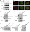
Interaction of IE2 with the SUMO-modified form of TAF12. (A) CoIP assays for IE2 and TAF12 in infected cells. HF in 100-mm-diameter dishes were first transfected with wild-type or K19R mutant TAF2 via electroporation and then infected with HCMV at an MOI of 5. At 8 h, whole-cell lysates were prepared, and CoIP assays were performed with an anti-IE2 Ab. (B) Colocalization of IE2 and TAF12 in infected cells. HF cultured in chamber slides were transfected and infected as described for panel A. Cells were fixed in cold methanol, and confocal double-label IFA was performed with anti-IE2 and anti-HA Abs. (C) In vitro interaction of IE2 with SUMO-modified TAF2 in a SIM-dependent manner. His-TAF12 and SUMO-1-modified His-TAF12 proteins, which were produced in E. coli cotransformed with plasmids expressing the SUMO conjugating enzymes (pT-E1E2-S1) (see Materials and Methods) and His-TAF12, were incubated with the control rabbit reticulocyte lysates or in vitro-translated myc-IE2(Wt) or myc-IE2(mSIM) proteins. After immunoprecipitation with an anti-myc Ab, the coprecipitated proteins were immunoblotted with an anti-His Ab (top). Ten percent (each) of the His-TAF12 proteins and myc-IE2 proteins is shown as an input control. The open circle at the top left indicates immunoglobulins. (D) An assay similar to that for which results are shown in panel C was conducted using GST-IE1 and SUMO-modified GST-IE1, which were produced in E. coli. (E) In vitro GST pulldown assays. GST, GST-TAF12, and GST-SUMO-1-TAF12 fusion proteins produced in bacteria were incubated with in vitro-translated myc-IE2(Wt) or myc-IE2(mSIM) proteins. The bound IE2 proteins were identified by immunoblotting with an anti-Myc Ab (left panel). The amounts of myc-IE2 and GST or GST fusion proteins used are shown as input controls (center and right panels).
FIG. 9.
FIG. 9.
Role of the interaction between IE2 and SUMO-modified TAF12 in the transactivation function of IE2. (A) HF (2 × 10⁵) were transfected by electroporation with a plasmid containing the Pol (UL54)-luciferase reporter gene and plasmids expressing wild-type or mutant HA-IE2 or HA-TAF12 as indicated. At 24 h after transfection, whole-cell lysates were prepared, and luciferase assays were performed. Luciferase activities are expressed as percentages of the level in cells transfected with both the wild-type IE2 and the wild-type TAF12 protein. The results shown are the mean values for three independent experiments with standard errors. The expression levels of HA-IE2 and HA-TAF12 proteins and β-actin in cell lysates are shown in immunoblots. (B) HF were transfected with plasmids encoding wild-type TAF12, TAF12(K19R), or the HA-SUMO-1-TAF12 fusion protein and were then infected with a recombinant virus containing a Pol-luciferase reporter gene (Pol-LUC) at an MOI of 1. At 24 h after infection, whole-cell lysates were prepared, and luciferase activities were assayed as for panel A. The expression levels of the HA-TAF12 and HA-SUMO-1-TAF12 proteins and β-actin in cell lysates are shown in immunoblots. (C) HF were transfected with an empty vector or a plasmid encoding HA-SUMO-1-TAF12 and were then infected with wild-type, mSIM mutant, or revertant (mSIM-R) virus at an MOI of 1. At 48 h after infection, cell lysates were prepared and analyzed by immunoblotting. (D) Hypothetical model showing the role of the covalent and noncovalent SUMO attachment of IE2 in transactivation activity. Both IE2 and TAF12 can be associated with TBP in the viral transcription domain. The binding of IE2 with SUMO-modified TAF12 through a SIM may contribute to the enhanced stabilization of the transcription initiation complex containing RNA Pol II. It is also likely that SUMO-modified IE2 may interact with other SIM-containing transcription factors, which may also promote the formation of the transcription initiation complex.

Similar articles

Cited by

References

    1. Ahn, J. H., C. J. Chiou, and G. S. Hayward. 1998. Evaluation and mapping of the DNA binding and oligomerization domains of the IE2 regulatory protein of human cytomegalovirus using yeast one and two hybrid interaction assays. Gene 210:25-36. - PubMed
    1. Ahn, J. H., and G. S. Hayward. 2000. Disruption of PML-associated nuclear bodies by IE1 correlates with efficient early stages of viral gene expression and DNA replication in human cytomegalovirus infection. Virology 274:39-55. - PubMed
    1. Ahn, J. H., and G. S. Hayward. 1997. The major immediate-early proteins IE1 and IE2 of human cytomegalovirus colocalize with and disrupt PML-associated nuclear bodies at very early times in infected permissive cells. J. Virol. 71:4599-4613. - PMC - PubMed
    1. Ahn, J. H., W. J. Jang, and G. S. Hayward. 1999. The human cytomegalovirus IE2 and UL112-113 proteins accumulate in viral DNA replication compartments that initiate from the periphery of promyelocytic leukemia protein-associated nuclear bodies (PODs or ND10). J. Virol. 73:10458-10471. - PMC - PubMed
    1. Ahn, J. H., Y. Xu, W. J. Jang, M. J. Matunis, and G. S. Hayward. 2001. Evaluation of interactions of human cytomegalovirus immediate-early IE2 regulatory protein with small ubiquitin-like modifiers and their conjugation enzyme Ubc9. J. Virol. 75:3859-3872. - PMC - PubMed

Publication types

LinkOut - more resources