. 2010 Aug;30(15):3737-48.
doi: 10.1128/MCB.01649-09. Epub 2010 Jun 1.

Wss1 is a SUMO-dependent isopeptidase that interacts genetically with the Slx5-Slx8 SUMO-targeted ubiquitin ligase

Affiliations

Wss1 is a SUMO-dependent isopeptidase that interacts genetically with the Slx5-Slx8 SUMO-targeted ubiquitin ligase

Janet R Mullen et al. Mol Cell Biol. 2010 Aug.

Abstract

Protein sumoylation plays an important but poorly understood role in controlling genome integrity. In Saccharomyces cerevisiae, the Slx5-Slx8 SUMO-targeted Ub ligase appears to be needed to ubiquitinate sumoylated proteins that arise in the absence of the Sgs1 DNA helicase. WSS1, a high-copy-number suppressor of a mutant SUMO, was implicated in this pathway because it shares phenotypes with SLX5-SLX8 mutants, including a wss1Delta sgs1Delta synthetic-fitness defect. Here we show that Wss1, a putative metalloprotease, physically binds SUMO and displays in vitro isopeptidase activity on poly-SUMO chains. Like that of SLX5, overexpression of WSS1 suppresses sgs1Delta slx5Delta lethality and the ulp1ts growth defect. Interestingly, although Wss1 is relatively inactive on ubiquitinated substrates and poly-Ub chains, it efficiently deubiquitinates a Ub-SUMO isopeptide conjugate and a Ub-SUMO fusion protein. Wss1 was further implicated in Ub metabolism on the basis of its physical association with proteasomal subunits. The results suggest that Wss1 is a SUMO-dependent isopeptidase that acts on sumoylated substrates as they undergo proteasomal degradation.


Figures

FIG. 1.
FIG. 1.
The wss1Δ mutation exhibits a SUMO-defective phenotype. (A) The indicated null mutants were crossed, and diploid strains were isolated and sporulated. The spores were then microdissected and allowed to germinate for 4 days at 30°C. The left-hand panel displays the phenotypes of sgs1Δ (squares), wss1Δ (diamonds), and the sgs1Δ wss1Δ double mutant (circles), in addition to wt spore clones. The right-hand panel displays the phenotype of slx5Δ (squares), wss1Δ (diamonds), and the slx5Δ wss1Δ double mutant (circles), in addition to wt spore clones. (B) The sgs1Δ wss1Δ cells were spread onto a YPD plate and photographed following 7 days of growth at 30°C. Arrows indicate colonies with a slightly nibbled phenotype. (C) A diploid strain heterozygous for sgs1Δ and slx5Δ was cured of the 2μm circle by the method of Tsalik and Gartenberg (50) and subjected to tetrad analysis as described above. The progeny obtained were sgs1Δ (squares), slx5Δ (diamonds), and the sgs1Δ slx5Δ double mutant (circles), in addition to wt spore clones. (D) Extracts from the indicated mutants were prepared under denaturing conditions and immunoblotted with anti-Smt3 antibody. The lower panel presents a pair of 45-kDa bands from the ponceau-stained membrane as a loading control. A bracket indicates the stacking gel. (E) The indicated strains were resuspended at an OD600 of 3.0 and serially diluted in 10-fold increments, and 5 μl was spotted onto YPD plates with or without 0.1 M hydroxyurea. The plates were photographed following 3 days of growth at 30°C.
FIG. 2.
FIG. 2.
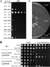
WSS1 interacts genetically with SLX5-SLX8. (A) The Y2H assay was used to detect physical interactions between Wss1, Slx5, and Slx8. Strain AH109, which contains the HIS3 reporter gene, was transformed with a GAL4 activation domain (AD) plasmid containing no insert (GAD) or the gene listed in the left-hand column, together with a GAL4 DNA binding domain (DB) plasmid containing no insert (GBK) or the gene listed in the right-hand column. Transformants were then spotted in 10-fold serial dilutions onto solid medium lacking histidine to detect expression of the reporter gene. Growth was recorded following 6 days at 30°C. (B) A wt yeast strain (NJY284) or the indicated double mutant strain (VCY1525 or NJY2481) was transformed with a multicopy vector containing WSS1 downstream of the GAL1-10 promoter (Wss1 OE; pJM7349) or vector alone (Vector). Transformants were first streaked onto medium lacking leucine but containing galactose to induce Wss1 expression and then spotted in 5-fold serial dilutions onto medium lacking leucine but containing galactose and 5-FOA to select against the balancer plasmid pJM500 (SGS1 URA3 CEN). Also spotted are strains that had previously undergone one round of selection for growth on 5-FOA (Wss1 OE - 2x). Plates were photographed following 2 (YPD) or 5 (−Leu FOA/Gal) days of growth at 30°C. (C) The 5-FOA-resistant sgs1Δ slx5Δ strain carrying pJM7349 was renamed JMY2775 and was retransformed with pJM500. Following growth in the absence of uracil, leucine auxotrophs (Leu) were identified and streaked onto 5-FOA.
FIG. 3.
FIG. 3.
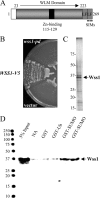
Wss1 contains an essential Zn-binding domain and physically interacts with SUMO. (A) Schematic diagram of the Wss1 protein illustrating its centrally located Zn-binding domain, two little finger (LF) domains, and putative type “a” (247-VVILDDDD-254) and type “b” (265-VIDLT-269) SIMs. (B) Strain NJY2953 (wss1Δ sgs1Δ) with complementing plasmid pJM500 (SGS1 URA3 ADE3) was transformed with a LEU2 vector containing wt WSS1-V5 (pJM7349), wss1-pd-V5 (pJM7350), or no insert (vector) under the control of the GAL1-10 promoter. Transformants were then streaked onto medium containing galactose and 5-FOA to select against pJM500. (C) One microgram of affinity-purified Wss1-HA (Ni pool) was fractionated by SDS-PAGE and stained with silver. (D) Wss1 was incubated with the indicated GST fusion protein, and the mixture was subjected to a glutathione bead pulldown assay. The precipitated protein was immunoblotted with anti-HA antibody to detect Wss1. NA, no addition.
FIG. 4.
FIG. 4.
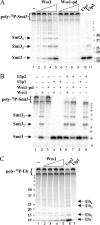
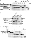
Poly-SUMO chains are proteolyzed by Wss1. (A) Siz2 sumoylation reactions were first performed in the presence of 32P-Smt3 to create poly-SUMO chains. The products of this reaction were then incubated for 1 h at 30°C with Wss1 or Wss1-pd (0, 8, 33, 130, or 520 nM), Ulp1UD (20 nM), or Ulp2 (20 nM). Following incubation, products were resolved by SDS-PAGE and autoradiography. (B) Cleavage of 32P-labeled poly-SUMO chains was performed as described above except that the substrate was incubated with either Wss1 or Wss1-pd (520 nM) for 45 min and then incubated with Ulp1 (20 nM) or Ulp2 (42.5 nM) for 15 min. (C) 32P-labeled poly-Ub chains were prepared and incubated for 1 h at 30°C with Wss1 (0, 8, 33, 130, or 520 nM), Usp2 (10 μM), or Ulp1UD (20 nM). Products were then analyzed as described above. A bracket indicates the stacking gel. The molecular weight markers are identical to those in Fig. 3D.
FIG. 5.
FIG. 5.
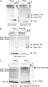
Wss1 cleaves isopeptide-linked Ub from the N terminus of a poly-SUMO chain. (A) Poly-SUMO chains were prepared as described for Fig. 4 using Siz2 and 32P-Smt3 [Siz2-(*Smt3)n] (lanes 1 to 7), or they were prepared using unlabeled Smt3 followed by ubiquitination by Slx5-Slx8 in the presence of 32P-Ub to introduce Ub onto the N terminus of the poly-SUMO chain [Siz2-(Smt3)n-*Ub] (lanes 8 to 14). Substrates (65 nM Siz2) were then incubated with Wss1 (3, 12, 50, or 200 nM), Ulp1 (20 nM), or protease 3C (20 nM) for 1 h at 30°C and analyzed by SDS-PAGE. (B) Poly-SUMO chains bearing an N-terminal 32P-Ub [Siz2-(Smt3)n-*Ub] were treated as described above with either Wss1 (0, 12.5, 50, 200, or 800 nM), Wss1-pd (0, 0.15, 0.6, 2.4, or 9.6 μM), Ulp1 (20 nM), or Ulp2 (2 μM). (C) Autoubiquitinated Slx5-Slx8 (lanes 1 to 5) or monosumoylated Siz2 (lanes 6 to 10) was treated as described for panel B with Wss1 (0, 12.5, 50, 200, or 800 nM). As a control, polysumoylated Siz2 was treated with or without 800 nM Wss1 (lanes 11 and 12).
FIG. 6.
FIG. 6.
Wss1 cleaves Ub from an alpha-peptide-linked Ub-Smt3 fusion protein. (A) A Ub-Smt3-V5 fusion protein was purified and labeled with 32P at two positions (*). This substrate was then either mock incubated (−) or incubated with Wss1 (2 μM), Ulp1 (20 nM), Usp2 (5 μM), or Ulp2 (4 μM) at 30°C for the indicated time points before analysis by SDS-PAGE (20%) and autoradiography. (B) The same substrate was mock incubated or treated with Ulp1 (20 nM) plus Usp2 (5 μM), Wss1 (2.5 μM), Usp2 (5 μM), or Ulp1 (20 nM) for 1 h at 30°C. The products were then analyzed by gradient gel electrophoresis (4 to 12%) in bis-Tris buffer. (C) The substrate was incubated with the indicated combinations of proteases in the presence or absence of Ub aldehyde. *Smt3 and *Ub indicate radiolabeled monomeric proteins as size markers.
FIG. 7.
FIG. 7.
WSS1 interacts genetically with ULP1 and ULP2. Cells of the indicated genotype were obtained from a cross between JMY2781 and MHY1614. Cells were resuspended and spotted onto YPD plates (30°C and 37°C) or onto a YPD plate containing 0.1 M HU as described for Fig. 1E. Plates were incubated at the indicated temperatures and photographed following 2 (37°C) or 3 days of growth.
FIG. 8.
FIG. 8.
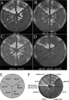
WSS1 is required for the growth of ulp1Δ ulp2Δ cells. (A to D) Four ulp1Δ yeast strains were constructed with the indicated WSS1 or ULP2 deletions, the complementing plasmid pNJ7352 (ULP1 CEN URA3 ADE3), and a multicopy plasmid expressing Smt3(G98). Each strain was then transformed with an additional multicopy plasmid, pRS424, containing either no insert or the SLX5 (pNJ6596) or slx5-8 (pNJ6595) gene under the control of the constitutive GPD1 promoter. As indicated in the schematic (E), the transformants were streaked in duplicate onto medium containing 5-FOA to select against the ULP1 plasmid. Colonies were photographed following 4 days of growth at 30°C. Note that the plate in panel A contains some large colonies among the slow-growing ulp1Δ strains. These fast-growing colonies arise occasionally due to mutations in the plasmid's URA3 gene. They are inconsequential, as they were shown to retain pNJ7352. Strains: (A) NJY2851-pNJ7340 [SMT3(G98) 2μm HIS3]; (B) NJY2857-pNJ7357 [SMT3(G98) 2μm LEU2]; (C) NJY2868-pNJ7340; (D) NJY2858-pNJ7357. (E) Schematic for complementing plasmids used for panels A to D. (F) Yeast strain JMY1803 (ulp1-333) was first transformed with either pNJ7340 [smt3(G98); upper half] or pRS423 (SMT3-Y101; lower half). Each strain was then transformed with the indicated multicopy TRP1 plasmid carrying WSS1 (pNJ7337), SLX5 (pNJ6596), SLX5 fused to the Gal4 DNA binding domain (pCS6513), or no insert (pRS424). Transformants were then streaked onto YPD medium and photographed following 3 days of growth at 37°C.
FIG. 9.
FIG. 9.
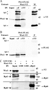
Wss1 physically associates with the proteasome. (A) Extracts were prepared from wt yeast cells expressing Pre1-FLAG and Wss1-V5 from galactose-inducible multicopy expression plasmids (KEp686 and pJM7349, respectively) or from wt cells expressing Wss1-V5 (pJM7349) alone (W). Extracts were subjected to IP with either anti-FLAG (αF), anti-V5 (αV5), or no antibody (−). The untreated extract (Input) or the indicated IP was then immunoblotted with antibody against the V5 epitope to detect Wss1. (B) The above-mentioned samples or extract from wt cells expressing Pre1-FLAG alone (P) was subjected to IP as described above and immunoblotted with antibody against the FLAG epitope to detect Pre1. (C) Extracts were prepared from wt yeast cells expressing Wss1-V5 and either a GST-Ubl fusion (+) or GST alone (−) from inducible multicopy expression plasmids (pJM7349 and either LEp17 or pGST, respectively) as indicated. Extracts were then subjected to precipitation with glutathione beads (GSH) or immunoprecipitated (IP) with antibody against Rpt1 (αRpt1). The untreated extract (Input) or the indicated IP was immunoblotted with antibody against V5 (upper), Rpt1 (middle), or Rpt6 (lower).

Similar articles

Cited by

References

    1. Alam, S. L., J. Sun, M. Payne, B. D. Welch, B. K. Blake, D. R. Davis, H. H. Meyer, S. D. Emr, and W. I. Sundquist. 2004. Ubiquitin interactions of NZF zinc fingers. EMBO J. 23:1411-1421. - PMC - PubMed
    1. Bachant, J., A. Alcasabas, Y. Blat, N. Kleckner, and S. J. Elledge. 2002. The SUMO-1 isopeptidase Smt4 is linked to centromeric cohesion through SUMO-1 modification of DNA topoisomerase II. Mol. Cell 9:1169-1182. - PubMed
    1. Biggins, S., N. Bhalla, A. Chang, D. L. Smith, and A. W. Murray. 2001. Genes involved in sister chromatid separation and segregation in the budding yeast Saccharomyces cerevisiae. Genetics 159:453-470. - PMC - PubMed
    1. Branzei, D., J. Sollier, G. Liberi, X. Zhao, D. Maeda, M. Seki, T. Enomoto, K. Ohta, and M. Foiani. 2006. Ubc9- and Mms21-mediated sumoylation counteracts recombinogenic events at damaged replication forks. Cell 127:509-522. - PubMed
    1. Burgess, R. C., S. Rahman, M. Lisby, R. Rothstein, and X. Zhao. 2007. The Slx5-Slx8 complex affects sumoylation of DNA repair proteins and negatively regulates recombination. Mol. Cell. Biol. 27:6153-6162. - PMC - PubMed

Publication types

MeSH terms