. 2010 Jul;28(7):1281-91.
doi: 10.1002/stem.447.

Decoded calreticulin-deficient embryonic stem cell transcriptome resolves latent cardiophenotype

Affiliations

Decoded calreticulin-deficient embryonic stem cell transcriptome resolves latent cardiophenotype

Randolph S Faustino et al. Stem Cells. 2010 Jul.

Abstract

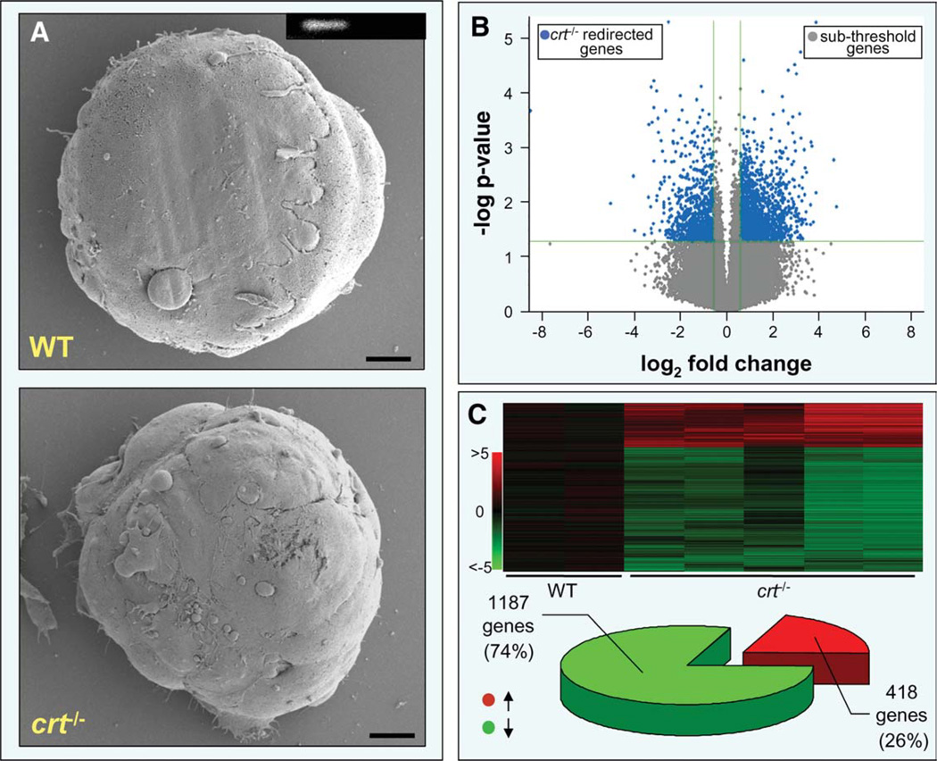
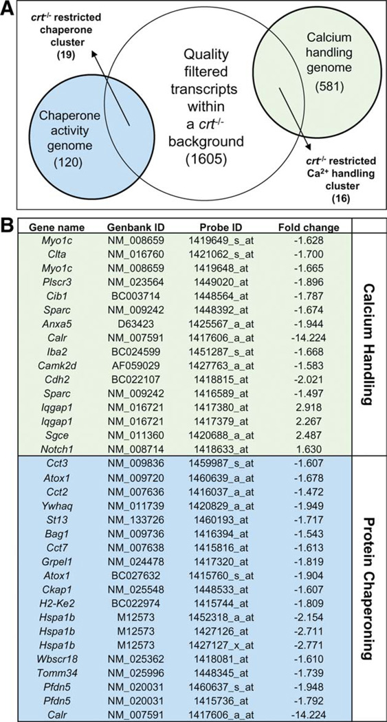
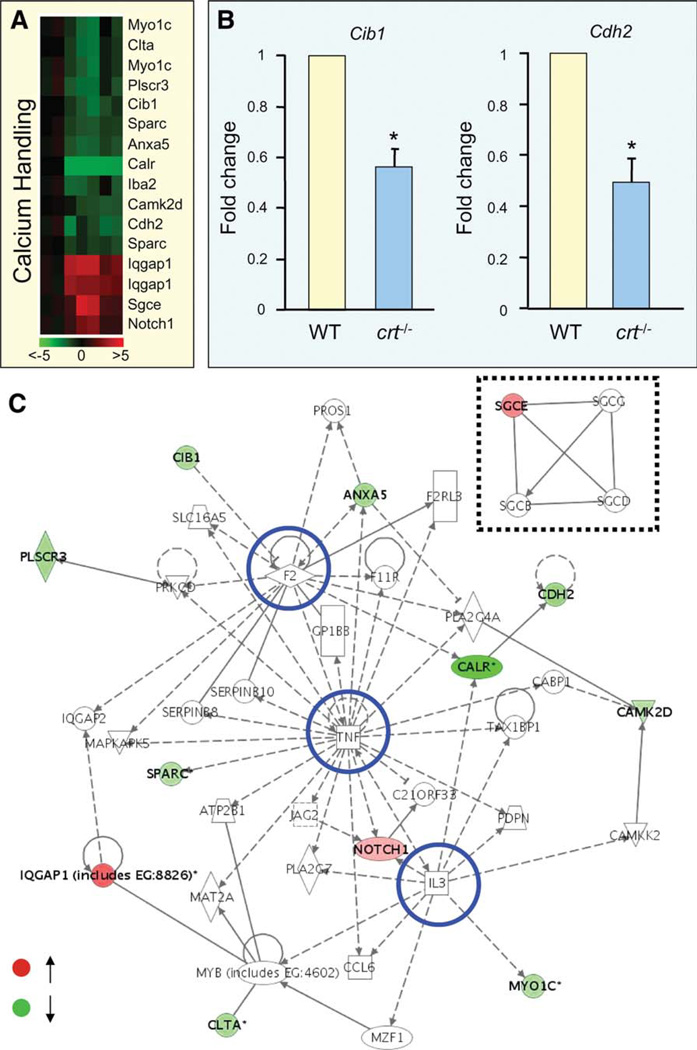
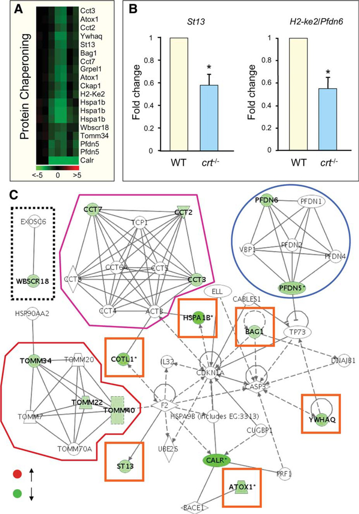
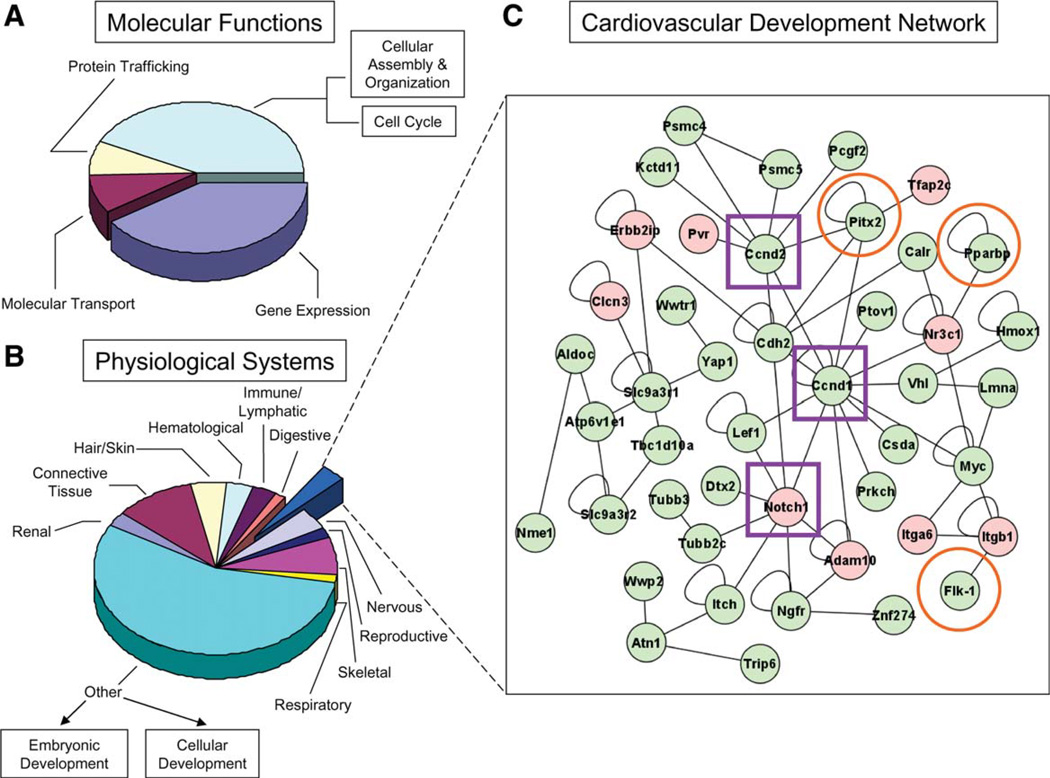
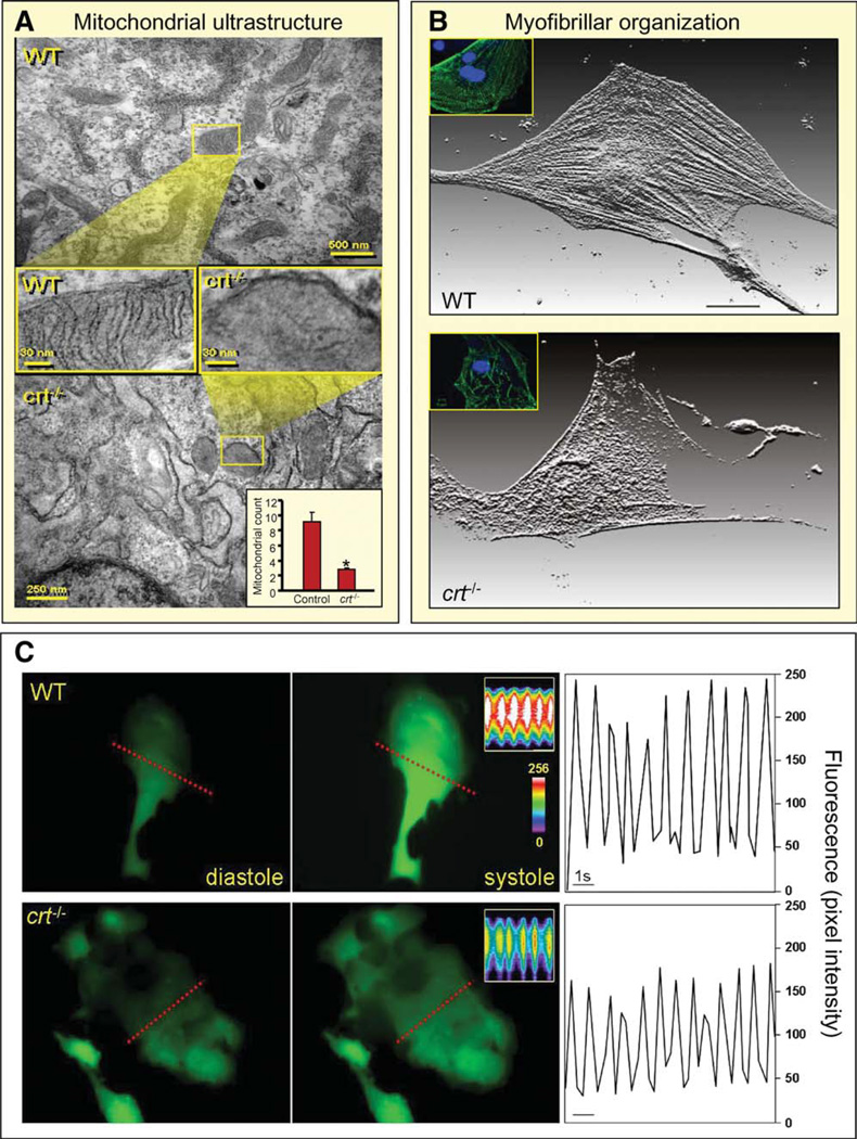
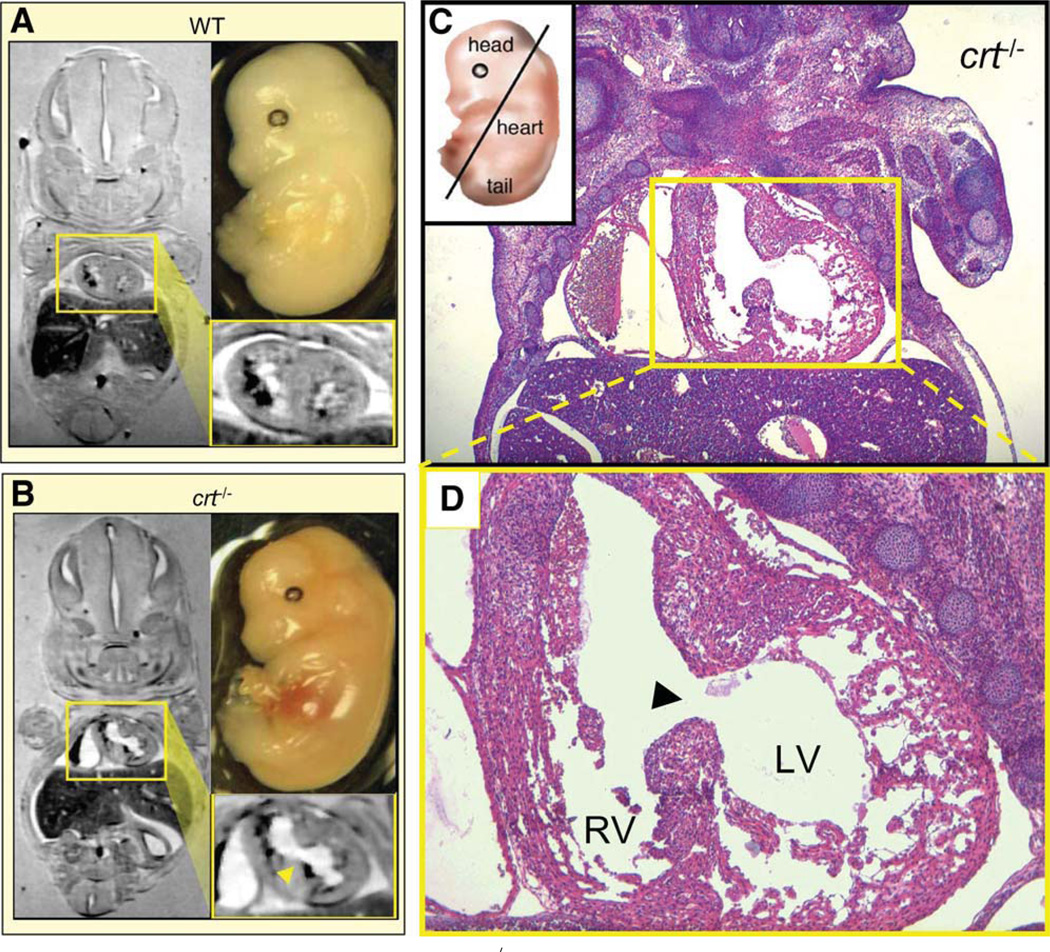
Genomic perturbations that challenge normal signaling at the pluripotent stage may trigger unforeseen ontogenic aberrancies. Anticipatory systems biology identification of transcriptome landscapes that underlie latent phenotypes would offer molecular diagnosis before the onset of symptoms. The purpose of this study was to assess the impact of calreticulin-deficient embryonic stem cell transcriptomes on molecular functions and physiological systems. Bioinformatic surveillance of calreticulin-null stem cells, a monogenic insult model, diagnosed a disruption in transcriptome dynamics, which re-prioritized essential cellular functions. Calreticulin-calibrated signaling axes were uncovered, and network-wide cartography of undifferentiated stem cell transcripts suggested cardiac manifestations. Calreticulin-deficient stem cell-derived cardiac cells verified disorganized sarcomerogenesis, mitochondrial paucity, and cytoarchitectural aberrations to validate calreticulin-dependent network forecasts. Furthermore, magnetic resonance imaging and histopathology detected a ventricular septal defect, revealing organogenic manifestation of calreticulin deletion. Thus, bioinformatic deciphering of a primordial calreticulin-deficient transcriptome decoded at the pluripotent stem cell stage a reconfigured multifunctional molecular registry to anticipate predifferentiation susceptibility toward abnormal cardiophenotype.


Conflict of interest statement

Disclosure of Potential Conflicts of Interest

The authors indicate no potential conflicts of interest.

Figures

Figure 1
Figure 1
Embryonic stem cells lacking calreticulin harbor significant transcriptome shifts. (A): Field-emission scanning electron microscopy, of WT and crt−/− embryonic stem cells, shows no significant morphological differences. Scale bar, 10 µm. Inset: Reverse transcription-polymerase chain reaction confirms absence of crt in knockout stem cells (left, WT; right, crt−/−). (B): Volcano plot analysis of total RNA changes between WT and crt−/− embryonic stem cells revealed 2,904 significant down- and upregulated genes (blue), distinct from genes falling below significance threshold or minimum 1.5-fold change cutoff (gray). (C): Sample reproducibility allowed construction of an expression heatmap of 1,605 quality restricted genes (upper panel). Composition of the shifted transcriptome was 74% downregulated (green) and 26% upregulated (red) (lower panel). Abbreviation: WT, wild type.
Figure 2
Figure 2
Compromised functions related to innate properties of calreticulin. (A): Cross-reference of quality-filtered genes within the calreticulin knockout background against Kyoto Encyclopedia of Genes and Genomes pathway lists for calcium handling and chaperone activity, identified 16 and 19 transcripts, respectively, significantly affected. (B): Identities of affected transcripts with Affymetrix Probe and Genbank IDs. Associated fold changes are listed on the right.
Figure 3
Figure 3
Loss of calreticulin impacts calcium handling processes. (A): Down- and upregulated calcium handling genes within a calreticulin-deficient background integrate into a heatmap and display pairwise correlation of gene expression changes. Color scale indicates range of fold change. (B): Calmyrin (Cib1) and cadherin 2 (Cdh2) as random examples of microarray-identified fold changes confirmed by reverse tran-scription-polymerase chain reaction. (C): Discrete sarcoglycan module (dashed box) and main network with highly connected hubs (blue circles) integrates downregulated (green) and upregulated (red) genes that together visualizes organization of transcripts affected by calreticulin knockout associated with calcium handling. Abbreviations: WT, wild type; TNF, tumor necrosis factor.
Figure 4
Figure 4
Affected protein chaperoning genes organize into a calreticulin inclusive network. (A): Calreticulin-impacted chaperoning genes were hierarchically clustered according to Pearson correlation. (B): Taqman validation of randomly selected chaperoning molecules, suppressor of tumorigenicity 13 (St13) and prefoldin six (Pfdn6). (C): Genes were downregulated (green) and organized into a network of associated modules. Clockwise from left: Discrete curated interaction of Exosc6 and Wbscr18 (boxed in dashed line); TRiC/ CCT module with Cct2 Cct3, and Cct7 downregulated (outlined in purple); prefoldin subnetwork showing downregulation in Pfdn5 and Pfdn6 (circled in blue); central network that integrated nonmodular genes (orange boxes); and the Tomm complex with affected genes highlighted in green (outlined in red). Abbreviation: WT, wild type.
Figure 5
Figure 5
Molecular attenuation of calreticulin impacts a transcriptome network with repercussions on cardiovascular development. (A): Onto-logical analysis of genes affected by calreticulin deficiency reveals transcripts that impact global processes of gene expression, protein trafficking, and molecular transport, with numerous accessory functions. (B): Re-analysis of the calreticulin-deficient transcriptome according to physiological development prioritized 11 distinct systems. (C): Genes associated with cardiac differentiation organized into a network of curated relationships within a calreticulin-deficient background. Composed of 45 network nodes, nine displayed increases in expression (red), while the remaining genes were downregulated (green), after calreticulin deletion. Within this network, Ccnd1 Ccnd2, and Notch1 are identified as network hubs (purple boxes), defined as nodes that possess the greatest number of connections to neighboring genes. Orange circles highlight genes with characterized effects on cardiac formation.
Figure 6
Figure 6
Embryonic transcriptome shift precipitated by calreticulin deletion translates into phenotype derangement. (A): Comparison of mitochondria, visualized by electron microscopy, in WT (upper) and calreticulin-deficient embryonic stem cells (lower). Cross-sectional magnification revealed absence of cristae in the latter. Inset, lower right: Enumeration of mitochondrial number in WT versus crt−/−. Mitochondrial count indicated on y-axis. *, p < .05, n = 5. (B): Individual stem cell-derived cardiomyocytes stained for α-actinin, displayed here as a three-dimensional reconstruction of fluorescence. Proper sarcomere formation in WT stem cell-derived cardiomyocytes is illustrated (upper), and disarrayed myofibrillogenesis or no sarcomere generation is present in cardiomyocytes from crt−/− stem cells. Inset: Stem cell-derived cardiomyocytes stained for MLC2V (green) and DAPI-counterstained nuclei (blue). (C): Calcium transient measurements. Left and middle panels: Ca2+ levels in diastole and systole in WT and crt−/− stem cell-derived cardiomyocytes loaded with the calcium indicator, Fluo-3. Inset: Line scan (indicated by dashed line) records calcium transients in contractile cells. Color scale indicates high (white) and low (blue/purple) calcium concentrations. Right: Analysis of calcium transients as a function of time demonstrate attenuated peak intensity in crt−/− knockouts (bottom) compared with WT counterparts (top). Bar = 1 second. Abbreviation: WT, wild type.
Figure 7
Figure 7
Magnetic resonance reveals ventricular septal defect in crt−/− embryo. Comparative in situ magnetic resonance imaging of coronal planes from 14.5 dpc-old WT and crt−/− embryos demonstrates normal (A) versus defective (B) cardiac structure, respectively, as a predicted manifestation of cardiac phenotype. Magnified cardiac digital cross-section (yellow boxes) shows prominent ventricular septal defect with interventricular communication in crt−/− (arrowhead) in contrast to distinct left and right ventricular lumens in WT hearts characteristic for this stage of cardiogenesis. (C): Pathoanatomical verification of crt−/− dysmorphic cardiac structure (right). Coronal hematoxylin-eosin stained thin section of embryo visualizes a direct communication (ventricular septal defect) between LV and RV through a patent ventricular septum in the crt-null mutant. (D): Magnification of cardiac section highlights incomplete septal development, a common manifestation of congenital heart disease. Histology confirms known defect of a thin left ventricular myocardium characterized by deep intertrabecular recesses with the predominance of large fenestrations. Right ventricle demonstrates normal muscularization without pathological thinning or gross abnormalities. Abbreviations: LV, left ventricle; RV, right ventricle; WT, wild type.

Similar articles

Cited by

References

    1. Suzuki A, Raya Á, Kawakami Y, et al. Maintenance of embryonic stem cell pluripotency by Nanog-mediated reversal of mesoderm specification. Nat Clin Pract Cardiovasc Med. 2006;3:S114–S122. - PubMed
    1. Boyer LA, Lee TI, Cole MF, et al. Core transcriptional regulatory circuitry in human embryonic stem cells. Cell. 2005;122:947–956. - PMC - PubMed
    1. Loh Y-H, Wu Q, Chew J-L, et al. The Oct4 and Nanog transcription network regulates pluripotency in mouse embryonic stem cells. Nat Genet. 2006;38:431–440. - PubMed
    1. Faustino RS, Terzic A. Interactome of a cardiopoietic precursor. J Cardiovasc Trans Res. 2008;1:120–126. - PubMed
    1. Komili S, Silver PA. Coupling and coordination in gene expression processes: A systems biology view. Nat Rev Genet. 2008;9:38–48. - PubMed

Publication types