Distinct FGFs promote differentiation of excitatory and inhibitory synapses
- PMID: 20505669
- PMCID: PMC4137042
- DOI: 10.1038/nature09041
Distinct FGFs promote differentiation of excitatory and inhibitory synapses
Abstract
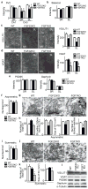
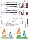
The differential formation of excitatory (glutamate-mediated) and inhibitory (GABA-mediated) synapses is a critical step for the proper functioning of the brain. An imbalance in these synapses may lead to various neurological disorders such as autism, schizophrenia, Tourette's syndrome and epilepsy. Synapses are formed through communication between the appropriate synaptic partners. However, the molecular mechanisms that mediate the formation of specific synaptic types are not known. Here we show that two members of the fibroblast growth factor (FGF) family, FGF22 and FGF7, promote the organization of excitatory and inhibitory presynaptic terminals, respectively, as target-derived presynaptic organizers. FGF22 and FGF7 are expressed by CA3 pyramidal neurons in the hippocampus. The differentiation of excitatory or inhibitory nerve terminals on dendrites of CA3 pyramidal neurons is specifically impaired in mutants lacking FGF22 or FGF7. These presynaptic defects are rescued by postsynaptic expression of the appropriate FGF. FGF22-deficient mice are resistant to epileptic seizures, and FGF7-deficient mice are prone to them, as expected from the alterations in excitatory/inhibitory balance. Differential effects of FGF22 and FGF7 involve both their distinct synaptic localizations and their use of different signalling pathways. These results demonstrate that specific FGFs act as target-derived presynaptic organizers and help to organize specific presynaptic terminals in the mammalian brain.
Figures


Similar articles
-
Distinct sets of FGF receptors sculpt excitatory and inhibitory synaptogenesis.Development. 2015 May 15;142(10):1818-30. doi: 10.1242/dev.115568. Epub 2015 Apr 29. Development. 2015. PMID: 25926357 Free PMC article.
-
Selective synaptic targeting of the excitatory and inhibitory presynaptic organizers FGF22 and FGF7.J Cell Sci. 2015 Jan 15;128(2):281-92. doi: 10.1242/jcs.158337. Epub 2014 Nov 27. J Cell Sci. 2015. PMID: 25431136 Free PMC article.
-
FGF22 and its close relatives are presynaptic organizing molecules in the mammalian brain.Cell. 2004 Jul 23;118(2):257-70. doi: 10.1016/j.cell.2004.06.025. Cell. 2004. PMID: 15260994
-
Cellular abnormalities and synaptic plasticity in seizure disorders of the immature nervous system.Ment Retard Dev Disabil Res Rev. 2000;6(4):258-67. doi: 10.1002/1098-2779(2000)6:4<258::AID-MRDD5>3.0.CO;2-H. Ment Retard Dev Disabil Res Rev. 2000. PMID: 11107191 Review.
-
The synaptic split of SNAP-25: different roles in glutamatergic and GABAergic neurons?Neuroscience. 2009 Jan 12;158(1):223-30. doi: 10.1016/j.neuroscience.2008.03.014. Epub 2008 Mar 20. Neuroscience. 2009. PMID: 18514426 Review. No abstract available.
Cited by
-
Distinct sets of FGF receptors sculpt excitatory and inhibitory synaptogenesis.Development. 2015 May 15;142(10):1818-30. doi: 10.1242/dev.115568. Epub 2015 Apr 29. Development. 2015. PMID: 25926357 Free PMC article.
-
Roles of FGFs As Paracrine or Endocrine Signals in Liver Development, Health, and Disease.Front Cell Dev Biol. 2016 Apr 13;4:30. doi: 10.3389/fcell.2016.00030. eCollection 2016. Front Cell Dev Biol. 2016. PMID: 27148532 Free PMC article. Review.
-
Fgf22 regulated by Fgf3/Fgf8 signaling is required for zebrafish midbrain development.Biol Open. 2013 Apr 10;2(5):515-24. doi: 10.1242/bio.20134226. Print 2013 May 15. Biol Open. 2013. PMID: 23789101 Free PMC article.
-
Genomic Targets of Positive Selection in Giant Mice from Gough Island.Mol Biol Evol. 2021 Mar 9;38(3):911-926. doi: 10.1093/molbev/msaa255. Mol Biol Evol. 2021. PMID: 33022034 Free PMC article.
-
The class 4 semaphorin Sema4D promotes the rapid assembly of GABAergic synapses in rodent hippocampus.J Neurosci. 2013 May 22;33(21):8961-73. doi: 10.1523/JNEUROSCI.0989-13.2013. J Neurosci. 2013. PMID: 23699507 Free PMC article.
References
-
- Wassef A, Baker J, Kochan LD. GABA and schizophrenia: a review of basic science and clinical studies. J Clin Psychopharmacol. 2003;23:601–640. - PubMed
-
- Singer HS, Minzer K. Neurobiology of Tourette’s syndrome: concepts of neuroanatomic localization and neurochemical abnormalities. Brain Dev. 2003;25:S70–S84. - PubMed
-
- Möhler H. GABAA receptors in central nervous system disease: anxiety, epilepsy, and insomnia. J Recept Signal Transduct Res. 2006;26:731–740. - PubMed
-
- Sanes JR, Lichtman JW. Development of the vertebrate neuromuscular junction. Annu Rev Neurosci. 1999;22:389–442. - PubMed
Publication types
MeSH terms
Substances
Grants and funding
LinkOut - more resources
Full Text Sources
Other Literature Sources
Molecular Biology Databases
Miscellaneous

