. 2010 Jun;120(6):2119-30.
doi: 10.1172/JCI40583. Epub 2010 May 10.

COMMD1 disrupts HIF-1alpha/beta dimerization and inhibits human tumor cell invasion

Affiliations

COMMD1 disrupts HIF-1alpha/beta dimerization and inhibits human tumor cell invasion

Bart van de Sluis et al. J Clin Invest. 2010 Jun.

Abstract

The gene encoding COMM domain-containing 1 (COMMD1) is a prototypical member of the COMMD gene family that has been shown to inhibit both NF-kappaB- and HIF-mediated gene expression. NF-kappaB and HIF are transcription factors that have been shown to play a role in promoting tumor growth, survival, and invasion. In this study, we demonstrate that COMMD1 expression is frequently suppressed in human cancer and that decreased COMMD1 expression correlates with a more invasive tumor phenotype. We found that direct repression of COMMD1 in human cell lines led to increased tumor invasion in a chick xenograft model, while increased COMMD1 expression in mouse melanoma cells led to decreased lung metastasis in a mouse model. Decreased COMMD1 expression also correlated with increased expression of genes known to promote cancer cell invasiveness, including direct targets of HIF. Mechanistically, our studies show that COMMD1 inhibits HIF-mediated gene expression by binding directly to the amino terminus of HIF-1alpha, preventing its dimerization with HIF-1beta and subsequent DNA binding and transcriptional activation. Altogether, our findings demonstrate a role for COMMD1 in tumor invasion and provide a detailed mechanism of how this factor regulates the HIF pathway in cancer cells.


Figures
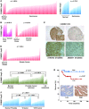
Figure 1
Figure 1. Decreased COMMD1 expression is found in several cancers.
(A and B) COMMD1 expression in individual samples from normal tissues or the corresponding cancer is shown (as normalized expression in log2 scale). Comparisons between groups were performed using the Student’s t test, with P values indicated in each panel. Panc duct, pancreatic duct. (C) Lack of detectable COMMD1 protein expression is frequent in ovarian cancer. Representative examples of COMMD1 immunohistochemical (IHC) staining of ovarian cancer specimens are shown (original magnification, ×200 [top panels]; ×600 [insets]; ×400 [bottom panels]). (D) COMMD1 expression in stromal cells from normal breast tissue and breast cancer is shown in a similar format as in A. (E) COMMD1 expression in normal prostate, prostate cancer, and metastatic prostate cancer specimens from lymph nodes is shown. Comparisons between groups were performed using the Student’s t test, with P values indicated. (F) COMMD1 expression in the same sample set, this time subdivided according to the local tumor T stage. Similar to A, the Student’s t test was performed to compare the groups, and P values are shown. (G) COMMD1 expression by immunohistochemical staining correlates with patient survival in endometrial cancer (n = 63). The survival curves of patients with intense cytoplasmic staining (high, blue curve) and weak/absent nucleocytoplasmic staining (low, red curve) are compared. Representative images (original magnification, ×400) are included.
Figure 2
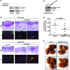
Figure 2. COMMD1 repression promotes tumor invasion.
(AC) HT29 or U2OS cells were infected with lentiviruses expressing short hairpin sequences targeting COMMD1 (shCOMMD1) or a control gene (shControl). (A) Western blot analysis demonstrating the decrease in COMMD1 levels in the cell lines used is shown. These cells were tested for invasion of the chick CAM, as described in the Methods section. (B) Representative images of H&E and immunofluorescence staining (IF) are shown (laminin, red; DAPI, blue; cancer cells, green). Cell invasion into the CAM is indicated with white arrows (original magnification, ×200 [HT29]; ×100 [U2OS]). (CE) B16.F10 mouse melanoma cells were stably transfected to express COMMD1-FLAG (as shown in the Western blot in C). These cells were subsequently injected into the tail vein of syngeneic C57BL6 mice (2 × 10⁵ cells/mouse), and (D) after 15 days, the mice were sacrificed, and the number of lung metastasis per mouse was counted (the horizontal bars represent the mean in each group). The Student’s t test was used for statistical analysis. (E) Representative images of the lungs of these mice are shown.
Figure 3
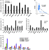
Figure 3. COMMD1 inhibits the expression of HIF-regulated genes.
(A) Gene expression for a panel of invasion-promoting genes was determined in HT29 cells by quantitative RT-PCR and expressed as fold over the shControl sample (mean ± SEM of triplicate samples are shown). (B) Normalized COMMD1 and CXCR4 expression in breast stromal cells (shown in Figure 1E) was plotted and a Pearson’s correlation value was calculated. (C and D) The expression of the same genes was examined in HT29 cells exposed to (C) hypoxia or to (D) the NF-κB activator, TNF. The respective mRNA levels were determined by quantitative RT-PCR and expressed as fold over the untreated sample (mean ± SEM of triplicate samples are shown). (E) Expression of selected HIF target genes was determined by quantitative RT-PCR analysis and expressed as fold over the shControl sample (mean ± SEM of triplicate samples are shown).
Figure 4
Figure 4. COMMD1 inhibits HIF-mediated transcription and DNA binding without affecting HIF-α expression.
(A) Protein expression of HIF-1α and HIF-2α was not affected by COMMD1 deficiency in HT29 and U2OS cells. Western blot analysis for HIF-1α, HIF-2α, COMMD1, and β-Actin from the indicated samples is shown. Long and short exposures of the HIF-1α Western blot are shown. (B) HEK 293T cells with a stable repression of COMMD1 or control cells were cotransfected with 5xHRE-firefly luciferase reporter and renilla luciferase control plasmid. Cells were incubated under normoxia or hypoxia for 16 hours. HRE reporter activity was determined by measuring firefly luciferase in the lysates and was corrected for transfection efficiency using renilla luciferase activities. Results are expressed as fold induction relative to normoxic conditions in shControl cells (mean ± SD of triplicate samples are shown). (C) The same cells examined in B were used for the HIF-1α HRE DNA binding assay. DNA binding is expressed as fold binding relative to normoxic conditions in shControl cells (mean ± SD of triplicate samples are shown). COMMD1 expression is shown by immunoblot analysis. (DF) HIF1A mRNA expression (encoding for HIF-1α) was determined by qRT-PCR in (D) HT29 and (E) U2OS cells and normalized to shControl transfected cells under normoxia. (F) Similarly, the effect of TNF treatment on HIF1A expression in HT29 was also examined (mean ± SEM of triplicate samples are shown).
Figure 5
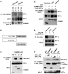
Figure 5. COMMD1 binds to HIF-1α preferentially under normoxic conditions.
(A) Endogenous COMMD1 immunoprecipitation using HEK 293T whole cell lysates incubated either under normoxia or hypoxia (for 1 hour). Whole cell lysates and immunoprecipitates were subjected to Western blot analysis. Supernatants after IP demonstrate immunodepletion of COMMD1. The lanes on the top panel were run on the same gel but were noncontiguous. (B) Truncation mutants of COMMD1. FL, full-length; ΔC, deletion of the carboxyl terminal COMM domain; ΔN, deletion of the amino terminus. (C) These proteins were coexpressed with HA-tagged HIF-1α in HEK293 cells and subsequently precipitated from cell lysates. Coprecipitation of HIF-1α with COMMD1 full-length and ΔN is shown. (D) HEK 293T cells expressing COMMD1-GST, GST, or FLAG–HIF-1α were cultured under normoxia (20% O2) or hypoxia (1% O2, 8 hours) or treated with DFO (0.1 mM, 8 hours), prior to glutahione-sepharose (GSH) precipitations and Western blot analysis as indicated. (E) HEK 293T cells expressing HA-VHL and FLAG–HIF-1α were cultured under normoxia, hypoxia, or treated with DFO as before. Immunoprecipitation of VHL (using anti-HA antibody) was followed by Western blot analysis. (F) HEK 293T cells were transfected with GST, COMMD1-GST, or GST-VHL, along with wild-type HIF-1α or HIF-1α P/A. Subsequently, GSH precipitations were followed by Western blot (WB) analysis as indicated. PD, pull down; PIS, preimmune serum.
Figure 6
Figure 6. COMMD1 binds directly to the amino terminus of HIF-1α and inhibits HIF-1β dimerization.
(A) Schematic representation of the domains in HIF-1α. AD, activation domain. (B) HEK 293T cells expressing COMMD1-GST or GST and the indicated truncated fragments of FLAG–HIF-1α were used for GSH precipitations and Western blot analysis as shown. (C) Recombinant COMMD-1-MBP was incubated with immobilized recombinant HIF-1α full-length, HIF-1α fragment (1–300), or uncoated wells. Binding of the proteins was detected by absorbance at 450 nm, as described in the Methods section. Data represent the mean of duplicate experiments. (D) HEK 293T cells were transfected to express COMMD1-HA, FLAG–HIF-1α, or HIF-1β as indicated. HIF-1α was immunoprecipitated using an anti-FLAG antibody, and the resulting samples were subjected to Western blot analysis. (E) HEK 293T cell lysates expressing GST, GST-HIF-1α, or COMMD1 were subjected to GSH precipitations. The amount of coprecipitated endogenous HIF-1β was identified by Western blot analysis. (F and G) Lysates from HEK293 cells transfected with HA–HIF-1α were incubated with the indicated recombinant proteins. (F) Immunoprecipitation of HIF-1α (HA antibody, FLAG antibody as a control) was performed. The amount of HIF-1β recovered was assessed by immunoblotting. (G) The lysates were subsequently mixed with tandem HRE oligonucleotide probes, which were then precipitated and immunoblotted for HIF-1α. 2xHRE represents a control oligonucleotide that was not biotinylated, biotin-2xHRE represents the biotinylated version. FL, COMMD1 full-length; SA, streptavidin.

Similar articles

Cited by

References

    1. Maine GN, Burstein E. COMMD proteins: COMMing to the scene. Cell Mol Life Sci. 2007;64(15):1997–2005. - PMC - PubMed
    1. Ganesh L, et al. The gene product Murr1 restricts HIV-1 replication in resting CD4+ lymphocytes. . Nature. 2003;426(6968):853–857. doi: 10.1038/nature02171. - DOI - PubMed
    1. van de Sluis B, Rothuizen J, Pearson PL, van Oost BA, Wijmenga C. Identification of a new copper metabolism gene by positional cloning in a purebred dog population. Hum Mol Genet. 2002;11(2):165–173. - PubMed
    1. van de Sluis B, et al. Increased activity of hypoxia-inducible factor 1 is associated with early embryonic lethality in Commd1 null mice. . Mol Cell Biol. 2007;27(11):4142–4156. doi: 10.1128/MCB.01932-06. - DOI - PMC - PubMed
    1. Gordan JD, Simon MC. Hypoxia-inducible factors: central regulators of the tumor phenotype. Curr Opin Genet Dev. 2007;17(1):71–77. doi: 10.1016/j.gde.2006.12.006. - DOI - PMC - PubMed

Publication types

MeSH terms