. 2010 Jun;298(6):C1445-56.
doi: 10.1152/ajpcell.00508.2009. Epub 2010 Mar 24.

Prostaglandin E2 induces interleukin-6 expression in human chondrocytes via cAMP/protein kinase A- and phosphatidylinositol 3-kinase-dependent NF-kappaB activation

Affiliations

Prostaglandin E2 induces interleukin-6 expression in human chondrocytes via cAMP/protein kinase A- and phosphatidylinositol 3-kinase-dependent NF-kappaB activation

Pu Wang et al. Am J Physiol Cell Physiol. 2010 Jun.

Abstract

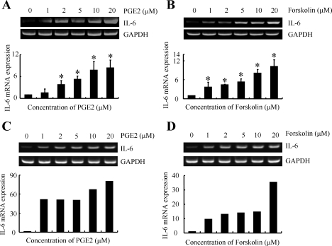
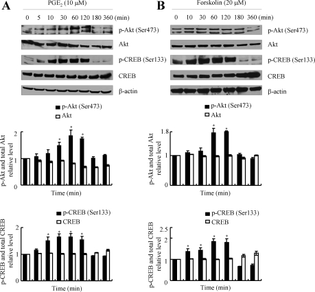
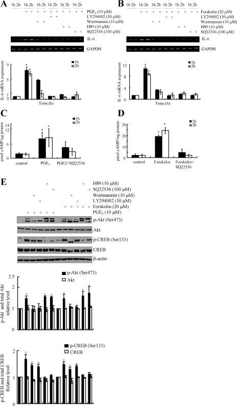
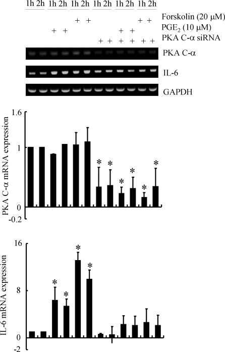
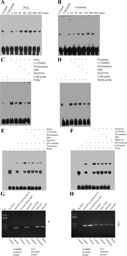
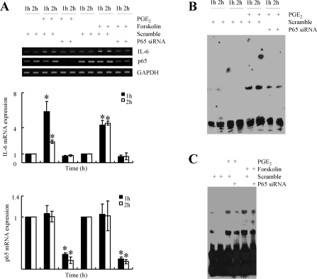
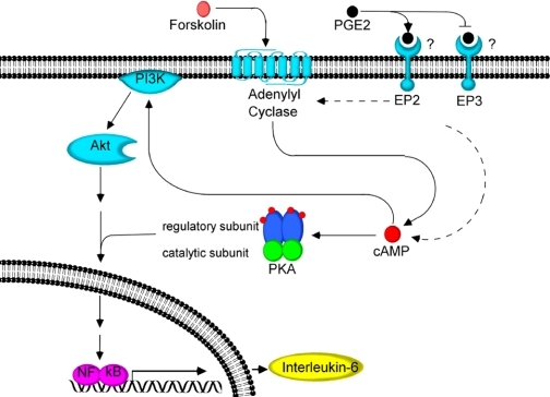
Elevated levels of prostaglandin (PG)E(2) and interleukin (IL)-6 have been reported in the cartilage and synovial fluid from patients with arthritic disorders. PGE(2) regulates IL-6 production in numerous different cells including macrophages and synovial fibroblasts. Although PGE(2) stimulates IL-6 expression in human chondrocytes, the underlying signaling pathway of this process has yet to be delineated. Here, we investigate the mechanism of IL-6 induction in human T/C-28a2 chondrocytes treated with exogenously added PGE(2). PGE(2) induces IL-6 mRNA and protein expression via a cAMP-dependent pathway, reaching maximal levels after 60 min of stimulation before declining to baseline levels at 6 h. Forskolin, an adenylyl cyclase activator, also stimulates IL-6 expression in human chondrocytes in a dose- and time-dependent fashion. Inhibition of downstream effectors of cAMP activity such as protein kinase A (PKA) or phosphatidylinositol 3 kinase (PI3K) blocks PGE(2)- and forskolin-induced IL-6 upregulation. Simultaneous inhibition of PKA and PI3K reduces IL-6 expression in stimulated chondrocytes well below the basal levels of untreated cells. Gel shift, supershift, and chromatin immunoprecipitation assays reveal the activation and binding of the nuclear factor (NF)-kappaB p65 subunit to the IL-6 promoter, which is markedly suppressed by selective PI3K or PKA pharmacological inhibitors. p65 knockdown completely abrogates IL-6 mRNA synthesis in PGE(2)- and forskolin-primed chondrocytes. Cumulatively, our data show that PGE(2) and forskolin induce IL-6 expression in human chondrocytes via cAMP/PKA and PI3K-dependent pathways, which in turn regulate the activation and binding of p65 to the IL-6 promoter.


Figures

Fig. 1.
Fig. 1.
Dose-dependent induction of interleukin (IL)-6 mRNA synthesis by prostaglandin (PG)E2 or forskolin in human chondrocytes. T/C-28a2 chondrocytes (A, B) or human primary articular chondrocytes (C, D) were treated with the indicated concentrations of either PGE2 (A, C) or forskolin (B, D) for 1 h. IL-6 mRNA expression was determined by quantitative RT-PCR. GAPDH served as internal control. Data are means ± SE of three independent experiments, except for the experiments performed using human primary chondrocytes (n = 1). *P < 0.05 with respect to no treatment (vehicle) control.
Fig. 2.
Fig. 2.
Time-dependent increases of IL-6 mRNA and protein synthesis and cAMP production induced by PGE2 or forskolin in human chondrocytes. T/C-28a2 chondrocytes or human primary articular chondrocytes (D, H) were treated with either PGE2 (10 μM) (AD) or forskolin (20 μM) (EH) for the indicated time intervals. IL-6 mRNA expression was determined by qRT-PCR (A, D, E, H). GAPDH served as internal control. IL-6 protein expression levels were determined by Western blotting using an anti-IL-6 mAb (B, F). β-Actin was used as loading control. cAMP formation was monitored by a cAMP enzyme immunoassay kit (C, G). Data are means ± SE of three independent experiments, except for the experiments performed using human primary chondrocytes (n = 1). *P < 0.05 with respect to no treatment (vehicle) control.
Fig. 3.
Fig. 3.
PGE2 and forskolin induce phosphorylation of Akt and cAMP responsive element binding protein (CREB) in human T/C-28a2 chondrocytes. T/C2–8a2 chondrocytes were treated with either PGE2 (10 μM) (A) or forskolin (20 μM) (B) for the indicated periods of time. Phosphorylated Akt (Ser473), total Akt, phosphorylated CREB (Ser133) and total CREB levels are shown by immunoblotting using specific Abs. Equal loading in each lane is shown by the similar intensities of total Akt, total CREB and β-actin. Immunoblots are representative of three independent experiments, all revealing similar results. The intensity of bands was quantified relative to β-actin for each treatment using the Bio-Rad gel image system and then normalized with respect to the value obtained for the untreated control. Data are means ± SE of three independent experiments. *P < 0.05 with respect to no treatment control.
Fig. 4.
Fig. 4.
Involvement of PI3K and cAMP/PKA signaling pathways in IL-6 mRNA synthesis and cAMP production induced by PGE2 or forskolin in human T/C28a2 chondrocytes. T/C-28a2 cells were incubated with either PGE2 (10 μM) (A, C) or forskolin (20 μM) (B, D) for 1 or 2 h in presence of the PI3K inhibitors [LY-294002 (30 μM) or wortmannin (10 μM)] or the PKA inhibitor H89 (10 μM) or the adenylyl cyclase inhibitor SQ-22536 (100 μM). IL-6 mRNA synthesis was determined by qRT-PCR (A, B). GAPDH served as internal control. cAMP formation was analyzed by a cAMP enzyme immunoassay kit (C, D). Data are means ± SE of three independent experiments. *P < 0.05 with respect to all pharmacological treatments and no treatment (vehicle) control. E: Western blots showing the effects of the phosphatidylinositol 3 kinase (PI3K) inhibitors (LY-294002; wortmannin), PKA inhibitor H89, or the adenylyl cyclase inhibitor SQ-22536 on PGE2- and forskolin-mediated phosphorylation of Akt (Ser473) and CREB (Ser133) using specific antibodies (Abs). Equal loading in each lane is shown by the similar intensities of Akt, CREB, and β-actin. The immunoblots shown are representative of three independent experiments with similar results. The intensity of bands was quantified relative to β-actin for each treatment using the Bio-Rad gel image system and then normalized with respect to the value obtained for the untreated control. Data are means ± SE of three independent experiments. *P < 0.05 with respect to no treatment control.
Fig. 5.
Fig. 5.
PKA C-α knockdown inhibits PGE2- and forskolin- induced IL-6 mRNA synthesis in human chondrocytes. T/C-28a2 chondrocytes were transfected with an small interfering RNA (siRNA) oligonucleotide sequence specific for PKA C-α before being treated with either PGE2 (10 μM) or forskolin (20 μM) for 1 or 2 h. PKA C-α (middle) and IL-6 mRNA (bottom) expressions were determined by quantitative RT-PCR. GAPDH was served as internal control. Data are means ± SE of three independent experiments. *P < 0.05 with respect to no treatment control.
Fig. 6.
Fig. 6.
The Epac-selective cAMP analog 8-(4-chlorophenylthio)-2′-O-methyladenosine-3′,5′-cyclic monophosphate (8-pCPT-2′-O-Me-cAMP·Na) does not induce IL-6 expression in human chondrocytes. T/C-28a2 chondrocytes were treated with 8-pCPT-2′-O-Me-cAMP·Na (10 μM) for indicated time intervals (A, C). Alternatively, cells were treated with the indicated concentrations of 8-pCPT-2′-O-Me-cAMP·Na for 1 h (B, D). Akt1, Akt2, Akt3, and IL-6 mRNA levels were determined by qRT-PCR (A, B). GAPDH served as internal control (A, B). Phosphorylated Akt and total Akt protein levels were monitored by immunoblotting using specific Abs. β-actin was used as loading control. The immunoblots are representative of three independent experiments, all revealing similar results.
Fig. 7.
Fig. 7.
CREB1 and ATF4 are not involved in PGE2- and forskolin-induced IL-6 synthesis in human T/C-28a2 chondrocytes. T/C-28a2 cells were transfected with an siRNA oligonucleotide sequence specific for CREB1 or ATF4, or a control siRNA, before being treated with forskolin (20 μM) for the indicated periods of time. IL-6, CREB1, and ATF4 mRNA levels were determined by qRT-PCR. GAPDH served as internal control. Data are means ± SE of three independent experiments. *P < 0.05 with respect to controls.
Fig. 8.
Fig. 8.
PGE2 and forskolin induce NF-κB activation in human T/C28a2 chondrocytes via cAMP/PKA and PI3K signaling pathways. T/C-28a2 cells were treated with PGE2 (10 μM) (A) or forskolin (20 μM) (B) for the indicated time intervals. Nuclear extracts were then isolated, and NF-κB-specific DNA-protein complex formation was determined by EMSA. T/C-28a2 chondrocytes were treated for 2 h with either PGE2 (C, E, G, H) or forskolin (D, F, G, H) in the presence or absence of the PI3K inhibitors [LY-294002 (30 μM) or wortmannin (10 μM)], the PKA inhibitor H89 (10 μM), or the adenylyl cyclase inhibitor SQ-22536 (100 μM). Nuclear extracts were prepared for the determination of NF-κB-specific DNA-protein complex formation by EMSA (C, D). Supershift (E, F) assays using an anti-p65 Ab were carried out as outlined in materials and methods. Results of a competition experiment using 50-fold unlabeled NF-κB oligonucleotide (cold probe) are shown. Cross-linked chromatin was immunoprecipitated using an anti-p65 antibody (G, H). In ChIP assays, the anti-RNA polymerase II antibody was used as positive control, whereas the normal mouse IgG and anti-TLR4 antibodies were used as negative controls. DNA purified from both the immunoprecipitated (IP) and preimmune (input) specimens was subjected to PCR amplification using primers for the GAPDH (control) and p65 promoter genes. All experiments are representative of three independent experiments, all revealing similar results.
Fig. 9.
Fig. 9.
Effect of NF-κB inhibition on IL-6 mRNA synthesis in human T/C-28a2 chondrocytes stimulated with PGE2 or forskolin. T/C-28a2 chondrocytes were treated with either PGE2 (10 μM) or forskolin (20 μM) for 2 h in the presence of an NF-κB-specific inhibitor QNZ (10 μM). IL-6 mRNA expression was determined by qRT-PCR (A, B). GAPDH served as internal control. Data are means ± SE of three independent experiments. *P < 0.05 with respect to QNZ treatment and no treatment control. Gel-shift (C) and super-shift (D) experiments using an anti-p65 Ab were carried out as described in materials and methods. These experiments are representative of three independent experiments, all revealing similar results.
Fig. 10.
Fig. 10.
Effect of p65 knockdown on IL-6 and p65 mRNA synthesis in human T/C-28a2 chondrocytes stimulated with PGE2 or forskolin. T/C-28a2 chondrocytes were transfected with an siRNA oligonucleotide sequence specific for p65 or an siRNA control before being treated with either PGE2 (10 μM) or forskolin (20 μM) for 1 or 2 h. IL-6 and p65 mRNA expression was determined by quantitative RT-PCR (A). GAPDH served as internal control. Data are means ± SE of three independent experiments. *P < 0.05 with respect to p65 knockdown and controls. Gel-shift (B) and super-shift (C) experiments using an anti-p65 Ab were carried out as described in materials and methods. These experiments are representative of three independent experiments, all revealing similar results.
Fig. 11.
Fig. 11.
Proposed cascade of signaling events in human T/C-28a2 chondrocytes stimulated with PGE2 or forskolin. PGE2 stimulates cAMP formation, which in turn upregulates PI3K/Akt and PKA activities, leading to NF-κB activation. Binding of NF-κB to IL-6 promoter induces IL-6 synthesis in human T/C28a2 chondrocytes.

Similar articles

Cited by

References

    1. Abulencia JP, Gaspard R, Healy ZR, Gaarde WA, Quackenbush J, Konstantopoulos K. Shear-induced cyclooxygenase-2 via a JNK2/c-Jun-dependent pathway regulates prostaglandin receptor expression in chondrocytic cells. J Biol Chem 278: 28388–28394, 2003 - PubMed
    1. Amin AR, Attur M, Patel RN, Thakker GD, Marshall PJ, Rediske J, Stuchin SA, Patel IR, Abramson SB. Superinduction of cyclooxygenase-2 activity in human osteoarthritis-affected cartilage. Influence of nitric oxide. J Clin Invest 99: 1231–1237, 1997 - PMC - PubMed
    1. Anderson GD, Hauser SD, McGarity KL, Bremer ME, Isakson PC, Gregory SA. Selective inhibition of cyclooxygenase (COX)-2 reverses inflammation and expression of COX-2 and interleukin 6 in rat adjuvant arthritis. J Clin Invest 97: 2672–2679, 1996 - PMC - PubMed
    1. Aoyama T, Liang B, Okamoto T, Matsusaki T, Nishijo K, Ishibe T, Yasura K, Nagayama S, Nakayama T, Nakamura T, Toguchida J. PGE2 signal through EP2 promotes the growth of articular chondrocytes. J Bone Miner Res 20: 377–389, 2005 - PubMed
    1. Attur M, Al-Mussawir HE, Patel J, Kitay A, Dave M, Palmer G, Pillinger MH, Abramson SB. Prostaglandin E2 exerts catabolic effects in osteoarthritis cartilage: evidence for signaling via the EP4 receptor. J Immunol 181: 5082–5088, 2008 - PubMed

Publication types

MeSH terms

Substances

LinkOut - more resources