A comparative study of gland cells implicated in the nerve dependence of salamander limb regeneration
- PMID: 20456522
- PMCID: PMC2913008
- DOI: 10.1111/j.1469-7580.2010.01239.x
A comparative study of gland cells implicated in the nerve dependence of salamander limb regeneration
Abstract
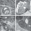
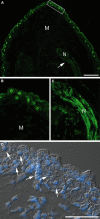
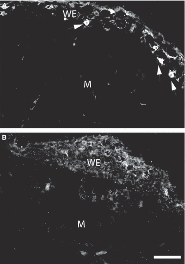
Limb regeneration in salamanders proceeds by formation of the blastema, a mound of proliferating mesenchymal cells surrounded by a wound epithelium. Regeneration by the blastema depends on the presence of regenerating nerves and in earlier work it was shown that axons upregulate the expression of newt anterior gradient (nAG) protein first in Schwann cells of the nerve sheath and second in dermal glands underlying the wound epidermis. The expression of nAG protein after plasmid electroporation was shown to rescue a denervated newt blastema and allow regeneration to the digit stage. We have examined the dermal glands by scanning and transmission electron microscopy combined with immunogold labelling of the nAG protein. It is expressed in secretory granules of ductless glands, which apparently discharge by a holocrine mechanism. No external ducts were observed in the wound epithelium of the newt and axolotl. The larval skin of the axolotl has dermal glands but these are absent under the wound epithelium. The nerve sheath was stained post-amputation in innervated but not denervated blastemas with an antibody to axolotl anterior gradient protein. This antibody reacted with axolotl Leydig cells in the wound epithelium and normal epidermis. Staining was markedly decreased in the wound epithelium after denervation but not in the epidermis. Therefore, in both newt and axolotl the regenerating axons induce nAG protein in the nerve sheath and subsequently the protein is expressed by gland cells, under (newt) or within (axolotl) the wound epithelium, which discharge by a holocrine mechanism. These findings serve to unify the nerve dependence of limb regeneration.
Figures


Similar articles
-
Neuregulin-1 signaling is essential for nerve-dependent axolotl limb regeneration.Development. 2016 Aug 1;143(15):2724-31. doi: 10.1242/dev.133363. Epub 2016 Jun 17. Development. 2016. PMID: 27317805
-
Cloning and neuronal expression of a type III newt neuregulin and rescue of denervated, nerve-dependent newt limb blastemas by rhGGF2.J Neurobiol. 2000 May;43(2):150-8. doi: 10.1002/(sici)1097-4695(200005)43:2<150::aid-neu5>3.0.co;2-g. J Neurobiol. 2000. PMID: 10770844
-
The role of peripheral nerves in urodele limb regeneration.Eur J Neurosci. 2011 Sep;34(6):908-16. doi: 10.1111/j.1460-9568.2011.07827.x. Eur J Neurosci. 2011. PMID: 21929624 Review.
-
Expression of fibroblast growth factors 4, 8, and 10 in limbs, flanks, and blastemas of Ambystoma.Dev Dyn. 2002 Mar;223(2):193-203. doi: 10.1002/dvdy.10049. Dev Dyn. 2002. PMID: 11836784
-
Putative epithelial-mesenchymal transitions during salamander limb regeneration: Current perspectives and future investigations.Ann N Y Acad Sci. 2024 Oct;1540(1):89-103. doi: 10.1111/nyas.15210. Epub 2024 Sep 13. Ann N Y Acad Sci. 2024. PMID: 39269330 Review.
Cited by
-
Now that We Got There, What Next?Methods Mol Biol. 2023;2562:471-479. doi: 10.1007/978-1-0716-2659-7_31. Methods Mol Biol. 2023. PMID: 36272095
-
The aneurogenic limb identifies developmental cell interactions underlying vertebrate limb regeneration.Proc Natl Acad Sci U S A. 2011 Aug 16;108(33):13588-93. doi: 10.1073/pnas.1108472108. Epub 2011 Aug 8. Proc Natl Acad Sci U S A. 2011. PMID: 21825124 Free PMC article.
-
Salamander-derived, human-optimized nAG protein suppresses collagen synthesis and increases collagen degradation in primary human fibroblasts.Biomed Res Int. 2013;2013:384091. doi: 10.1155/2013/384091. Epub 2013 Oct 31. Biomed Res Int. 2013. PMID: 24288677 Free PMC article.
-
Denervation affects regenerative responses in MRL/MpJ and repair in C57BL/6 ear wounds.J Anat. 2012 Jan;220(1):3-12. doi: 10.1111/j.1469-7580.2011.01452.x. Epub 2011 Nov 8. J Anat. 2012. PMID: 22066944 Free PMC article.
-
Hydrogels derived from central nervous system extracellular matrix.Biomaterials. 2013 Jan;34(4):1033-40. doi: 10.1016/j.biomaterials.2012.10.062. Epub 2012 Nov 16. Biomaterials. 2013. PMID: 23158935 Free PMC article.
References
-
- Aberger F, Weidinger G, Grunz H, et al. Anterior specification of embryonic ectoderm: the role of the Xenopus cement gland-specific gene XAG-2. Mech Dev. 1998;72:115–130. - PubMed
-
- Brockes JP. Mitogenic growth factors and nerve dependence of limb regeneration. Science. 1984;225:1280–1287. - PubMed
-
- Brockes JP. Amphibian limb regeneration: rebuilding a complex structure. Science. 1997;276:81–87. - PubMed
-
- Brockes JP, Kumar A. Comparative aspects of animal regeneration. Annu Rev Cell Dev Biol. 2008;24:525–549. - PubMed
Publication types
MeSH terms
Substances
Grants and funding
LinkOut - more resources
Full Text Sources

