. 2010 May;24(9):904-15.
doi: 10.1101/gad.1908710.

Argonaute quenching and global changes in Dicer homeostasis caused by a pathogen-encoded GW repeat protein

Affiliations

Argonaute quenching and global changes in Dicer homeostasis caused by a pathogen-encoded GW repeat protein

Jacinthe Azevedo et al. Genes Dev. 2010 May.

Erratum in

Abstract

In plants and invertebrates, viral-derived siRNAs processed by the RNaseIII Dicer guide Argonaute (AGO) proteins as part of antiviral RNA-induced silencing complexes (RISC). As a counterdefense, viruses produce suppressor proteins (VSRs) that inhibit the host silencing machinery, but their mechanisms of action and cellular targets remain largely unknown. Here, we show that the Turnip crinckle virus (TCV) capsid, the P38 protein, acts as a homodimer, or multiples thereof, to mimic host-encoded glycine/tryptophane (GW)-containing proteins normally required for RISC assembly/function in diverse organisms. The P38 GW residues bind directly and specifically to Arabidopsis AGO1, which, in addition to its role in endogenous microRNA-mediated silencing, is identified as a major effector of TCV-derived siRNAs. Point mutations in the P38 GW residues are sufficient to abolish TCV virulence, which is restored in Arabidopsis ago1 hypomorphic mutants, uncovering both physical and genetic interactions between the two proteins. We further show how AGO1 quenching by P38 profoundly impacts the cellular availability of the four Arabidopsis Dicers, uncovering an AGO1-dependent, homeostatic network that functionally connects these factors together. The likely widespread occurrence and expected consequences of GW protein mimicry on host silencing pathways are discussed in the context of innate and adaptive immunity in plants and metazoans.


Figures
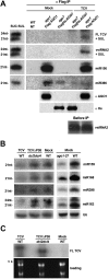
Figure 1.
Figure 1.
Preferential loading of TCV 22-nt siRNAs into AGO1 and effects of P38 on accumulation of AGO1-dependent miRNAs in infected plants. (A) Small RNA fraction associated with Flag-AGO1 and Flag-HA-AGO7 immunoprecipitates (IP anti-Flag) in mock- or TCV-inoculated crude inflorescence extracts. RNAs and proteins were extracted directly from α-Flag immunoprecipitates (αFlag IP). sRNA blots were hybridized with either DNA oligonucleotide probes complementary to the indicated miRNAs or TCV-derived siRNA2 or a full-length virus probe (FL TCV). The panel on the left contains RNA from the SUC:SUL transgenic line that expresses an RNAi hairpin targeting the SUL endogenous mRNA (Dunoyer et al. 2007). The resulting dsRNA is processed into 21-nt and 24-nt siRNAs, used here as size references. The SUL RNA signals were detected on the same blots upon stripping. miR156 and miR390 are used as qualitative controls for their established loading preferences into AGO1 and AGO7, respectively. AGO1 was detected using an anti-AGO1 antibody, and AGO7 was detected using an anti-HA antibody. (B, left panel) miRNA accumulation in inflorescences of mock-, TCVΔP38-, or TCV-inoculated plants with a dcl2dcl4 or wild-type background. (Right panel) miRNA levels in noninfected ago1-27 versus wild-type inflorescences. The U6 RNA was used as a loading control. (C) Compared levels of TCV and TCVΔP38 in wild-type or dcl2dcl4 mutant plants. The virus genomic RNA (v) can be visualized by ethidium bromide staining; rRNA serves as a loading control.
Figure 2.
Figure 2.
AGO1 is a physical target of P38. (A) Coimmunoprecipitation of P38 with AGO proteins in flower extracts from the indicated transgenic lines. Anti-Flag or anti-Myc immunoprecipitates were analyzed by immunodetection using anti-P38 serum. AGO7 and AGO4 were detected using anti-HA- and anti-Myc antibodies, respectively, and AGO1 was detected with anti-AGO1 antibodies. All immunoprecipitates were washed with high salt concentrations (see the Materials and Methods). (B) Polypeptides elution profile of AGO1 immunoprecipitates from inflorescence extracts of mock- or TCV-inoculated Flag-AGO1 plants. Flag-AGO1 immunoprecipitate (IP anti-Flag) products were released through three stepwise elutions by competition (Flag peptide), and were analyzed by either silver nitrate staining or immunodetection for AGO1 and P38. AGO1 and P38 positions in the stained gel are indicated with arrows. (C) Separation, by gel filtration, of Flag-AGO1 immunoprecipitate products (IP anti-Flag) from TCV-infected inflorescences under high stringency conditions. Fractions eluted from Superose 6 column were analyzed by either silver nitrate staining or protein immunodetection for AGO1 and P38. Void volume and Ferritine (440 kDa) are marked with arrows. The elution peak of the AGO1 complex is centered on 14 mL (asterisks).
Figure 3.
Figure 3.
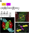
P38 VSR function likely requires C:C homodimerization. (A) Schematic of P38 structural domains with the position of the amino acids mutated in the m1 and m2 alleles impaired for VSR function (in yellow and pink, respectively). The same colors are used throughout the figure. The m1 and m2 proteins are stable, as visualized by immunodetection using total extracts from transgenic lines expressing mutant and wild-type P38. (B) Representation of a P38 hexamer made of two A–B–C trimers that interact through C:C dimerization at the twofold axis, as viewed from the inside of the capsid. (C) A close-up view of B in which the C backbone and all side chains are represented with simple lines. This allows visualization of m2 (E122) embedded within the β barrels that form the S domain, and unravels the sterical proximity of m1 and m2. The C backbone is represented by a green line, and the two amino acids of interest are represented by space-filling.
Figure 4.
Figure 4.
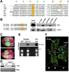
The physical interaction between P38 and AGO1 is mediated by two GW motifs that are essential for VSR activity. (A) Amino acid alignment of the P38 proteins from various Carmoviruses. The N-terminal GW residues (in red and yellow) are highly conserved in an otherwise poorly conserved domain (other conserved residues are shown in green). (B) Equimolar amounts of biotin GW peptides (P38wt) or biotin mutated peptides (P38mu) were coupled to streptavidin beads and subjected to inflorescence extracts from Myc-AGO4 or Flag-AGO1 transgenic plants. Bound proteins were detected by immunoblotting using anti-Myc or anti-AGO1 antibodies. (C) Effect of dual GW mutations on the VSR activity of P38, as assayed by cotransient expression of a reporter GFP transgene together with P38 or P38GA2 in N. benthamiana leaves. Photographs were taken 5 d post-infiltration (5 dpi) under ultraviolet light to show green fluorescence from GFP and red fluorescence from chlorophyll. (D) Accumulation of P38 and P38GA2 in the leaves depicted in C, as assayed by immunoblotting using the P38 serum. (E) RNA blot hybridizations showing accumulation levels of TCVGA2 and wild-type TCV in inoculated leaves of wild-type or ago1-27 plants. The virus genomic RNA (v) can be visualized by ethidium bromide staining; rRNA serves as a loading control. (F) GW motifs are exposed to the surface and are positioned away from the C:C dimerization interface. R, S, and P domains of P38 C/C homodimer viewed normal to the dyad. In capsid context, the center of the particle (S) would lie on the left of the model and the outside of the particle (P), at the right. The R domain and the fold-back region of the arm, where R74 lies, point to the interior of the capsid. The N-terminal and C-terminal GW motifs are indicated in white, although the former cannot be modeled in the structure presented. The other W residues that normally facilitate AGO binding are shown in light blue, alongside the m1 and m2 mutations, as depicted in Figure 3.
Figure 5.
Figure 5.
Restoration of TCVGA2 virus level in dcl2dcl4 mutants and global changes in DCL levels caused by P38 interference with AGO1. (A) RNA blot hybridizations showing accumulation of TCVGA2 and wild-type TCV in inoculated leaves (top panel) and systemic inflorescences (bottom panel) of wild-type or dcl2–dcl4 plants. The virus genomic RNA (v) can be visualized by ethidium bromide staining of rRNA. Coomassie staining was used as a protein loading control. (B) DCL1 levels were evaluated by immunodetection using an anti-DCL1 antibody (see the Materials and Methods) in total inflorescence extracts. Samples from mock-inoculated plants (wild type [WT] or dcl2–dcl4), TCVΔP38-infected dcl2–dcl4; TCVGA2-infected plants (wild type [WT] or dcl2–dcl4), and TCV-infected wild-type plants were analyzed. (C). Expression of GFP under the control of the DCL1 promoter (see the Materials and Methods) was monitored by quantitative RT–PCR analysis of three biological replicates involving three TCV infections each. Systemic leaf tissue was harvested at 21 dpi, and cDNA inputs were normalized using Actin2 abundances; expression ratios are given relative to levels in mock-inoculated plants (set to 1). Data are displayed as averages ± standard deviation (three PCR replicates). (D, left and middle panels) The tissues from wild-type Arabidopsis inflorescences used in B were employed for immunodetection of DCL4 and DCL3, respectively (see the Materials and Methods). Coomassie staining was used as a protein loading control. (Right panel) DCL2 accumulation was analyzed by semiquantitative RT–PCR on total RNA from the inflorescence tissues used in B. Ubiquitin (Ub) was used as a loading control.
Figure 6.
Figure 6.
A three-step model for the molecular effects of TCV infection on Arabidopsis RNA silencing pathways. See the text for details.

Comment in

Similar articles

Cited by

References

    1. Baulcombe D 2004. RNA silencing in plants. Nature 431: 356–363 - PubMed
    1. Baumberger N, Baulcombe DC 2005. Arabidopsis ARGONAUTE1 is an RNA Slicer that selectively recruits microRNAs and short interfering RNAs. Proc Natl Acad Sci 102: 11928–11933 - PMC - PubMed
    1. Bechtold N, Pelletier G 1998. In planta Agrobacterium-mediated transformation of adult Arabidopsis thaliana plants by vacuum infiltration. Methods Mol Biol 82: 259–266 - PubMed
    1. Blevins T, Rajeswaran R, Shivaprasad PV, Beknazariants D, Si-Ammour A, Park HS, Vazquez F, Robertson D, Meins F Jr, Hohn T, et al. 2006. Four plant Dicers mediate viral small RNA biogenesis and DNA virus induced silencing. Nucleic Acids Res 34: 6233–6246 - PMC - PubMed
    1. Cabanes D, Dehoux P, Dussurget O, Frangeul L, Cossart P 2002. Surface proteins and the pathogenic potential of Listeria monocytogenes. Trends Microbiol 10: 238–245 - PubMed

Publication types

MeSH terms