

New device for high-throughput viability screening of flow biofilms
- PMID: 20435763
- PMCID: PMC2897429
- DOI: 10.1128/AEM.03065-09
New device for high-throughput viability screening of flow biofilms
Abstract
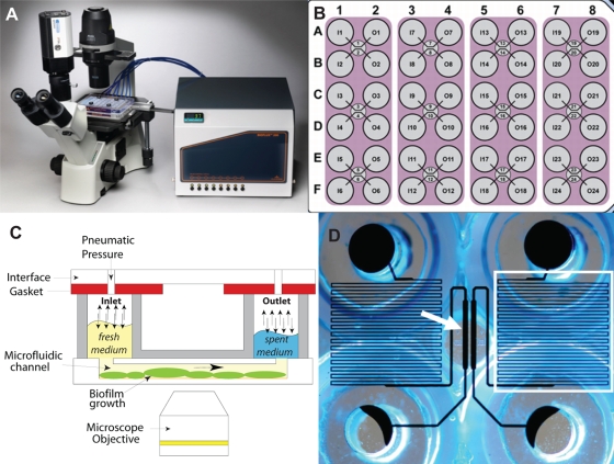
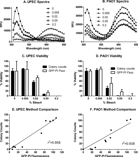
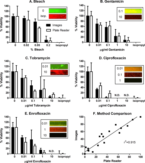
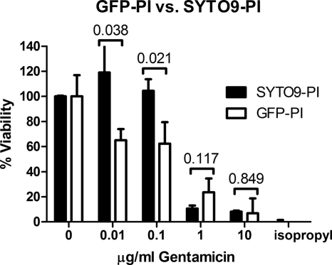
Control of biofilms requires rapid methods to identify compounds effective against them and to isolate resistance-compromised mutants for identifying genes involved in enhanced biofilm resistance. While rapid screening methods for microtiter plate well ("static") biofilms are available, there are no methods for such screening of continuous flow biofilms ("flow biofilms"). Since the latter biofilms more closely approximate natural biofilms, development of a high-throughput (HTP) method for screening them is desirable. We describe here a new method using a device comprised of microfluidic channels and a distributed pneumatic pump (BioFlux) that provides fluid flow to 96 individual biofilms. This device allows fine control of continuous or intermittent fluid flow over a broad range of flow rates, and the use of a standard well plate format provides compatibility with plate readers. We show that use of green fluorescent protein (GFP)-expressing bacteria, staining with propidium iodide, and measurement of fluorescence with a plate reader permit rapid and accurate determination of biofilm viability. The biofilm viability measured with the plate reader agreed with that determined using plate counts, as well as with the results of fluorescence microscope image analysis. Using BioFlux and the plate reader, we were able to rapidly screen the effects of several antimicrobials on the viability of Pseudomonas aeruginosa PAO1 flow biofilms.
Figures
Similar articles
-
Optimization of a High-Throughput 384-Well Plate-Based Screening Platform with Staphylococcus aureus ATCC 25923 and Pseudomonas aeruginosa ATCC 15442 Biofilms.Int J Mol Sci. 2020 Apr 25;21(9):3034. doi: 10.3390/ijms21093034. Int J Mol Sci. 2020. PMID: 32344836 Free PMC article.
-
Image-based 384-well high-throughput screening method for the discovery of skyllamycins A to C as biofilm inhibitors and inducers of biofilm detachment in Pseudomonas aeruginosa.Antimicrob Agents Chemother. 2014;58(2):1092-9. doi: 10.1128/AAC.01781-13. Epub 2013 Dec 2. Antimicrob Agents Chemother. 2014. PMID: 24295976 Free PMC article.
-
Real-Time Monitoring of nfxB Mutant Occurrence and Dynamics in Pseudomonas aeruginosa Biofilm Exposed to Subinhibitory Concentrations of Ciprofloxacin.Antimicrob Agents Chemother. 2017 Feb 23;61(3):e02292-16. doi: 10.1128/AAC.02292-16. Print 2017 Mar. Antimicrob Agents Chemother. 2017. PMID: 27993856 Free PMC article.
-
Anti-biofilm and resistance suppression activities of CXA-101 against chronic respiratory infection phenotypes of Pseudomonas aeruginosa strain PAO1.J Antimicrob Chemother. 2010 Jul;65(7):1399-404. doi: 10.1093/jac/dkq143. Epub 2010 Apr 30. J Antimicrob Chemother. 2010. PMID: 20435779
-
[Biofilms and Oxidizing Biocides; Evaluation of Disinfection and Removal Effects by Using Established Microbial Systems].Yakugaku Zasshi. 2017;137(6):707-717. doi: 10.1248/yakushi.16-00254. Yakugaku Zasshi. 2017. PMID: 28566577 Review. Japanese.
Cited by
-
Time-resolved, single-cell analysis of induced and programmed cell death via non-invasive propidium iodide and counterstain perfusion.Sci Rep. 2016 Sep 1;6:32104. doi: 10.1038/srep32104. Sci Rep. 2016. PMID: 27580964 Free PMC article.
-
Biofilm Formation As a Response to Ecological Competition.PLoS Biol. 2015 Jul 9;13(7):e1002191. doi: 10.1371/journal.pbio.1002191. eCollection 2015 Jul. PLoS Biol. 2015. PMID: 26158271 Free PMC article.
-
Prevention of Initial Bacterial Attachment by Osteopontin and Other Bioactive Milk Proteins.Biomedicines. 2022 Aug 9;10(8):1922. doi: 10.3390/biomedicines10081922. Biomedicines. 2022. PMID: 36009469 Free PMC article.
-
Life under flow: A novel microfluidic device for the assessment of anti-biofilm technologies.Biomicrofluidics. 2013 Dec 23;7(6):64118. doi: 10.1063/1.4850796. eCollection 2013 Dec 23. Biomicrofluidics. 2013. PMID: 24454610 Free PMC article.
-
Machine-assisted cultivation and analysis of biofilms.Sci Rep. 2019 Jun 20;9(1):8933. doi: 10.1038/s41598-019-45414-6. Sci Rep. 2019. PMID: 31222095 Free PMC article.
References
-
- Anderson, G. G., J. J. Palermo, J. D. Schilling, R. Roth, J. Heuser, and S. J. Hultgren. 2003. Intracellular bacterial biofilm-like pods in urinary tract infections. Science 301:105-107. - PubMed
-
- Beech, I. B., and J. Sunner. 2004. Biocorrosion: towards understanding interactions between biofilms and metals. Curr. Opin. Biotechnol. 15:181-186. - PubMed
-
- Burton, E., N. Yakandawala, K. LoVetri, and M. S. Madhyastha. 2007. A microplate spectrofluorometric assay for bacterial biofilms. J. Ind. Microbiol. Biotechnol. 34:1-4. - PubMed
Publication types
MeSH terms
Substances
Grants and funding
LinkOut - more resources
Full Text Sources
Other Literature Sources
Medical
