Review
. 2010 Jun;31(6):284-95.
doi: 10.1016/j.tips.2010.03.001.

Retinoids for treatment of retinal diseases

Affiliations
Review

Retinoids for treatment of retinal diseases

Krzysztof Palczewski. Trends Pharmacol Sci. 2010 Jun.

Abstract

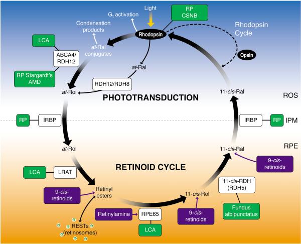
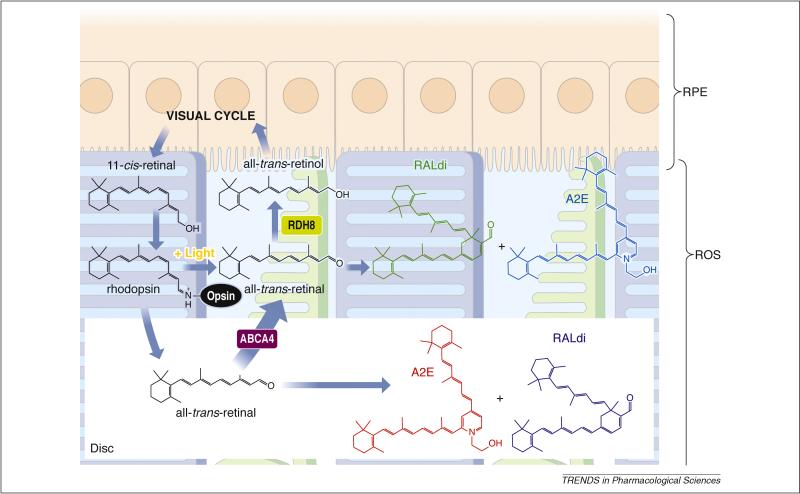
Knowledge about retinal photoreceptor signal transduction and the visual cycle required for normal eyesight has increased exponentially over the past decade. Substantial progress in human genetics has facilitated the identification of candidate genes and complex networks underlying inherited retinal diseases. Natural mutations in animal models that mimic human diseases have been characterized and advanced genetic manipulation can now be used to generate small mammalian models of human retinal diseases. Pharmacological repair of defective visual processes in animal models not only validates their involvement in vision, but also provides great promise for the development of improved therapies for millions who are progressing towards blindness or are almost completely robbed of their eyesight.


Figures

Figure 1
Figure 1. Phototransduction and the visual (retinoid) cycle
In vertebrates vision is triggered by light-dependent activation of rhodopsin or other visual pigments. In rod cells, this chromophore couples to a protein opsin, forming rhodopsin. Absorption of a photon of light by rhodopsin causes photoisomerization of 11-cis-retinal to all-trans-retinal. In turn, photoactivated rhodopsin generates activation of hundreds of heterotrimeric G proteins, called transducin or Gt, in photoreceptors. This G protein-coupled receptor cascade is a classic cyclic nucleotide pathway that results in lowering cGMP levels (not depicted on the figure), and consequently hyperpolarization of the plasma membranes and ultimately reduction of glutamate secretion to the secondary neurons. The visual cycle regenerates 11-cis-retinal from released all-trans-retinal from the chromophore binding pocket of opsin. All-trans-retinal is reduced to all-trans-retinol in a reversible reaction catalyzed by RDH12 and RDH8, which are NADPH-dependent all-trans-retinol dehydrogenases. All-trans-retinol diffuses into the RPE where it is esterified in a reaction catalyzed by lecithin:retinol acyl transferase to long-chain fatty acids. As a consequence of the propensity of retinyl esters to aggregate, these esters are stored in lipid droplet-like structures called retinosomes. The all-trans-retinyl esters appear to be the substrate for RPE65 that converts it to 11-cis-retinol, which then is further oxidized back to 11-cis-retinal by retinol dehydrogenases, RDH5, RDH11, and other NAD-dependent retinol dehydrogenases. 11-cis-retinal formed in the RPE diffuses back into the ROS and COS, where it completes the cycle by recombining with opsins to form rhodopsin and cone pigments. Mutations in genes encoding proteins of phototransduction and the retinoid cycle are associated with various retinal diseases, some of which are indicated by the green boxes. Pharmacological intervention has been successful in animal models in a few instances, as indicated by the compounds in the blue boxes. IPM, interphotoreceptor matrix; IRBP, interphotoreceptor retinoid-binding protein; CSNB, congenital stationary nightblindness, ROS, rod outer segments; RPE, retinal pigment epithelium; Ral, retinal, RDH, retinol dehydrogenase; Rol, retinol; LCA, Leber congenital amaurosis; RP, retinitis pigmentosa, ABCR, ATP-binding transporter 4 .
Figure 2
Figure 2. Retinoid flow in the visual cycle and condensation of all-trans-retinal
After 11-cis-retinal binds to opsin, forming rhodopsin, the resulting visual chromophore, 11-cis-retinylidene, is photoisomerized to all-trans-retinylidene, the precursor of all-trans-retinal that is subsequently released. Most of the all-trans-retinal dissociates from opsin into the cytoplasm where it is reduced to all-trans-retinol by RDHs including RDH8. The fraction of all-trans-retinal that dissociates into the disc lumens is transported by ABCA4 back into the cytoplasm before it is reduced. Thus, condensation products can be generated both within the disc lumens and the cytoplasm. Loss of ABCA4 and RDH8 exacerbates this condensation reminiscent of an accelerated aging process. In humans, as a result of daily phagocytosis of the part of rod outer segments, lipofuscin fluorophores accumulate with age in the RPE, especially in RPE cells underlying the cone-rich macula , . Such accumulation has been considered to constitute one of the major risk factors for AMD, the predominant cause of legal blindness in developed countries . Lipofuscin fluorophores also are especially abundant in Stargardt disease, the most common juvenile form of macular degeneration . Di-retinoid-pyridinium-ethanolamine (A2E) and all-trans-retinal dimer (RALdi), the major fluorophores of lipofuscin, is formed by condensation of phosphatidylethanolamine with two molecules of all-trans-retinal followed by oxidation and hydrolysis of the phosphate ester . Various mechanisms have been proposed to explain the toxicity of A2E. These include its cationic detergent properties , physiological interference with RPE function , , and radical reaction products induced by light-dependent oxidation .
Figure 3
Figure 3. Transformations of visual cycle retinoids in the RPE
All-trans-retinol diffuses from photoreceptor cells into the RPE, where it is esterified by LRAT to all-trans-retinyl esters. Hydrophobic retinyl esters then form retinosomes (RESTs). All-trans-retinyl esters are isomerized to 11-cis-retinol (reaction a) in a reaction that involves an RPE-abundant protein, termed RPE65. 11-cis-Retinol is then oxidized by 11-cis-RDH to 11-cis-retinal (reaction b). 11-cis-Retinal diffuses back into the rod and cone outer segments, where it completes the retinoid cycle by recombining with opsins to reform rhodopsin and cone pigments. A. Retinosomes imaged in RPE cells by two-photon microscopy (courtesy of Grazyna Palczewska, Polgenix, Inc., Cleveland). Fluorescence emission from the isolated intact mouse eye at 560–700 nm in green pseudocolor was observed after excitation by a 730-nm mode-locked Ti:Sapphire laser. Scale bar 5 μm. B. Flash-dependent changes in fluorescence and all-trans-retinol/all-trans-retinyl esters in the RPE cell layer of isolated mouse eyes. Top: A row of images showing optical sections of the retina, perpendicular to the ocular tissue. RPE fluorescence (a.u., arbitrary unit) was quantified as a function of time. Numbers refer to minutes after the flash. Middle and bottom graphs show quantified fluorescence from retinoids and retinoid analyses by HPLC (all-trans-retinol and all-trans-retinyl esters; mean ± SD, n = 3), respectively. Dashed lines indicate half-times for formation of RESTs and the increase in all-trans-retinol and all-trans-retinyl esters. On the right, light-dependent changes in the fluorescent signal in different subcellular compartments are shown. (copied from ref. with permission from the Rockefeller University Press).
Figure 4
Figure 4. Delivery and action of 9-cis-R-Ac
This retinyl ester is effective when taken orally. In the small intestine, 9-cis-R-Ac is either hydrolyzed and esterified with fatty acyl coenzyme A, or trans-esterified with phospholipids before being transported in chylomicrons to the liver, where these intermediate products are found in lipid droplets. This cycle of hydrolysis and esterification may occur several times before storage in hepatic stellate cells. Fatty acid (mostly palmitate) esters and free 9-cis-retinol are then secreted into the systemic circulation, either bound to RBP or albumin or incorporated into chylomicrons. In the eye, these retinoids are likely hydrolyzed again as they pass from the choroid capillaries into the RPE in both a STRA6-dependent and independent manner. Fatty acid esters of 9-cis-retinol in the RPE are stored in specific lipid droplets called retinosomes. When required, these esters are hydrolyzed and oxidized to the drug, 9-cis-retinal, which is then delivered to opsins in photoreceptors to form light-sensitive visual pigments.
Figure 5
Figure 5. Delivery and action of retinylamide
This amide ester can be taken orally. Once in the intestine it is hydrolyzed and re-amidated with fatty acid coenzyme A, and transported in chylomicrons to the liver, where it is stored as lipid droplets in hepatic stellate cells. This cycle of hydrolysis and re-amidation can occur several times. Fatty acid (mostly palmitate) amide and free retinylamine are then secreted into the systemic circulation, either bound to RBP or albumin, or incorporated into chylomicrons. The amides likely are hydrolyzed again as they pass from the choroid capillaries into the RPE in a STRA6-independent manner. There, retinylamine is stored in specific lipid droplets called retinosomes. When required, retinylamine is released and acts as a very potent transition state inhibitor of RPE65, which catalyses the hydrolytic isomerization of retinyl esters. Suppression of this isomerization can last for weeks because of long-term storage. Retinylamine also conjugates with free retinal, preventing accumulation of other toxic retinal condensation products. Eventually retinylamine is metabolized to retinol.

Similar articles

Cited by

References

    1. Thompson DA, Gal A. Vitamin A metabolism in the retinal pigment epithelium: genes, mutations, and diseases. Progress in retinal and eye research. 2003;22:683–703. - PubMed
    1. Travis GH, et al. Diseases caused by defects in the visual cycle: retinoids as potential therapeutic agents. Annu Rev Pharmacol Toxicol. 2007;47:469–512. - PMC - PubMed
    1. Maeda T, et al. Effects of long-term administration of 9-cis-retinyl acetate on visual function in mice. Investigative ophthalmology & visual science. 2009;50:322–333. - PMC - PubMed
    1. Jackson GR, et al. Impact of aging and age-related maculopathy on inactivation of the a-wave of the rod-mediated electroretinogram. Vision research. 2006;46:1422–1431. - PubMed
    1. Yau KW, Hardie RC. Phototransduction motifs and variations. Cell. 2009;139:246–264. - PMC - PubMed

Publication types

MeSH terms